+データを開く
-基本情報
登録情報 | データベース: EMDB / ID: EMD-8163 | |||||||||
---|---|---|---|---|---|---|---|---|---|---|
タイトル | Structure of the F-actin-tropomyosin complex (reprocessed) | |||||||||
マップデータ | None | |||||||||
試料 |
| |||||||||
機能・相同性 | 機能・相同性情報 cytoskeletal motor activator activity / tropomyosin binding / mesenchyme migration / troponin I binding / myosin heavy chain binding / filamentous actin / actin filament bundle / skeletal muscle thin filament assembly / striated muscle thin filament / actin filament bundle assembly ...cytoskeletal motor activator activity / tropomyosin binding / mesenchyme migration / troponin I binding / myosin heavy chain binding / filamentous actin / actin filament bundle / skeletal muscle thin filament assembly / striated muscle thin filament / actin filament bundle assembly / skeletal muscle myofibril / actin monomer binding / skeletal muscle fiber development / stress fiber / titin binding / actin filament polymerization / filopodium / actin filament / 加水分解酵素; 酸無水物に作用; 酸無水物に作用・細胞または細胞小器官の運動に関与 / calcium-dependent protein binding / lamellipodium / cell body / hydrolase activity / protein domain specific binding / calcium ion binding / positive regulation of gene expression / magnesium ion binding / ATP binding / identical protein binding / cytoplasm 類似検索 - 分子機能 | |||||||||
生物種 | Rabbit (ウサギ) / Mus musculus (ハツカネズミ) | |||||||||
手法 | 単粒子再構成法 / クライオ電子顕微鏡法 / 解像度: 3.6 Å | |||||||||
データ登録者 | von der Ecken J / Raunser S | |||||||||
資金援助 | ドイツ, 2件
| |||||||||
引用 | 
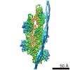
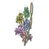
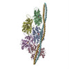
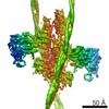
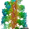
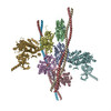
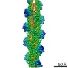
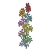
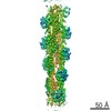
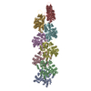
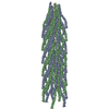
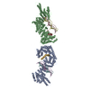
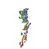
ジャーナル: Nature / 年: 2015 タイトル: Structure of the F-actin-tropomyosin complex. 著者: Julian von der Ecken / Mirco Müller / William Lehman / Dietmar J Manstein / Pawel A Penczek / Stefan Raunser / 要旨: Filamentous actin (F-actin) is the major protein of muscle thin filaments, and actin microfilaments are the main component of the eukaryotic cytoskeleton. Mutations in different actin isoforms lead ...Filamentous actin (F-actin) is the major protein of muscle thin filaments, and actin microfilaments are the main component of the eukaryotic cytoskeleton. Mutations in different actin isoforms lead to early-onset autosomal dominant non-syndromic hearing loss, familial thoracic aortic aneurysms and dissections, and multiple variations of myopathies. In striated muscle fibres, the binding of myosin motors to actin filaments is mainly regulated by tropomyosin and troponin. Tropomyosin also binds to F-actin in smooth muscle and in non-muscle cells and stabilizes and regulates the filaments there in the absence of troponin. Although crystal structures for monomeric actin (G-actin) are available, a high-resolution structure of F-actin is still missing, hampering our understanding of how disease-causing mutations affect the function of thin muscle filaments and microfilaments. Here we report the three-dimensional structure of F-actin at a resolution of 3.7 Å in complex with tropomyosin at a resolution of 6.5 Å, determined by electron cryomicroscopy. The structure reveals that the D-loop is ordered and acts as a central region for hydrophobic and electrostatic interactions that stabilize the F-actin filament. We clearly identify map density corresponding to ADP and Mg(2+) and explain the possible effect of prominent disease-causing mutants. A comparison of F-actin with G-actin reveals the conformational changes during filament formation and identifies the D-loop as their key mediator. We also confirm that negatively charged tropomyosin interacts with a positively charged groove on F-actin. Comparison of the position of tropomyosin in F-actin-tropomyosin with its position in our previously determined F-actin-tropomyosin-myosin structure reveals a myosin-induced transition of tropomyosin. Our results allow us to understand the role of individual mutations in the genesis of actin- and tropomyosin-related diseases and will serve as a strong foundation for the targeted development of drugs. | |||||||||
履歴 |
|
-構造の表示
ムービー |
ムービービューア |
---|---|
構造ビューア | EMマップ: SurfViewMolmilJmol/JSmol |
添付画像 |
-ダウンロードとリンク
-EMDBアーカイブ
マップデータ | emd_8163.map.gz | 45.8 MB | EMDBマップデータ形式 | |
---|---|---|---|---|
ヘッダ (付随情報) | emd-8163-v30.xml emd-8163.xml | 18.1 KB 18.1 KB | 表示 表示 | EMDBヘッダ |
画像 | emd_8163.png | 134.7 KB | ||
アーカイブディレクトリ | http://ftp.pdbj.org/pub/emdb/structures/EMD-8163
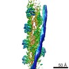
 ftp://ftp.pdbj.org/pub/emdb/structures/EMD-8163 | HTTPS FTP |
-検証レポート
文書・要旨 | emd_8163_validation.pdf.gz | 273.8 KB | 表示 | EMDB検証レポート |
---|---|---|---|---|
文書・詳細版 | emd_8163_full_validation.pdf.gz | 273 KB | 表示 | |
XML形式データ | emd_8163_validation.xml.gz | 6.1 KB | 表示 | |
アーカイブディレクトリ | https://ftp.pdbj.org/pub/emdb/validation_reports/EMD-8163 ftp://ftp.pdbj.org/pub/emdb/validation_reports/EMD-8163 | HTTPS FTP |
-関連構造データ
-リンク
EMDBのページ | EMDB (EBI/PDBe) / EMDataResource |
---|---|
「今月の分子」の関連する項目 |
-マップ
ファイル | ダウンロード / ファイル: emd_8163.map.gz / 形式: CCP4 / 大きさ: 64 MB / タイプ: IMAGE STORED AS FLOATING POINT NUMBER (4 BYTES) | ||||||||||||||||||||||||||||||||||||||||||||||||||||||||||||||||||||
---|---|---|---|---|---|---|---|---|---|---|---|---|---|---|---|---|---|---|---|---|---|---|---|---|---|---|---|---|---|---|---|---|---|---|---|---|---|---|---|---|---|---|---|---|---|---|---|---|---|---|---|---|---|---|---|---|---|---|---|---|---|---|---|---|---|---|---|---|---|
注釈 | None | ||||||||||||||||||||||||||||||||||||||||||||||||||||||||||||||||||||
ボクセルのサイズ | X=Y=Z: 1.1 Å | ||||||||||||||||||||||||||||||||||||||||||||||||||||||||||||||||||||
密度 |
| ||||||||||||||||||||||||||||||||||||||||||||||||||||||||||||||||||||
対称性 | 空間群: 1 | ||||||||||||||||||||||||||||||||||||||||||||||||||||||||||||||||||||
詳細 | EMDB XML:
CCP4マップ ヘッダ情報:
|
-添付データ
-試料の構成要素
-全体 : F-actin-tropomyosin complex
全体 | 名称: F-actin-tropomyosin complex |
---|---|
要素 |
|
-超分子 #1: F-actin-tropomyosin complex
超分子 | 名称: F-actin-tropomyosin complex / タイプ: complex / ID: 1 / 親要素: 0 / 含まれる分子: all / 詳細: Filament |
---|
-分子 #1: ACTIN, ALPHA SKELETAL MUSCLE
分子 | 名称: ACTIN, ALPHA SKELETAL MUSCLE / タイプ: protein_or_peptide / ID: 1 / 光学異性体: LEVO |
---|---|
由来(天然) | 生物種: Rabbit (ウサギ) |
配列 | 文字列: DEDETTALVC DNGSGLVKAG FAGDDAPRAV FPSIVGRPRH QGVMVGMGQK DSYVGDEAQS KRGILTLKYP IE(HIC)GII TNW DDMEKIWHHT FYNELRVAPE EHPTLLTEAP LNPKANREKM TQIMFETFNV PAMYVAIQAV LSLYASGRTT GIVLDSG DG ...文字列: DEDETTALVC DNGSGLVKAG FAGDDAPRAV FPSIVGRPRH QGVMVGMGQK DSYVGDEAQS KRGILTLKYP IE(HIC)GII TNW DDMEKIWHHT FYNELRVAPE EHPTLLTEAP LNPKANREKM TQIMFETFNV PAMYVAIQAV LSLYASGRTT GIVLDSG DG VTHNVPIYEG YALPHAIMRL DLAGRDLTDY LMKILTERGY SFVTTAEREI VRDIKEKLCY VALDFENEMA TAASSSSL E KSYELPDGQV ITIGNERFRC PETLFQPSFI GMESAGIHET TYNSIMKCDI DIRKDLYANN VMSGGTTMYP GIADRMQKE ITALAPSTMK IKIIAPPERK YSVWIGGSIL ASLSTFQQMW ITKQEYDEAG PSIVHRKCF |
-分子 #2: TROPOMYOSIN ALPHA-1
分子 | 名称: TROPOMYOSIN ALPHA-1 / タイプ: protein_or_peptide / ID: 2 / 光学異性体: LEVO |
---|---|
由来(天然) | 生物種: Mus musculus (ハツカネズミ) |
組換発現 | 生物種: Escherichia coli (大腸菌) |
配列 | 文字列: (UNK)(UNK)(UNK)(UNK)(UNK)(UNK)(UNK)(UNK)(UNK)(UNK) (UNK)(UNK)(UNK)(UNK)(UNK)(UNK) (UNK)(UNK)(UNK) (UNK)(UNK)(UNK)(UNK)(UNK)(UNK)(UNK)(UNK)(UNK)(UNK) (UNK)(UNK)(UNK) (UNK)(UNK)(UNK)(UNK) ...文字列: (UNK)(UNK)(UNK)(UNK)(UNK)(UNK)(UNK)(UNK)(UNK)(UNK) (UNK)(UNK)(UNK)(UNK)(UNK)(UNK) (UNK)(UNK)(UNK) (UNK)(UNK)(UNK)(UNK)(UNK)(UNK)(UNK)(UNK)(UNK)(UNK) (UNK)(UNK)(UNK) (UNK)(UNK)(UNK)(UNK)(UNK)(UNK) (UNK)(UNK)(UNK)(UNK)(UNK)(UNK)(UNK)(UNK)(UNK)(UNK) (UNK)(UNK)(UNK)(UNK)(UNK)(UNK)(UNK)(UNK)(UNK) (UNK)(UNK)(UNK)(UNK)(UNK)(UNK)(UNK) (UNK)(UNK) (UNK)(UNK)(UNK)(UNK)(UNK)(UNK)(UNK)(UNK)(UNK)(UNK) (UNK)(UNK)(UNK)(UNK) (UNK)(UNK)(UNK)(UNK)(UNK) (UNK)(UNK)(UNK)(UNK)(UNK)(UNK)(UNK)(UNK)(UNK)(UNK) (UNK) (UNK)(UNK)(UNK)(UNK)(UNK)(UNK)(UNK)(UNK) (UNK)(UNK)(UNK)(UNK)(UNK)(UNK)(UNK)(UNK) (UNK) (UNK)(UNK)(UNK)(UNK)(UNK)(UNK)(UNK)(UNK)(UNK)(UNK) (UNK)(UNK)(UNK)(UNK)(UNK) (UNK)(UNK)(UNK)(UNK) (UNK)(UNK)(UNK) |
-実験情報
-構造解析
手法 | クライオ電子顕微鏡法 |
---|---|
解析 | 単粒子再構成法 |
試料の集合状態 | filament |
-試料調製
緩衝液 | pH: 7.5 構成要素:
| ||||||||||
---|---|---|---|---|---|---|---|---|---|---|---|
糖包埋 | 材質: I | ||||||||||
グリッド | モデル: C-flat-2/1 / 材質: COPPER / メッシュ: 300 / 支持フィルム - 材質: CARBON / 支持フィルム - トポロジー: HOLEY / 前処理 - タイプ: GLOW DISCHARGE | ||||||||||
凍結 | 凍結剤: ETHANE / チャンバー内湿度: 90 % / 装置: GATAN CRYOPLUNGE 3 詳細: Sample was applied to a glow-discharged holey carbon grid, incubated for 10 s and manually blotted for 3 s from the backside with filter paper.. |
-電子顕微鏡法
顕微鏡 | FEI TITAN KRIOS |
---|---|
詳細 | Cs corrected microscope |
撮影 | フィルム・検出器のモデル: FEI FALCON II (4k x 4k) 検出モード: COUNTING / デジタル化 - 画像ごとのフレーム数: 2-8 / 平均露光時間: 0.475 sec. / 平均電子線量: 16.0 e/Å2 |
電子線 | 加速電圧: 300 kV / 電子線源: FIELD EMISSION GUN |
電子光学系 | 最大 デフォーカス(補正後): 2.6 µm / 最小 デフォーカス(補正後): 0.8 µm / 照射モード: OTHER / 撮影モード: BRIGHT FIELD / 倍率(公称値): 59000 |
実験機器 | モデル: Titan Krios / 画像提供: FEI Company |
+画像解析
-原子モデル構築 1
初期モデル | PDB ID: Chain - Chain ID -: Chain - Chain ID - 1: B / Chain - Chain ID - 2: C / Chain - Chain ID - 3: D / Chain - Chain ID - 4: E |
---|---|
精密化 | 空間: RECIPROCAL / プロトコル: BACKBONE TRACE / 温度因子: 98 |
得られたモデル | PDB-5jlf: |