 Movie
Movie Controller

[English] 日本語
 Yorodumi
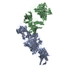
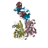
- PDB-6h8q: Structural basis for Scc3-dependent cohesin recruitment to chromatin -
+ Open data

- Basic information
| Entry | Database: PDB / ID: 6h8q | ||||||
|---|---|---|---|---|---|---|---|
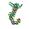
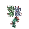
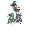
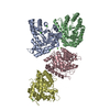
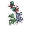
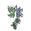
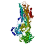
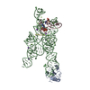
| Title | Structural basis for Scc3-dependent cohesin recruitment to chromatin | ||||||
 Components Components |
| ||||||
 Keywords Keywords | CELL CYCLE / Cohesin cell proliferation Scc3 DNA binding | ||||||
| Function / homology |  Function and homology information Function and homology informationEstablishment of Sister Chromatid Cohesion / Resolution of Sister Chromatid Cohesion / mitotic cohesin complex / cohesin complex / establishment of meiotic sister chromatid cohesion / replication-born double-strand break repair via sister chromatid exchange / SUMOylation of DNA damage response and repair proteins / establishment of mitotic sister chromatid cohesion / mitotic chromosome condensation / mitotic sister chromatid cohesion ...Establishment of Sister Chromatid Cohesion / Resolution of Sister Chromatid Cohesion / mitotic cohesin complex / cohesin complex / establishment of meiotic sister chromatid cohesion / replication-born double-strand break repair via sister chromatid exchange / SUMOylation of DNA damage response and repair proteins / establishment of mitotic sister chromatid cohesion / mitotic chromosome condensation / mitotic sister chromatid cohesion / protein acetylation / sister chromatid cohesion / chromosome, centromeric region / condensed nuclear chromosome / chromosome segregation / G2/M transition of mitotic cell cycle / double-strand break repair / cell division / chromatin binding / apoptotic process / DNA damage response / protein kinase binding / chromatin / mitochondrion / nucleus / cytosol / cytoplasm Similarity search - Function | ||||||
| Biological species |   Homo sapiens (human) Homo sapiens (human) | ||||||
| Method |  X-RAY DIFFRACTION / SYNCHROTRON / MOLECULAR REPLACEMENT / Resolution: 3.631 Å X-RAY DIFFRACTION / SYNCHROTRON / MOLECULAR REPLACEMENT / Resolution: 3.631 Å | ||||||
 Authors Authors | Li, Y. / Muir, K. / Panne, D. | ||||||
 Citation Citation | Journal: Elife / Year: 2018 Title: Structural basis for Scc3-dependent cohesin recruitment to chromatin. Authors: Li, Y. / Muir, K. / Bowler, M.W. / Metz, J. / Haering, C.H. / Panne, D. | ||||||
| History |
|
- Structure visualization
| Structure viewer | Molecule: MolmilJmol/JSmol |
|---|
- Downloads & links
-Download
| PDBx/mmCIF format | 6h8q.cif.gz | 428.8 KB | Display | PDBx/mmCIF format |
|---|---|---|---|---|
| PDB format | pdb6h8q.ent.gz | 334.8 KB | Display | PDB format |
| PDBx/mmJSON format | 6h8q.json.gz | Tree view | PDBx/mmJSON format | |
| Others |  Other downloads Other downloads |
-Validation report
| Arichive directory | https://data.pdbj.org/pub/pdb/validation_reports/h8/6h8qftp://data.pdbj.org/pub/pdb/validation_reports/h8/6h8q | HTTPS FTP |
|---|
-Related structure data


-Links
 PDBj
PDBj


- Assembly
| Deposited unit | 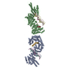
| ||||||||
|---|---|---|---|---|---|---|---|---|---|
| 1 | 
| ||||||||
| 2 | 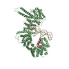
| ||||||||
| Unit cell |
|
-Components
-Protein , 2 types, 4 molecules ABGH
| #1: Protein | Mass: 133172.359 Da / Num. of mol.: 2 Source method: isolated from a genetically manipulated source Source: (gene. exp.) Strain: ATCC 204508 / S288c / Gene: IRR1, SCC3, YIL026C / Production host:  #2: Protein | Mass: 11583.825 Da / Num. of mol.: 2 Source method: isolated from a genetically manipulated source Source: (gene. exp.) Strain: ATCC 204508 / S288c / Gene: MCD1, PDS3, RHC21, SCC1, YDL003W, YD8119.04 / Production host: |
|---|
-DNA (5'-D(P*CP*TP*TP*TP*CP*GP*TP*TP*TP*CP*CP*TP*TP*GP*AP*AP*AP*AP*A)- ... , 3 types, 3 molecules EFD
| #3: DNA chain | Mass: 5861.837 Da / Num. of mol.: 1 / Source method: obtained synthetically / Source: (synth.) Homo sapiens (human) |
|---|---|
| #4: DNA chain | Mass: 5785.758 Da / Num. of mol.: 1 / Source method: obtained synthetically / Source: (synth.) Homo sapiens (human) |
| #6: DNA chain | Mass: 5769.759 Da / Num. of mol.: 1 / Source method: obtained synthetically / Source: (synth.) Homo sapiens (human) |
-DNA chain , 1 types, 1 molecules C
| #5: DNA chain | Mass: 5876.848 Da / Num. of mol.: 1 / Source method: obtained synthetically / Source: (synth.) Homo sapiens (human) |
|---|
-Experimental details
-Experiment
| Experiment | Method: X-RAY DIFFRACTION / Number of used crystals: 1 |
|---|
- Sample preparation
| Crystal | Density Matthews: 2.63 Å3/Da / Density % sol: 53.25 % |
|---|---|
| Crystal grow | Temperature: 277 K / Method: vapor diffusion / Details: 10% PEG 8000, 0.1M Bis-TRIS, pH 6.5 |
-Data collection
| Diffraction | Mean temperature: 100 K |
|---|---|
| Diffraction source | Source: SYNCHROTRON / Site: ESRF  / Beamline: MASSIF-1 / Wavelength: 0.966 Å / Beamline: MASSIF-1 / Wavelength: 0.966 Å |
| Detector | Type: DECTRIS PILATUS3 2M / Detector: PIXEL / Date: Mar 21, 2017 |
| Radiation | Protocol: SINGLE WAVELENGTH / Monochromatic (M) / Laue (L): M / Scattering type: x-ray |
| Radiation wavelength | Wavelength: 0.966 Å / Relative weight: 1 |
| Reflection | Resolution: 3.6→50 Å / Num. obs: 20963 / % possible obs: 91.4 % / Redundancy: 4.4 % / Rmerge(I) obs: 0.058 / Net I/σ(I): 11.9 |
| Reflection shell | Resolution: 3.6→3.8 Å |
- Processing
| Software |
| ||||||||||||||||||||||||
|---|---|---|---|---|---|---|---|---|---|---|---|---|---|---|---|---|---|---|---|---|---|---|---|---|---|
| Refinement | Method to determine structure: MOLECULAR REPLACEMENT Starting model: 4UVK, 4UVL Resolution: 3.631→50 Å / SU ML: 0.73 / Cross valid method: FREE R-VALUE / σ(F): 1.36 / Phase error: 50.54 / Stereochemistry target values: ML
| ||||||||||||||||||||||||
| Solvent computation | Shrinkage radii: 0.9 Å / VDW probe radii: 1.11 Å / Solvent model: FLAT BULK SOLVENT MODEL | ||||||||||||||||||||||||
| Refinement step | Cycle: LAST / Resolution: 3.631→50 Å
| ||||||||||||||||||||||||
| Refine LS restraints |
|
