 Movie
Movie Controller


+ Open data

- Basic information
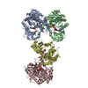
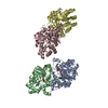
| Entry | Database: PDB / ID: 4uvk | ||||||
|---|---|---|---|---|---|---|---|
| Title | Cohesin subunit Scc3 from Z. rouxii, 88-1035 | ||||||
 Components Components | ZYRO0D15994P | ||||||
 Keywords Keywords | CELL CYCLE / COHESIN / MITOSIS / HEAT REPEATS / SMC PROTEINS | ||||||
| Function / homology |  Function and homology information Function and homology informationcohesin complex / sister chromatid cohesion / chromatin binding / chromatin / nucleus Similarity search - Function | ||||||
| Biological species |  ZYGOSACCHAROMYCES ROUXII (yeast) ZYGOSACCHAROMYCES ROUXII (yeast) | ||||||
| Method |  X-RAY DIFFRACTION / SYNCHROTRON / SAD / Resolution: 2.6 Å X-RAY DIFFRACTION / SYNCHROTRON / SAD / Resolution: 2.6 Å | ||||||
 Authors Authors | Roig, M.B. / Nasmyth, K. / Lowe, J. | ||||||
 Citation Citation | Journal: FEBS Lett. / Year: 2014 Title: Structure and Function of Cohesins Scc3/Sa Regulatory Subunit Authors: Roig, M.B. / Lowe, J. / Chan, K.L. / Beckouet, F. / Metson, J. / Nasmyth, K. | ||||||
| History |
|
- Structure visualization
| Structure viewer | Molecule: MolmilJmol/JSmol |
|---|
- Downloads & links
-Download
| PDBx/mmCIF format | 4uvk.cif.gz | 193.7 KB | Display | PDBx/mmCIF format |
|---|---|---|---|---|
| PDB format | pdb4uvk.ent.gz | 152 KB | Display | PDB format |
| PDBx/mmJSON format | 4uvk.json.gz | Tree view | PDBx/mmJSON format | |
| Others |  Other downloads Other downloads |
-Validation report
| Arichive directory | https://data.pdbj.org/pub/pdb/validation_reports/uv/4uvkftp://data.pdbj.org/pub/pdb/validation_reports/uv/4uvk | HTTPS FTP |
|---|
-Related structure data


-Links
 PDBj
PDBj- Assembly
| Deposited unit | 
| ||||||||
|---|---|---|---|---|---|---|---|---|---|
| 1 |
| ||||||||
| Unit cell |
|
-Components
| #1: Protein | Mass: 109749.078 Da / Num. of mol.: 1 / Fragment: SCC3 HIS-TAGGED, UNP RESIDUES 88-1035 Source method: isolated from a genetically manipulated source Source: (gene. exp.) ZYGOSACCHAROMYCES ROUXII (yeast) / Production host:  |
|---|---|
| #2: Water | ChemComp-HOH /  Mass: 18.015 Da / Num. of mol.: 209 / Source method: isolated from a natural source / Formula: H2O Mass: 18.015 Da / Num. of mol.: 209 / Source method: isolated from a natural source / Formula: H2O |
-Experimental details
-Experiment
| Experiment | Method: X-RAY DIFFRACTION |
|---|
- Sample preparation
| Crystal | Density Matthews: 2.9 Å3/Da / Density % sol: 65 % / Description: NONE |
|---|
-Data collection
| Diffraction | Mean temperature: 100 K |
|---|---|
| Diffraction source | Source: SYNCHROTRON / Site: Diamond  / Beamline: I04 / Wavelength: 1.0332 / Beamline: I04 / Wavelength: 1.0332 |
| Detector | Type: DECTRIS PILATUS / Detector: PIXEL |
| Radiation | Protocol: SINGLE WAVELENGTH / Monochromatic (M) / Laue (L): M / Scattering type: x-ray |
| Radiation wavelength | Wavelength: 1.0332 Å / Relative weight: 1 |
| Reflection | Resolution: 2.6→50 Å / Num. obs: 39907 / % possible obs: 99.7 % / Observed criterion σ(I): 0 / Redundancy: 3.7 % / Biso Wilson estimate: 52 Å2 / Rmerge(I) obs: 0.08 / Net I/σ(I): 14.3 |
| Reflection shell | Resolution: 2.6→2.74 Å / Redundancy: 3.7 % / Rmerge(I) obs: 0.4 / Mean I/σ(I) obs: 3.5 / % possible all: 99.9 |
- Processing
| Software |
| ||||||||||||||||||||||||||||||||||||||||||||||||||||||||||||||||||||||||||||||||||||||||||||||||||||||||||||||||||||||||||||||||||||||||||||||||||||||||||||||||||||||||||||||||||||||||||||||||||||
|---|---|---|---|---|---|---|---|---|---|---|---|---|---|---|---|---|---|---|---|---|---|---|---|---|---|---|---|---|---|---|---|---|---|---|---|---|---|---|---|---|---|---|---|---|---|---|---|---|---|---|---|---|---|---|---|---|---|---|---|---|---|---|---|---|---|---|---|---|---|---|---|---|---|---|---|---|---|---|---|---|---|---|---|---|---|---|---|---|---|---|---|---|---|---|---|---|---|---|---|---|---|---|---|---|---|---|---|---|---|---|---|---|---|---|---|---|---|---|---|---|---|---|---|---|---|---|---|---|---|---|---|---|---|---|---|---|---|---|---|---|---|---|---|---|---|---|---|---|---|---|---|---|---|---|---|---|---|---|---|---|---|---|---|---|---|---|---|---|---|---|---|---|---|---|---|---|---|---|---|---|---|---|---|---|---|---|---|---|---|---|---|---|---|---|---|---|---|
| Refinement | Method to determine structure: SAD Starting model: NONE Resolution: 2.6→48.347 Å / SU ML: 0.35 / σ(F): 1 / Phase error: 24.13 / Stereochemistry target values: ML
| ||||||||||||||||||||||||||||||||||||||||||||||||||||||||||||||||||||||||||||||||||||||||||||||||||||||||||||||||||||||||||||||||||||||||||||||||||||||||||||||||||||||||||||||||||||||||||||||||||||
| Solvent computation | Shrinkage radii: 0.98 Å / VDW probe radii: 1.2 Å / Solvent model: FLAT BULK SOLVENT MODEL / Bsol: 34.01 Å2 / ksol: 0.348 e/Å3 | ||||||||||||||||||||||||||||||||||||||||||||||||||||||||||||||||||||||||||||||||||||||||||||||||||||||||||||||||||||||||||||||||||||||||||||||||||||||||||||||||||||||||||||||||||||||||||||||||||||
| Displacement parameters |
| ||||||||||||||||||||||||||||||||||||||||||||||||||||||||||||||||||||||||||||||||||||||||||||||||||||||||||||||||||||||||||||||||||||||||||||||||||||||||||||||||||||||||||||||||||||||||||||||||||||
| Refinement step | Cycle: LAST / Resolution: 2.6→48.347 Å
| ||||||||||||||||||||||||||||||||||||||||||||||||||||||||||||||||||||||||||||||||||||||||||||||||||||||||||||||||||||||||||||||||||||||||||||||||||||||||||||||||||||||||||||||||||||||||||||||||||||
| Refine LS restraints |
| ||||||||||||||||||||||||||||||||||||||||||||||||||||||||||||||||||||||||||||||||||||||||||||||||||||||||||||||||||||||||||||||||||||||||||||||||||||||||||||||||||||||||||||||||||||||||||||||||||||
| LS refinement shell |
|
