National Health and Medical Research Council (NHMRC, Australia)
1061044
オーストラリア
National Health and Medical Research Council (NHMRC, Australia)
1126857
オーストラリア
National Health and Medical Research Council (NHMRC, Australia)
1065410
オーストラリア
National Health and Medical Research Council (NHMRC, Australia)
1055134
オーストラリア
引用
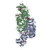
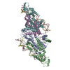
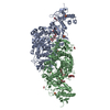
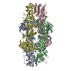
ジャーナル: Nature / 年: 2018 タイトル: Phase-plate cryo-EM structure of a biased agonist-bound human GLP-1 receptor-Gs complex. 著者: Yi-Lynn Liang / Maryam Khoshouei / Alisa Glukhova / Sebastian G B Furness / Peishen Zhao / Lachlan Clydesdale / Cassandra Koole / Tin T Truong / David M Thal / Saifei Lei / Mazdak Radjainia / ...著者: Yi-Lynn Liang / Maryam Khoshouei / Alisa Glukhova / Sebastian G B Furness / Peishen Zhao / Lachlan Clydesdale / Cassandra Koole / Tin T Truong / David M Thal / Saifei Lei / Mazdak Radjainia / Radostin Danev / Wolfgang Baumeister / Ming-Wei Wang / Laurence J Miller / Arthur Christopoulos / Patrick M Sexton / Denise Wootten / 要旨: The class B glucagon-like peptide-1 (GLP-1) G protein-coupled receptor is a major target for the treatment of type 2 diabetes and obesity. Endogenous and mimetic GLP-1 peptides exhibit biased agonism- ...The class B glucagon-like peptide-1 (GLP-1) G protein-coupled receptor is a major target for the treatment of type 2 diabetes and obesity. Endogenous and mimetic GLP-1 peptides exhibit biased agonism-a difference in functional selectivity-that may provide improved therapeutic outcomes. Here we describe the structure of the human GLP-1 receptor in complex with the G protein-biased peptide exendin-P5 and a Gα heterotrimer, determined at a global resolution of 3.3 Å. At the extracellular surface, the organization of extracellular loop 3 and proximal transmembrane segments differs between our exendin-P5-bound structure and previous GLP-1-bound GLP-1 receptor structure. At the intracellular face, there was a six-degree difference in the angle of the Gαs-α5 helix engagement between structures, which was propagated across the G protein heterotrimer. In addition, the structures differed in the rate and extent of conformational reorganization of the Gα protein. Our structure provides insights into the molecular basis of biased agonism.
全体 : Complex of a full-length, active-state glucagon-like peptide-1 re...
全体
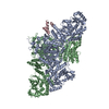
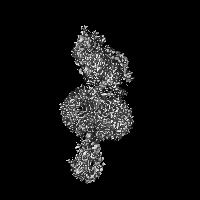
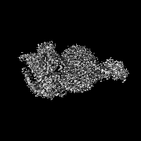
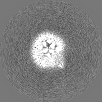
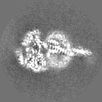
名称: Complex of a full-length, active-state glucagon-like peptide-1 receptor with exendin-P5 ligand, heterotrimeric Gs protein and nanobody 35.
要素
複合体: Complex of a full-length, active-state glucagon-like peptide-1 receptor with exendin-P5 ligand, heterotrimeric Gs protein and nanobody 35.
タンパク質・ペプチド: Glucagon-like peptide 1 receptor
タンパク質・ペプチド: Exendin-P5
タンパク質・ペプチド: Guanine nucleotide-binding protein G(s) subunit alpha isoforms short
タンパク質・ペプチド: Guanine nucleotide-binding protein G(I)/G(S)/G(T) subunit beta-1
タンパク質・ペプチド: Guanine nucleotide-binding protein G(I)/G(S)/G(O) subunit gamma-2
タンパク質・ペプチド: Nanobody 35
-
超分子 #1: Complex of a full-length, active-state glucagon-like peptide-1 re...
超分子
名称: Complex of a full-length, active-state glucagon-like peptide-1 receptor with exendin-P5 ligand, heterotrimeric Gs protein and nanobody 35. タイプ: complex / ID: 1 / 親要素: 0 / 含まれる分子: all
3C5T was rigid body fitted with minimal manual adjustments. The rest of the chain R and chains A,B,G,N were extensively remodeled. Chain P was built de novo.
得られたモデル
PDB-6b3j: 3.3 angstrom phase-plate cryo-EM structure of a biased agonist-bound human GLP-1 receptor-Gs complex
 ムービー
ムービー コントローラー


 データを開く
 基本情報
基本情報 マップデータ
 試料
試料 機能・相同性情報
 Homo sapiens (ヒト) / synthetic construct (人工物) /
 データ登録者
 オーストラリア, 4件
  引用
引用


 構造の表示
構造の表示
 ダウンロードとリンク
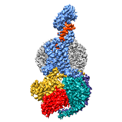 emd_7039.png
 http://ftp.pdbj.org/pub/emdb/structures/EMD-7039


 Z (Sec.)
 Y (Row.)
 X (Col.)


 試料の構成要素
 Trichoplusia ni (イラクサキンウワバ)
 解析
解析 電子顕微鏡法
 FIELD EMISSION GUN