 Movie
Movie Controller

[English] 日本語
 Yorodumi
- EMDB-6184: Cryo-EM reconstruction of quasi-HPV16 complexed with H263.A2 Fab -
+ Open data

- Basic information
| Entry | Database: EMDB / ID: EMD-6184 | |||||||||
|---|---|---|---|---|---|---|---|---|---|---|
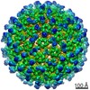
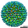
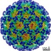
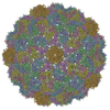
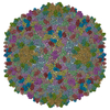
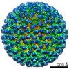
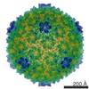
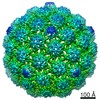
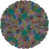
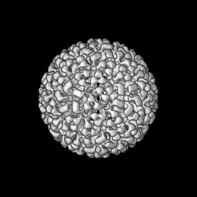
| Title | Cryo-EM reconstruction of quasi-HPV16 complexed with H263.A2 Fab | |||||||||
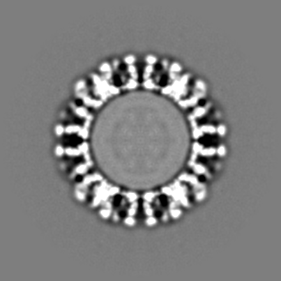
 Map data Map data | Reconstruction of quasi-HPV16 complexed with H263.A2 Fabs | |||||||||
 Sample Sample |
| |||||||||
 Keywords Keywords | quasi-HPV16 / L1 capsomer / H263.A2 Fab | |||||||||
| Function / homology |  Function and homology information Function and homology informationT=7 icosahedral viral capsid / endocytosis involved in viral entry into host cell / host cell nucleus / virion attachment to host cell / structural molecule activity Similarity search - Function | |||||||||
| Biological species |   Human papillomavirus 16 Human papillomavirus 16 | |||||||||
| Method | single particle reconstruction / cryo EM / Resolution: 13.0 Å | |||||||||
 Authors Authors | Guan J / Bywaters SM / Brendle SA / Lee H / Ashley R / Conway JF / Makhov AM / Christensen ND / Hafenstein S | |||||||||
 Citation Citation | Journal: Virology / Year: 2015 Title: Structural comparison of four different antibodies interacting with human papillomavirus 16 and mechanisms of neutralization. Authors: Jian Guan / Stephanie M Bywaters / Sarah A Brendle / Hyunwook Lee / Robert E Ashley / Alexander M Makhov / James F Conway / Neil D Christensen / Susan Hafenstein /  Abstract: Cryo-electron microscopy (cryo-EM) was used to solve the structures of human papillomavirus type 16 (HPV16) complexed with fragments of antibody (Fab) from three different neutralizing monoclonals ...Cryo-electron microscopy (cryo-EM) was used to solve the structures of human papillomavirus type 16 (HPV16) complexed with fragments of antibody (Fab) from three different neutralizing monoclonals (mAbs): H16.1A, H16.14J, and H263.A2. The structure-function analysis revealed predominantly monovalent binding of each Fab with capsid interactions that involved multiple loops from symmetry related copies of the major capsid protein. The residues identified in each Fab-virus interface map to a conformational groove on the surface of the capsomer. In addition to the known involvement of the FG and HI loops, the DE loop was also found to constitute the core of each epitope. Surprisingly, the epitope mapping also identified minor contributions by EF and BC loops. Complementary immunological assays included mAb and Fab neutralization. The specific binding characteristics of mAbs correlated with different neutralizing behaviors in pre- and post-attachment neutralization assays. | |||||||||
| History |
|
- Structure visualization
| Movie |
Movie viewer |
|---|---|
| Structure viewer | EM map: SurfViewMolmilJmol/JSmol |
| Supplemental images |
 UCSF Chimera
- Downloads & links
-EMDB archive
| Map data | emd_6184.map.gz | 78.8 MB | EMDB map data format | |
|---|---|---|---|---|
| Header (meta data) | emd-6184-v30.xmlemd-6184.xml | 11.9 KB 11.9 KB | Display Display | EMDB header |
| Images | 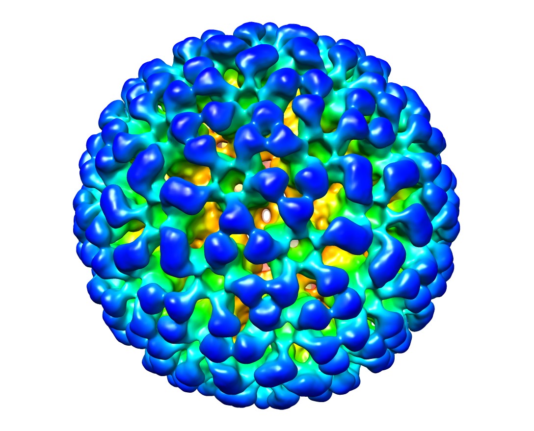 emd_6184.jpg emd_6184.jpg | 172.9 KB | ||
| Archive directory |  http://ftp.pdbj.org/pub/emdb/structures/EMD-6184ftp://ftp.pdbj.org/pub/emdb/structures/EMD-6184 http://ftp.pdbj.org/pub/emdb/structures/EMD-6184ftp://ftp.pdbj.org/pub/emdb/structures/EMD-6184 | HTTPS FTP |
-Validation report
| Summary document | emd_6184_validation.pdf.gz | 317.5 KB | Display | EMDB validaton report |
|---|---|---|---|---|
| Full document | emd_6184_full_validation.pdf.gz | 317.1 KB | Display | |
| Data in XML | emd_6184_validation.xml.gz | 7.4 KB | Display | |
| Arichive directory | https://ftp.pdbj.org/pub/emdb/validation_reports/EMD-6184ftp://ftp.pdbj.org/pub/emdb/validation_reports/EMD-6184 | HTTPS FTP |
-Related structure data
| Related structure data |  3j8wMC  5990C  6121C 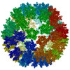 3j8vC  3j8zC M: atomic model generated by this map C: citing same article ( |
|---|---|
| Similar structure data |
-Links
| EMDB pages | EMDB (EBI/PDBe) / EMDataResource |
|---|---|
| Related items in Molecule of the Month |


-Map
| File | Download / File: emd_6184.map.gz / Format: CCP4 / Size: 243.8 MB / Type: IMAGE STORED AS FLOATING POINT NUMBER (4 BYTES) | ||||||||||||||||||||||||||||||||||||||||||||||||||||||||||||
|---|---|---|---|---|---|---|---|---|---|---|---|---|---|---|---|---|---|---|---|---|---|---|---|---|---|---|---|---|---|---|---|---|---|---|---|---|---|---|---|---|---|---|---|---|---|---|---|---|---|---|---|---|---|---|---|---|---|---|---|---|---|
| Annotation | Reconstruction of quasi-HPV16 complexed with H263.A2 Fabs | ||||||||||||||||||||||||||||||||||||||||||||||||||||||||||||
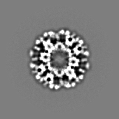
| Projections & slices | Image control
Images are generated by Spider. | ||||||||||||||||||||||||||||||||||||||||||||||||||||||||||||
| Voxel size | X=Y=Z: 2.86 Å | ||||||||||||||||||||||||||||||||||||||||||||||||||||||||||||
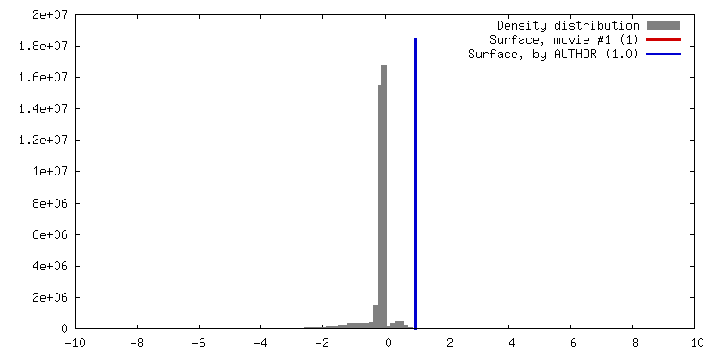
| Density |
| ||||||||||||||||||||||||||||||||||||||||||||||||||||||||||||
| Symmetry | Space group: 1 | ||||||||||||||||||||||||||||||||||||||||||||||||||||||||||||
| Details | EMDB XML:
CCP4 map header:
| ||||||||||||||||||||||||||||||||||||||||||||||||||||||||||||
 Z (Sec.)
 Y (Row.)
 X (Col.)


-Supplemental data
- Sample components
-Entire : quasi-HPV16 complexed with H263.A2 Fabs
| Entire | Name: quasi-HPV16 complexed with H263.A2 Fabs |
|---|---|
| Components |
|
-Supramolecule #1000: quasi-HPV16 complexed with H263.A2 Fabs
| Supramolecule | Name: quasi-HPV16 complexed with H263.A2 Fabs / type: sample / ID: 1000 / Number unique components: 2 |
|---|---|
| Molecular weight | Theoretical: 44.7 MDa |
-Supramolecule #1: Human papillomavirus 16
| Supramolecule | Name: Human papillomavirus 16 / type: virus / ID: 1 / Details: isolated by gradient centrifugation / NCBI-ID: 337041 / Sci species name: Human papillomavirus 16 / Database: NCBI / Virus type: VIRION / Virus isolate: OTHER / Virus enveloped: No / Virus empty: No |
|---|---|
| Host (natural) | Organism:  Homo sapiens (human) / synonym: VERTEBRATES Homo sapiens (human) / synonym: VERTEBRATES |
| Virus shell | Shell ID: 1 / Name: L1 L2 / Diameter: 600 Å / T number (triangulation number): 7 |
-Macromolecule #1: H263.A2 Fab
| Macromolecule | Name: H263.A2 Fab / type: protein_or_peptide / ID: 1 / Recombinant expression: No / Database: NCBI |
|---|---|
| Source (natural) | Organism: |
-Experimental details
-Structure determination
| Method | cryo EM |
|---|---|
 Processing Processing | single particle reconstruction |
| Aggregation state | particle |
-Sample preparation
| Concentration | 1.2 mg/mL |
|---|---|
| Buffer | pH: 7.4 / Details: 1 M NaCl, 200 nM Tris |
| Grid | Details: glow-discharged holey carbon supported grid |
| Vitrification | Cryogen name: ETHANE / Chamber humidity: 90 % / Chamber temperature: 102 K / Instrument: GATAN CRYOPLUNGE 3 |
- Electron microscopy
| Microscope | JEOL 2100 |
|---|---|
| Temperature | Average: 95 K |
| Date | Jul 31, 2014 |
| Image recording | Category: CCD / Film or detector model: GATAN ULTRASCAN 4000 (4k x 4k) / Number real images: 264 / Average electron dose: 15 e/Å2 |
| Electron beam | Acceleration voltage: 200 kV / Electron source: LAB6 |
| Electron optics | Illumination mode: SPOT SCAN / Imaging mode: BRIGHT FIELD / Cs: 2 mm / Nominal defocus max: 5.52 µm / Nominal defocus min: 1.49 µm / Nominal magnification: 40000 |
| Sample stage | Specimen holder model: GATAN LIQUID NITROGEN |
-Image processing
| Details | The particles were selected using semi-automatic program e2boxer.py (EMAN2). |
|---|---|
| CTF correction | Details: Each particle |
| Final reconstruction | Algorithm: OTHER / Resolution.type: BY AUTHOR / Resolution: 13.0 Å / Resolution method: OTHER / Software - Name: auto3dem Details: Semi-automatic particle selection was performed using e2boxer.py to obtain the particle coordinates, followed by particle boxing, linearization, normalization, and apodization of the images ...Details: Semi-automatic particle selection was performed using e2boxer.py to obtain the particle coordinates, followed by particle boxing, linearization, normalization, and apodization of the images using Robem. Defocus and astigmatism values used to perform contrast transfer function (CTF) correction were assessed using Robem for the extracted particles. The icosahedrally averaged reconstruction was initiated using a random model generated with setup_rmc and reached 14 A resolution estimated at a Fourier Shell Correlation (FSC) of 0.5. For the last step of refinement, the final maps were CTF-corrected using a B factor of 200 A2. Number images used: 8908 |
-Atomic model buiding 1
| Initial model | PDB ID:  3oae Chain - #0 - Chain ID: A / Chain - #1 - Chain ID: B / Chain - #2 - Chain ID: C / Chain - #3 - Chain ID: D / Chain - #4 - Chain ID: E |
|---|---|
| Software | Name: Chimera, Situs |
| Refinement | Space: REAL / Protocol: RIGID BODY FIT |
| Output model | PDB-3j8w: |
