 Movie
Movie Controller

[English] 日本語
 Yorodumi
- EMDB-48918: CryoEM structure of the Azotobacter vinelandii flagellar filament -
+ Open data

- Basic information
| Entry |  | |||||||||
|---|---|---|---|---|---|---|---|---|---|---|
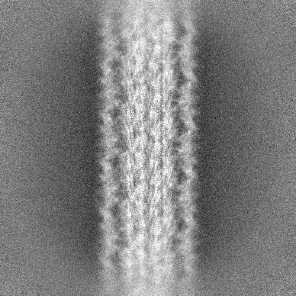
| Title | CryoEM structure of the Azotobacter vinelandii flagellar filament | |||||||||
 Map data Map data | ||||||||||
 Sample Sample |
| |||||||||
 Keywords Keywords | Flagellum / STRUCTURAL PROTEIN | |||||||||
| Function / homology |  Function and homology information Function and homology informationbacterial-type flagellum / structural molecule activity / extracellular region Similarity search - Function | |||||||||
| Biological species |  Azotobacter vinelandii (bacteria) Azotobacter vinelandii (bacteria) | |||||||||
| Method | helical reconstruction / cryo EM / Resolution: 2.82 Å | |||||||||
 Authors Authors | Warmack RA | |||||||||
| Funding support |  United States, 2 items United States, 2 items
| |||||||||
 Citation Citation | Journal: Structure / Year: 2025 Title: CryoEM-enabled visual proteomics reveals de novo structures of oligomeric protein complexes. Authors: Yuanbo Shen / Ailiena O Maggiolo / Tianzheng Zhang / Rebeccah A Warmack / Abstract: Single particle cryoelectron microscopy (cryoEM) and cryoelectron tomography (cryoET) are powerful methods for unveiling unique and functionally relevant structural states. Aided by mass spectrometry ...Single particle cryoelectron microscopy (cryoEM) and cryoelectron tomography (cryoET) are powerful methods for unveiling unique and functionally relevant structural states. Aided by mass spectrometry and machine learning, they promise to facilitate the visual exploration of proteomes. Leveraging visual proteomics, we interrogate structures isolated from a complex cellular milieu by cryoEM to identify and classify molecular structures and complexes de novo. By comparing three automated model building programs, CryoID, DeepTracer, and ModelAngelo, we determine the identity of six distinct oligomeric protein complexes from partially purified extracts of the nitrogen-fixing bacterium Azotobacter vinelandii using both anaerobic and aerobic cryoEM, including two original oligomeric structures. Overall, by allowing the study of near-native oligomeric protein states, cryoEM-enabled visual proteomics reveals unique structures that correspond to relevant species observed in situ. | |||||||||
| History |
|
- Structure visualization
| Supplemental images |
|---|

- Downloads & links
-EMDB archive
| Map data | emd_48918.map.gz | 3.3 GB | EMDB map data format | |
|---|---|---|---|---|
| Header (meta data) | emd-48918-v30.xmlemd-48918.xml | 16.4 KB 16.4 KB | Display Display | EMDB header |
| FSC (resolution estimation) | emd_48918_fsc.xml | 32.5 KB | Display | FSC data file |
| Images |  emd_48918.png emd_48918.png | 73.2 KB | ||
| Filedesc metadata | emd-48918.cif.gz | 5.4 KB | ||
| Others | emd_48918_half_map_1.map.gzemd_48918_half_map_2.map.gz | 3.3 GB 3.3 GB | ||
| Archive directory |  http://ftp.pdbj.org/pub/emdb/structures/EMD-48918ftp://ftp.pdbj.org/pub/emdb/structures/EMD-48918 http://ftp.pdbj.org/pub/emdb/structures/EMD-48918ftp://ftp.pdbj.org/pub/emdb/structures/EMD-48918 | HTTPS FTP |
-Related structure data
| Related structure data |  9n59MC  9n4vC  9n4wC  9n4xC  9n4yC  9n5aC  9nsvC M: atomic model generated by this map C: citing same article ( |
|---|---|
| Similar structure data |


-Links
| EMDB pages | EMDB (EBI/PDBe) / EMDataResource |
|---|
-Map
| File | Download / File: emd_48918.map.gz / Format: CCP4 / Size: 3.5 GB / Type: IMAGE STORED AS FLOATING POINT NUMBER (4 BYTES) | ||||||||||||||||||||||||||||||||||||
|---|---|---|---|---|---|---|---|---|---|---|---|---|---|---|---|---|---|---|---|---|---|---|---|---|---|---|---|---|---|---|---|---|---|---|---|---|---|
| Projections & slices | Image control
Images are generated by Spider. | ||||||||||||||||||||||||||||||||||||
| Voxel size | X=Y=Z: 0.65 Å | ||||||||||||||||||||||||||||||||||||
| Density |
| ||||||||||||||||||||||||||||||||||||
| Symmetry | Space group: 1 | ||||||||||||||||||||||||||||||||||||
| Details | EMDB XML:
|
 Z (Sec.)
 Y (Row.)
 X (Col.)


-Supplemental data
-Half map: #1
| File | emd_48918_half_map_1.map | ||||||||||||
|---|---|---|---|---|---|---|---|---|---|---|---|---|---|
| Projections & Slices |
| ||||||||||||
| Density Histograms |


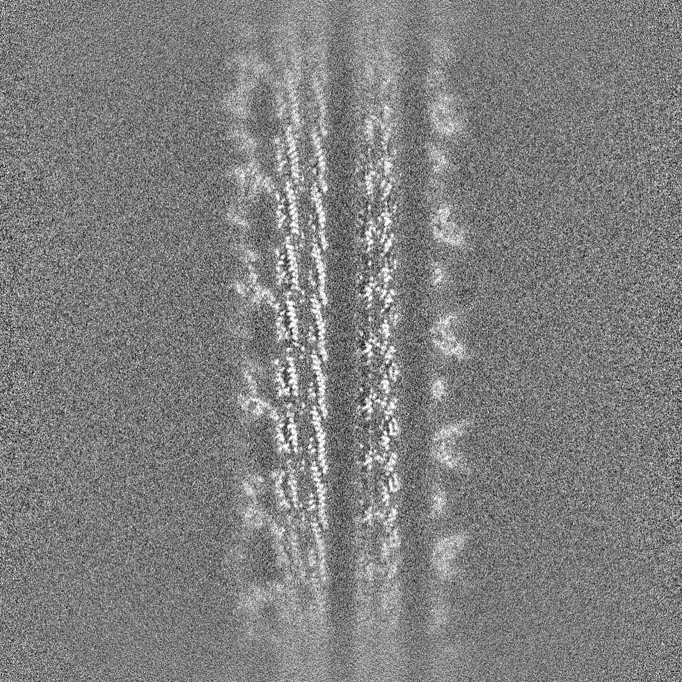


-Half map: #2
| File | emd_48918_half_map_2.map | ||||||||||||
|---|---|---|---|---|---|---|---|---|---|---|---|---|---|
| Projections & Slices |
| ||||||||||||
| Density Histograms |


- Sample components
-Entire : Filamentous assembly of the Azotobacter vinelandii flagellar filament
| Entire | Name: Filamentous assembly of the Azotobacter vinelandii flagellar filament |
|---|---|
| Components |
|
-Supramolecule #1: Filamentous assembly of the Azotobacter vinelandii flagellar filament
| Supramolecule | Name: Filamentous assembly of the Azotobacter vinelandii flagellar filament type: complex / ID: 1 / Parent: 0 / Macromolecule list: all |
|---|---|
| Source (natural) | Organism: Azotobacter vinelandii (bacteria) / Strain: DJ |
-Macromolecule #1: Flagellin
| Macromolecule | Name: Flagellin / type: protein_or_peptide / ID: 1 / Number of copies: 2 / Enantiomer: LEVO |
|---|---|
| Source (natural) | Organism: Azotobacter vinelandii (bacteria) / Strain: DJ |
| Molecular weight | Theoretical: 58.040414 KDa |
| Sequence | String: MPVINTNITS LIAQKNLSSS QSALTTAMER LSSGMRINSA KDDAAGQAIA NRMSSQITGL TQAQRNANDG ISVAQTTEGA LDQINDNLQ RIRELTVQAQ NGTNSSEDLT SIQNEIAQRL DEIDRISQET EFNGVKVLSK DTNLNIQVGA NDGQSIGISL K QINAKTLG ...String: MPVINTNITS LIAQKNLSSS QSALTTAMER LSSGMRINSA KDDAAGQAIA NRMSSQITGL TQAQRNANDG ISVAQTTEGA LDQINDNLQ RIRELTVQAQ NGTNSSEDLT SIQNEIAQRL DEIDRISQET EFNGVKVLSK DTNLNIQVGA NDGQSIGISL K QINAKTLG LDGFNVDGSG ATQNSTATVT SLEGAGAVKN STTGNYVLTT TFDEASLERM LGEMKDGDVV TVGSGASTAV EY TFNTASG SFTYTANATN AAAYGDLADE LIPPTGSSIT GTYEFADGNV ATFAVDASGK LTLDGQAAYV TATGELTNNA TSG ATQATL EDLFATVGAG AAGTDTSARS LTVGGVTYTG AGTTAGLSYT DTATSSDVLA ASASTVASIE MHNGITSATI DFDN TGAQS STGTQVYVDE DGQLTTVGSY TTTFAVNADT GEVTVVDNSD TAGDYALEEG ATVYVGSNGR LTTSTTSKGD VTEDP LAAL DAALASVDAL RSDLGAIQNR FDSAINNLST TTTNLSAARS RIEDADYAVE VANMTKAQIL QQAGTSVLAQ ANQVPQ SVL SLLG UniProtKB: Flagellin |
-Experimental details
-Structure determination
| Method | cryo EM |
|---|---|
 Processing Processing | helical reconstruction |
| Aggregation state | helical array |
-Sample preparation
| Concentration | 0.75 mg/mL |
|---|---|
| Buffer | pH: 7.5 |
| Vitrification | Cryogen name: ETHANE-PROPANE |
- Electron microscopy
| Microscope | TFS KRIOS |
|---|---|
| Image recording | Film or detector model: GATAN K3 (6k x 4k) / Average electron dose: 60.0 e/Å2 |
| Electron beam | Acceleration voltage: 300 kV / Electron source:  FIELD EMISSION GUN FIELD EMISSION GUN |
| Electron optics | Illumination mode: FLOOD BEAM / Imaging mode: BRIGHT FIELD / Nominal defocus max: -2.5 µm / Nominal defocus min: -0.8 µm |
| Experimental equipment |  Model: Titan Krios / Image courtesy: FEI Company |

