 Movie
Movie Controller

[English] 日本語
 Yorodumi
- EMDB-48907: Decameric Glucose-6-phosphate isomerase from Azotobacter vinelandii -
+ Open data

- Basic information
| Entry |  | |||||||||
|---|---|---|---|---|---|---|---|---|---|---|
| Title | Decameric Glucose-6-phosphate isomerase from Azotobacter vinelandii | |||||||||
 Map data Map data | ||||||||||
 Sample Sample |
| |||||||||
 Keywords Keywords | Glycolysis / decamer / ISOMERASE | |||||||||
| Function / homology |  Function and homology information Function and homology informationglucose-6-phosphate isomerase / glucose-6-phosphate isomerase activity / glucose 6-phosphate metabolic process / carbohydrate derivative binding / monosaccharide binding / glycolytic process / gluconeogenesis / cytosol Similarity search - Function | |||||||||
| Biological species |  Azotobacter vinelandii (bacteria) Azotobacter vinelandii (bacteria) | |||||||||
| Method | single particle reconstruction / cryo EM / Resolution: 2.5 Å | |||||||||
 Authors Authors | Warmack RA / Maggiolo AO | |||||||||
| Funding support |  United States, 2 items United States, 2 items
| |||||||||
 Citation Citation | Journal: Structure / Year: 2025 Title: CryoEM-enabled visual proteomics reveals de novo structures of oligomeric protein complexes. Authors: Yuanbo Shen / Ailiena O Maggiolo / Tianzheng Zhang / Rebeccah A Warmack / Abstract: Single particle cryoelectron microscopy (cryoEM) and cryoelectron tomography (cryoET) are powerful methods for unveiling unique and functionally relevant structural states. Aided by mass spectrometry ...Single particle cryoelectron microscopy (cryoEM) and cryoelectron tomography (cryoET) are powerful methods for unveiling unique and functionally relevant structural states. Aided by mass spectrometry and machine learning, they promise to facilitate the visual exploration of proteomes. Leveraging visual proteomics, we interrogate structures isolated from a complex cellular milieu by cryoEM to identify and classify molecular structures and complexes de novo. By comparing three automated model building programs, CryoID, DeepTracer, and ModelAngelo, we determine the identity of six distinct oligomeric protein complexes from partially purified extracts of the nitrogen-fixing bacterium Azotobacter vinelandii using both anaerobic and aerobic cryoEM, including two original oligomeric structures. Overall, by allowing the study of near-native oligomeric protein states, cryoEM-enabled visual proteomics reveals unique structures that correspond to relevant species observed in situ. | |||||||||
| History |
|
- Structure visualization
| Supplemental images |
|---|

- Downloads & links
-EMDB archive
| Map data | emd_48907.map.gz | 373.9 MB | EMDB map data format | |
|---|---|---|---|---|
| Header (meta data) | emd-48907-v30.xmlemd-48907.xml | 16.4 KB 16.4 KB | Display Display | EMDB header |
| FSC (resolution estimation) | emd_48907_fsc.xml | 19 KB | Display | FSC data file |
| Images |  emd_48907.png emd_48907.png | 96.8 KB | ||
| Filedesc metadata | emd-48907.cif.gz | 5.5 KB | ||
| Others | emd_48907_half_map_1.map.gzemd_48907_half_map_2.map.gz | 676.9 MB 676.9 MB | ||
| Archive directory |  http://ftp.pdbj.org/pub/emdb/structures/EMD-48907ftp://ftp.pdbj.org/pub/emdb/structures/EMD-48907 http://ftp.pdbj.org/pub/emdb/structures/EMD-48907ftp://ftp.pdbj.org/pub/emdb/structures/EMD-48907 | HTTPS FTP |
-Related structure data
| Related structure data |  9n4wMC  9n4vC  9n4xC  9n4yC  9n59C  9n5aC  9nsvC M: atomic model generated by this map C: citing same article ( |
|---|---|
| Similar structure data |


-Links
| EMDB pages | EMDB (EBI/PDBe) / EMDataResource |
|---|---|
| Related items in Molecule of the Month |


-Map
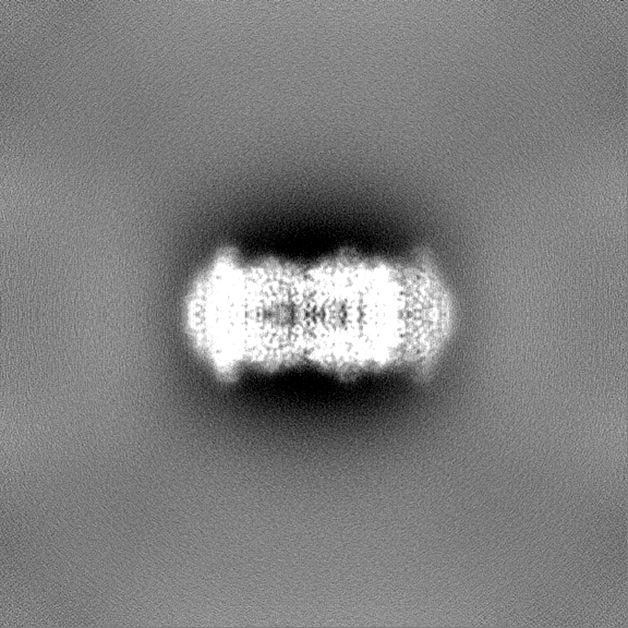
| File | Download / File: emd_48907.map.gz / Format: CCP4 / Size: 729 MB / Type: IMAGE STORED AS FLOATING POINT NUMBER (4 BYTES) | ||||||||||||||||||||||||||||||||||||
|---|---|---|---|---|---|---|---|---|---|---|---|---|---|---|---|---|---|---|---|---|---|---|---|---|---|---|---|---|---|---|---|---|---|---|---|---|---|
| Projections & slices | Image control
Images are generated by Spider. | ||||||||||||||||||||||||||||||||||||
| Voxel size | X=Y=Z: 0.65 Å | ||||||||||||||||||||||||||||||||||||
| Density |
| ||||||||||||||||||||||||||||||||||||
| Symmetry | Space group: 1 | ||||||||||||||||||||||||||||||||||||
| Details | EMDB XML:
|
 Z (Sec.)
 Y (Row.)
 X (Col.)


-Supplemental data
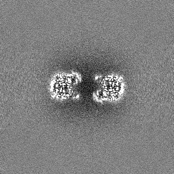
-Half map: #1
| File | emd_48907_half_map_1.map | ||||||||||||
|---|---|---|---|---|---|---|---|---|---|---|---|---|---|
| Projections & Slices |
| ||||||||||||
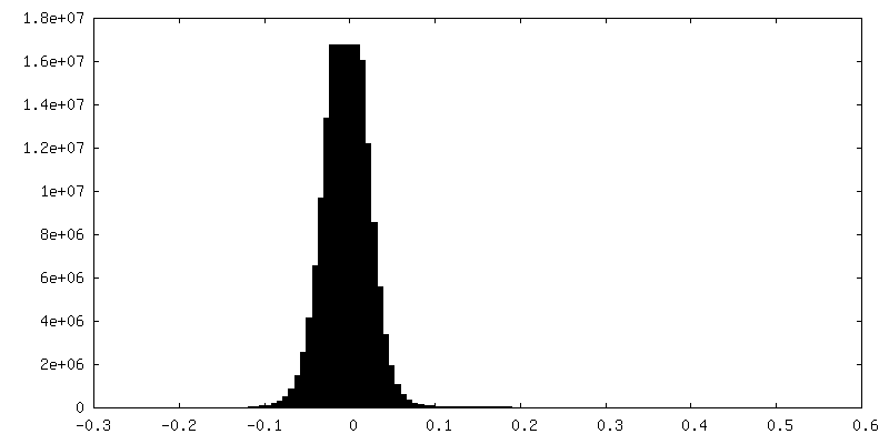
| Density Histograms |


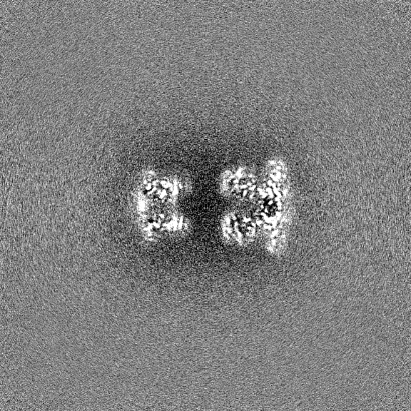
-Half map: #2
| File | emd_48907_half_map_2.map | ||||||||||||
|---|---|---|---|---|---|---|---|---|---|---|---|---|---|
| Projections & Slices |
| ||||||||||||
| Density Histograms |


- Sample components
-Entire : Decameric complex of Glucose-6-phosphate isomerase
| Entire | Name: Decameric complex of Glucose-6-phosphate isomerase |
|---|---|
| Components |
|
-Supramolecule #1: Decameric complex of Glucose-6-phosphate isomerase
| Supramolecule | Name: Decameric complex of Glucose-6-phosphate isomerase / type: complex / ID: 1 / Parent: 0 / Macromolecule list: all |
|---|---|
| Source (natural) | Organism: Azotobacter vinelandii (bacteria) / Strain: DJ |
-Macromolecule #1: Glucose-6-phosphate isomerase
| Macromolecule | Name: Glucose-6-phosphate isomerase / type: protein_or_peptide / ID: 1 / Number of copies: 10 / Enantiomer: LEVO / EC number: glucose-6-phosphate isomerase |
|---|---|
| Source (natural) | Organism: Azotobacter vinelandii (bacteria) / Strain: DJ |
| Molecular weight | Theoretical: 61.978125 KDa |
| Sequence | String: MSYYQQAFDV TSLPSWRALQ EHRLAMQNFH MREAFLSDPG RFDEFSTSSC GLFLDYSKNL ITPETRDLLV NLAREAGVEQ AARAMFDGE PVNASERRPA LHTALRRPVG DSLLIDGHNI MRDVHAALAQ MTDIVGRIHN KLWRGYSDRA ITDVVNIGIG G SYLGPELV ...String: MSYYQQAFDV TSLPSWRALQ EHRLAMQNFH MREAFLSDPG RFDEFSTSSC GLFLDYSKNL ITPETRDLLV NLAREAGVEQ AARAMFDGE PVNASERRPA LHTALRRPVG DSLLIDGHNI MRDVHAALAQ MTDIVGRIHN KLWRGYSDRA ITDVVNIGIG G SYLGPELV SEALLPYTQH GVRCHYLANI DGSEFHELTA RLNAETTLFI ISSKTFGTLE TLKNAQAART WYLASGGSEE RL YRHFIAV TSNIPAAIEF GIREKNIFPM WDWVGGRYSL WSAIGLPTAL AIGMANFKDL LSGAYAMDCH FRREPFENNM PVL LAMLGV WYGNFWGAQS HAILPYDHYL RNFVKHLQQM DMESNGKSVR QDGSPVSCET GPVIWGGVGC NGQHAYHQLL HQGT LLIPA DFIVPVVSHN PVADHHQWLY ANCLSQSQAL MLGKSREEAE AELRAKGLPE AEVKRLAPHK VIPGNRPSNT LVMET LNPS RLGALIALYE HKVFVQGVIW GINSFDQWGV ELGKELGKNV YGRLTSYEAP PAEDSSTQGL IDYFRGRHRG UniProtKB: Glucose-6-phosphate isomerase |
-Experimental details
-Structure determination
| Method | cryo EM |
|---|---|
 Processing Processing | single particle reconstruction |
| Aggregation state | particle |
-Sample preparation
| Buffer | pH: 7.8 |
|---|---|
| Vitrification | Cryogen name: ETHANE-PROPANE |
- Electron microscopy
| Microscope | TFS KRIOS |
|---|---|
| Image recording | Film or detector model: GATAN K3 (6k x 4k) / Average electron dose: 60.0 e/Å2 |
| Electron beam | Acceleration voltage: 300 kV / Electron source:  FIELD EMISSION GUN FIELD EMISSION GUN |
| Electron optics | Illumination mode: FLOOD BEAM / Imaging mode: BRIGHT FIELD / Nominal defocus max: -3.0 µm / Nominal defocus min: -0.8 µm |
| Experimental equipment |  Model: Titan Krios / Image courtesy: FEI Company |

