+ データを開く
- 基本情報
基本情報
| 登録情報 |  | |||||||||
|---|---|---|---|---|---|---|---|---|---|---|
| タイトル | Intact V-ATPase State 2 and synaptophysin complex in mouse brain isolated synaptic vesicles | |||||||||
 マップデータ マップデータ | Full map of V-ATPase State 2 synaptophysin complex in wild-type ISVs. | |||||||||
 試料 試料 |
| |||||||||
 キーワード キーワード | V-ATPase / synaptic vesicle / MEMBRANE PROTEIN | |||||||||
| 機能・相同性 |  機能・相同性情報 機能・相同性情報Ion channel transport / regulation of opioid receptor signaling pathway / Amino acids regulate mTORC1 / clathrin-sculpted glutamate transport vesicle membrane / Transferrin endocytosis and recycling / Insulin receptor recycling / eye pigmentation / central nervous system maturation / Metabolism of Angiotensinogen to Angiotensins / transporter activator activity ...Ion channel transport / regulation of opioid receptor signaling pathway / Amino acids regulate mTORC1 / clathrin-sculpted glutamate transport vesicle membrane / Transferrin endocytosis and recycling / Insulin receptor recycling / eye pigmentation / central nervous system maturation / Metabolism of Angiotensinogen to Angiotensins / transporter activator activity / negative regulation of autophagic cell death / plasma membrane proton-transporting V-type ATPase complex / rostrocaudal neural tube patterning / RHOA GTPase cycle / positive regulation of transforming growth factor beta1 production / cellular response to increased oxygen levels / regulation of synaptic vesicle priming / proton-transporting V-type ATPase, V1 domain / ROS and RNS production in phagocytes / proton-transporting V-type ATPase, V0 domain / synaptic vesicle lumen acidification / extrinsic component of synaptic vesicle membrane / P-type proton-exporting transporter activity / intracellular organelle / endosome to plasma membrane protein transport / lysosomal lumen acidification / clathrin-coated vesicle membrane / vacuolar transport / endosomal lumen acidification / vacuolar proton-transporting V-type ATPase, V0 domain / vacuolar proton-transporting V-type ATPase, V1 domain / vacuolar proton-transporting V-type ATPase complex / proton-transporting V-type ATPase complex / neuron spine / head morphogenesis / vacuolar acidification / osteoclast development / protein localization to cilium / regulation of short-term neuronal synaptic plasticity / neuron projection terminus / dendritic spine membrane / regulation of cellular pH / syntaxin-1 binding / cholesterol binding / ATPase complex / regulation of MAPK cascade / regulation of neuronal synaptic plasticity / presynaptic active zone / microvillus / ATPase activator activity / excitatory synapse / autophagosome membrane / positive regulation of Wnt signaling pathway / synaptic vesicle endocytosis / cilium assembly / transmembrane transporter complex / endomembrane system / regulation of macroautophagy / angiotensin maturation / ATP metabolic process / H+-transporting two-sector ATPase / axon terminus / ruffle / endoplasmic reticulum-Golgi intermediate compartment membrane / proton-transporting ATPase activity, rotational mechanism / RNA endonuclease activity / proton-transporting ATP synthase activity, rotational mechanism / Neutrophil degranulation / SH2 domain binding / receptor-mediated endocytosis / proton transmembrane transport / SNARE binding / secretory granule / regulation of long-term neuronal synaptic plasticity / modulation of chemical synaptic transmission / neuromuscular junction / Schaffer collateral - CA1 synapse / terminal bouton / transmembrane transport / cilium / small GTPase binding / synaptic vesicle membrane / endocytosis / positive regulation of canonical Wnt signaling pathway / melanosome / synaptic vesicle / apical part of cell / myelin sheath / presynapse / signaling receptor activity / presynaptic membrane / ATPase binding / cell body / chemical synaptic transmission / postsynaptic membrane / intracellular iron ion homeostasis / 加水分解酵素; エステル加水分解酵素 / receptor-mediated endocytosis of virus by host cell / lysosome / positive regulation of ERK1 and ERK2 cascade 類似検索 - 分子機能 | |||||||||
| 生物種 |  | |||||||||
| 手法 | 単粒子再構成法 / クライオ電子顕微鏡法 / 解像度: 4.3 Å | |||||||||
 データ登録者 データ登録者 | Wang C / Jiang W / Yang K / Wang X / Guo Q / Brunger AT | |||||||||
| 資金援助 |  米国, 2件 米国, 2件
| |||||||||
 引用 引用 |  ジャーナル: Nature / 年: 2024 ジャーナル: Nature / 年: 2024タイトル: Structure and topography of the synaptic V-ATPase-synaptophysin complex. 著者: Chuchu Wang / Wenhong Jiang / Jeremy Leitz / Kailu Yang / Luis Esquivies / Xing Wang / Xiaotao Shen / Richard G Held / Daniel J Adams / Tamara Basta / Lucas Hampton / Ruiqi Jian / Lihua Jiang ...著者: Chuchu Wang / Wenhong Jiang / Jeremy Leitz / Kailu Yang / Luis Esquivies / Xing Wang / Xiaotao Shen / Richard G Held / Daniel J Adams / Tamara Basta / Lucas Hampton / Ruiqi Jian / Lihua Jiang / Michael H B Stowell / Wolfgang Baumeister / Qiang Guo / Axel T Brunger /     要旨: Synaptic vesicles are organelles with a precisely defined protein and lipid composition, yet the molecular mechanisms for the biogenesis of synaptic vesicles are mainly unknown. Here we discovered a ...Synaptic vesicles are organelles with a precisely defined protein and lipid composition, yet the molecular mechanisms for the biogenesis of synaptic vesicles are mainly unknown. Here we discovered a well-defined interface between the synaptic vesicle V-ATPase and synaptophysin by in situ cryo-electron tomography and single-particle cryo-electron microscopy of functional synaptic vesicles isolated from mouse brains. The synaptic vesicle V-ATPase is an ATP-dependent proton pump that establishes the proton gradient across the synaptic vesicle, which in turn drives the uptake of neurotransmitters. Synaptophysin and its paralogues synaptoporin and synaptogyrin belong to a family of abundant synaptic vesicle proteins whose function is still unclear. We performed structural and functional studies of synaptophysin-knockout mice, confirming the identity of synaptophysin as an interaction partner with the V-ATPase. Although there is little change in the conformation of the V-ATPase upon interaction with synaptophysin, the presence of synaptophysin in synaptic vesicles profoundly affects the copy number of V-ATPases. This effect on the topography of synaptic vesicles suggests that synaptophysin assists in their biogenesis. In support of this model, we observed that synaptophysin-knockout mice exhibit severe seizure susceptibility, suggesting an imbalance of neurotransmitter release as a physiological consequence of the absence of synaptophysin. | |||||||||
| 履歴 |
|
- 構造の表示
構造の表示
| 添付画像 |
|---|
- ダウンロードとリンク
-EMDBアーカイブ
| マップデータ |  emd_44839.map.gz emd_44839.map.gz | 322.4 MB |  EMDBマップデータ形式 EMDBマップデータ形式 | |
|---|---|---|---|---|
| ヘッダ (付随情報) |  emd-44839-v30.xml emd-44839-v30.xml emd-44839.xml emd-44839.xml | 38.9 KB 38.9 KB | 表示 表示 |  EMDBヘッダ EMDBヘッダ |
| 画像 |  emd_44839.png emd_44839.png | 128.9 KB | ||
| Filedesc metadata |  emd-44839.cif.gz emd-44839.cif.gz | 9.9 KB | ||
| その他 |  emd_44839_additional_1.map.gz emd_44839_additional_1.map.gz emd_44839_additional_2.map.gz emd_44839_additional_2.map.gz emd_44839_half_map_1.map.gz emd_44839_half_map_1.map.gz emd_44839_half_map_2.map.gz emd_44839_half_map_2.map.gz | 318.5 MB 274 MB 318.1 MB 318.1 MB | ||
| アーカイブディレクトリ |  http://ftp.pdbj.org/pub/emdb/structures/EMD-44839 http://ftp.pdbj.org/pub/emdb/structures/EMD-44839 ftp://ftp.pdbj.org/pub/emdb/structures/EMD-44839 ftp://ftp.pdbj.org/pub/emdb/structures/EMD-44839 | HTTPS FTP |
-検証レポート
| 文書・要旨 |  emd_44839_validation.pdf.gz emd_44839_validation.pdf.gz | 1.1 MB | 表示 |  EMDB検証レポート EMDB検証レポート |
|---|---|---|---|---|
| 文書・詳細版 |  emd_44839_full_validation.pdf.gz emd_44839_full_validation.pdf.gz | 1.1 MB | 表示 | |
| XML形式データ |  emd_44839_validation.xml.gz emd_44839_validation.xml.gz | 16.9 KB | 表示 | |
| CIF形式データ |  emd_44839_validation.cif.gz emd_44839_validation.cif.gz | 19.9 KB | 表示 | |
| アーカイブディレクトリ |  https://ftp.pdbj.org/pub/emdb/validation_reports/EMD-44839 https://ftp.pdbj.org/pub/emdb/validation_reports/EMD-44839 ftp://ftp.pdbj.org/pub/emdb/validation_reports/EMD-44839 ftp://ftp.pdbj.org/pub/emdb/validation_reports/EMD-44839 | HTTPS FTP |
-関連構造データ
| 関連構造データ |  9braMC 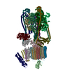 9brqC 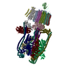 9brrC 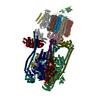 9brsC 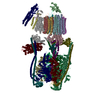 9brtC 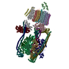 9bruC 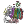 9bryC 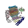 9brzC M: このマップから作成された原子モデル C: 同じ文献を引用 ( |
|---|---|
| 類似構造データ | 類似検索 - 機能・相同性  F&H 検索 F&H 検索 |
- リンク
リンク
| EMDBのページ |  EMDB (EBI/PDBe) / EMDB (EBI/PDBe) /  EMDataResource EMDataResource |
|---|---|
| 「今月の分子」の関連する項目 |
- マップ
マップ
| ファイル |  ダウンロード / ファイル: emd_44839.map.gz / 形式: CCP4 / 大きさ: 343 MB / タイプ: IMAGE STORED AS FLOATING POINT NUMBER (4 BYTES) ダウンロード / ファイル: emd_44839.map.gz / 形式: CCP4 / 大きさ: 343 MB / タイプ: IMAGE STORED AS FLOATING POINT NUMBER (4 BYTES) | ||||||||||||||||||||||||||||||||||||
|---|---|---|---|---|---|---|---|---|---|---|---|---|---|---|---|---|---|---|---|---|---|---|---|---|---|---|---|---|---|---|---|---|---|---|---|---|---|
| 注釈 | Full map of V-ATPase State 2 synaptophysin complex in wild-type ISVs. | ||||||||||||||||||||||||||||||||||||
| 投影像・断面図 | 画像のコントロール
画像は Spider により作成 | ||||||||||||||||||||||||||||||||||||
| ボクセルのサイズ | X=Y=Z: 1.11138 Å | ||||||||||||||||||||||||||||||||||||
| 密度 |
| ||||||||||||||||||||||||||||||||||||
| 対称性 | 空間群: 1 | ||||||||||||||||||||||||||||||||||||
| 詳細 | EMDB XML:
|
-添付データ
-追加マップ: EMhancer map of V-ATPase State 2 synaptophysin complex...
| ファイル | emd_44839_additional_1.map | ||||||||||||
|---|---|---|---|---|---|---|---|---|---|---|---|---|---|
| 注釈 | EMhancer map of V-ATPase State 2 synaptophysin complex in wild-type ISVs. | ||||||||||||
| 投影像・断面図 |
| ||||||||||||
| 密度ヒストグラム |
-追加マップ: Unsharpened map of V-ATPase State 2
| ファイル | emd_44839_additional_2.map | ||||||||||||
|---|---|---|---|---|---|---|---|---|---|---|---|---|---|
| 注釈 | Unsharpened map of V-ATPase State 2 | ||||||||||||
| 投影像・断面図 |
| ||||||||||||
| 密度ヒストグラム |
-ハーフマップ: Half map of V-ATPase State 2 synaptophysin complex in wild-type ISVs.
| ファイル | emd_44839_half_map_1.map | ||||||||||||
|---|---|---|---|---|---|---|---|---|---|---|---|---|---|
| 注釈 | Half map of V-ATPase State 2 synaptophysin complex in wild-type ISVs. | ||||||||||||
| 投影像・断面図 |
| ||||||||||||
| 密度ヒストグラム |
-ハーフマップ: Half map of V-ATPase State 2 synaptophysin complex in wild-type ISVs.
| ファイル | emd_44839_half_map_2.map | ||||||||||||
|---|---|---|---|---|---|---|---|---|---|---|---|---|---|
| 注釈 | Half map of V-ATPase State 2 synaptophysin complex in wild-type ISVs. | ||||||||||||
| 投影像・断面図 |
| ||||||||||||
| 密度ヒストグラム |
- 試料の構成要素
+全体 : Mouse brain isolated glutamatergic synaptic vesicles
+超分子 #1: Mouse brain isolated glutamatergic synaptic vesicles
+分子 #1: V-type proton ATPase subunit E 1
+分子 #2: V-type proton ATPase subunit G 2
+分子 #3: V-type proton ATPase catalytic subunit A
+分子 #4: V-type proton ATPase subunit B, brain isoform
+分子 #5: V-type proton ATPase subunit C 1
+分子 #6: V-type proton ATPase subunit D
+分子 #7: V-type proton ATPase subunit H
+分子 #8: V-type proton ATPase subunit F
+分子 #9: V-type proton ATPase 116 kDa subunit a 1
+分子 #10: V-type proton ATPase 21 kDa proteolipid subunit c''
+分子 #11: V-type proton ATPase subunit d 1
+分子 #12: V-type proton ATPase 16 kDa proteolipid subunit c
+分子 #13: Renin receptor cytoplasmic fragment
+分子 #14: V-type proton ATPase subunit S1
+分子 #15: Ribonuclease kappa
+分子 #16: Synaptophysin
+分子 #17: V-type proton ATPase subunit e 2
-実験情報
-構造解析
| 手法 | クライオ電子顕微鏡法 |
|---|---|
 解析 解析 | 単粒子再構成法 |
| 試料の集合状態 | cell |
- 試料調製
試料調製
| 緩衝液 | pH: 7.4 |
|---|---|
| 凍結 | 凍結剤: ETHANE |
| 詳細 | The specimen state should be an intact subcellular component. |
- 電子顕微鏡法
| 顕微鏡 | TFS KRIOS |
|---|---|
| 撮影 | フィルム・検出器のモデル: GATAN K3 BIOQUANTUM (6k x 4k) 平均電子線量: 50.0 e/Å2 |
| 電子線 | 加速電圧: 300 kV / 電子線源:  FIELD EMISSION GUN FIELD EMISSION GUN |
| 電子光学系 | 照射モード: FLOOD BEAM / 撮影モード: BRIGHT FIELD / 最大 デフォーカス(公称値): 4.0 µm / 最小 デフォーカス(公称値): 1.0 µm |
| 実験機器 |  モデル: Titan Krios / 画像提供: FEI Company |
- 画像解析
画像解析
| 初期モデル | モデルのタイプ: OTHER |
|---|---|
| 最終 再構成 | 解像度のタイプ: BY AUTHOR / 解像度: 4.3 Å / 解像度の算出法: FSC 0.143 CUT-OFF / 使用した粒子像数: 25667 |
| 初期 角度割当 | タイプ: RANDOM ASSIGNMENT |
| 最終 角度割当 | タイプ: MAXIMUM LIKELIHOOD |
-原子モデル構築 1
| 精密化 | プロトコル: FLEXIBLE FIT |
|---|---|
| 得られたモデル |  PDB-9bra: |
 ムービー
ムービー コントローラー


 Z (Sec.)
 Y (Row.)
 X (Col.)


