 Movie
Movie Controller

[English] 日本語
 Yorodumi
- PDB-9bra: Intact V-ATPase State 2 and synaptophysin complex in mouse brain ... -
+ Open data

- Basic information
| Entry | Database: PDB / ID: 9bra | |||||||||
|---|---|---|---|---|---|---|---|---|---|---|
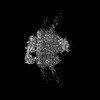
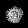
| Title | Intact V-ATPase State 2 and synaptophysin complex in mouse brain isolated synaptic vesicles | |||||||||
 Components Components |
| |||||||||
 Keywords Keywords | MEMBRANE PROTEIN / V-ATPase / synaptic vesicle | |||||||||
| Function / homology |  Function and homology information Function and homology informationIon channel transport / regulation of opioid receptor signaling pathway / Amino acids regulate mTORC1 / clathrin-sculpted glutamate transport vesicle membrane / Transferrin endocytosis and recycling / plasma membrane proton-transporting V-type ATPase complex / Insulin receptor recycling / Metabolism of Angiotensinogen to Angiotensins / negative regulation of autophagic cell death / eye pigmentation ...Ion channel transport / regulation of opioid receptor signaling pathway / Amino acids regulate mTORC1 / clathrin-sculpted glutamate transport vesicle membrane / Transferrin endocytosis and recycling / plasma membrane proton-transporting V-type ATPase complex / Insulin receptor recycling / Metabolism of Angiotensinogen to Angiotensins / negative regulation of autophagic cell death / eye pigmentation / central nervous system maturation / proton-transporting V-type ATPase, V1 domain / ROS and RNS production in phagocytes / positive regulation of transforming growth factor beta1 production / rostrocaudal neural tube patterning / RHOA GTPase cycle / synaptic vesicle lumen acidification / regulation of synaptic vesicle priming / P-type proton-exporting transporter activity / proton-transporting V-type ATPase, V0 domain / osteoclast development / cellular response to increased oxygen levels / extrinsic component of synaptic vesicle membrane / vacuolar transport / vacuolar proton-transporting V-type ATPase, V1 domain / endosome to plasma membrane protein transport / vacuolar proton-transporting V-type ATPase, V0 domain / lysosomal lumen acidification / clathrin-coated vesicle membrane / endosomal lumen acidification / proton-transporting V-type ATPase complex / protein localization to cilium / head morphogenesis / neuron spine / vacuolar proton-transporting V-type ATPase complex / regulation of short-term neuronal synaptic plasticity / vacuolar acidification / : / dendritic spine membrane / syntaxin-1 binding / presynaptic active zone / cholesterol binding / neuron projection terminus / ATPase activator activity / microvillus / regulation of neuronal synaptic plasticity / autophagosome membrane / cilium assembly / regulation of MAPK cascade / proton-transporting ATPase activity, rotational mechanism / regulation of macroautophagy / positive regulation of Wnt signaling pathway / H+-transporting two-sector ATPase / ATP metabolic process / angiotensin maturation / receptor-mediated endocytosis of virus by host cell / ruffle / Neutrophil degranulation / RNA endonuclease activity / endoplasmic reticulum-Golgi intermediate compartment membrane / endomembrane system / proton transmembrane transport / excitatory synapse / secretory granule / receptor-mediated endocytosis / SH2 domain binding / axon terminus / molecular function activator activity / SNARE binding / regulation of long-term neuronal synaptic plasticity / neuromuscular junction / protein homooligomerization / modulation of chemical synaptic transmission / Schaffer collateral - CA1 synapse / small GTPase binding / transmembrane transport / endocytosis / apical part of cell / terminal bouton / positive regulation of canonical Wnt signaling pathway / melanosome / myelin sheath / synaptic vesicle / synaptic vesicle membrane / ATPase binding / presynapse / signaling receptor activity / cell body / presynaptic membrane / chemical synaptic transmission / Hydrolases; Acting on ester bonds / intracellular iron ion homeostasis / early endosome / positive regulation of ERK1 and ERK2 cascade / lysosome / cilium / endosome / apical plasma membrane / endosome membrane / nuclear speck Similarity search - Function | |||||||||
| Biological species |  | |||||||||
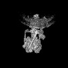
| Method | ELECTRON MICROSCOPY / single particle reconstruction / cryo EM / Resolution: 4.3 Å | |||||||||
 Authors Authors | Wang, C. / Jiang, W. / Yang, K. / Wang, X. / Guo, Q. / Brunger, A.T. | |||||||||
| Funding support |  United States, 2items United States, 2items
| |||||||||
 Citation Citation | Journal: Nature / Year: 2024 Title: Structure and topography of the synaptic V-ATPase-synaptophysin complex. Authors: Chuchu Wang / Wenhong Jiang / Jeremy Leitz / Kailu Yang / Luis Esquivies / Xing Wang / Xiaotao Shen / Richard G Held / Daniel J Adams / Tamara Basta / Lucas Hampton / Ruiqi Jian / Lihua ...Authors: Chuchu Wang / Wenhong Jiang / Jeremy Leitz / Kailu Yang / Luis Esquivies / Xing Wang / Xiaotao Shen / Richard G Held / Daniel J Adams / Tamara Basta / Lucas Hampton / Ruiqi Jian / Lihua Jiang / Michael H B Stowell / Wolfgang Baumeister / Qiang Guo / Axel T Brunger /    Abstract: Synaptic vesicles are organelles with a precisely defined protein and lipid composition, yet the molecular mechanisms for the biogenesis of synaptic vesicles are mainly unknown. Here we discovered a ...Synaptic vesicles are organelles with a precisely defined protein and lipid composition, yet the molecular mechanisms for the biogenesis of synaptic vesicles are mainly unknown. Here we discovered a well-defined interface between the synaptic vesicle V-ATPase and synaptophysin by in situ cryo-electron tomography and single-particle cryo-electron microscopy of functional synaptic vesicles isolated from mouse brains. The synaptic vesicle V-ATPase is an ATP-dependent proton pump that establishes the proton gradient across the synaptic vesicle, which in turn drives the uptake of neurotransmitters. Synaptophysin and its paralogues synaptoporin and synaptogyrin belong to a family of abundant synaptic vesicle proteins whose function is still unclear. We performed structural and functional studies of synaptophysin-knockout mice, confirming the identity of synaptophysin as an interaction partner with the V-ATPase. Although there is little change in the conformation of the V-ATPase upon interaction with synaptophysin, the presence of synaptophysin in synaptic vesicles profoundly affects the copy number of V-ATPases. This effect on the topography of synaptic vesicles suggests that synaptophysin assists in their biogenesis. In support of this model, we observed that synaptophysin-knockout mice exhibit severe seizure susceptibility, suggesting an imbalance of neurotransmitter release as a physiological consequence of the absence of synaptophysin. | |||||||||
| History |
|
- Structure visualization
| Structure viewer | Molecule: MolmilJmol/JSmol |
|---|
- Downloads & links
-Download
| PDBx/mmCIF format | 9bra.cif.gz | 1.4 MB | Display | PDBx/mmCIF format |
|---|---|---|---|---|
| PDB format | pdb9bra.ent.gz | Display | PDB format | |
| PDBx/mmJSON format | 9bra.json.gz | Tree view | PDBx/mmJSON format | |
| Others |  Other downloads Other downloads |
-Validation report
| Arichive directory | https://data.pdbj.org/pub/pdb/validation_reports/br/9braftp://data.pdbj.org/pub/pdb/validation_reports/br/9bra | HTTPS FTP |
|---|
-Related structure data
| Related structure data |  44839MC  9brqC  9brrC  9brsC  9brtC 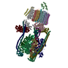 9bruC 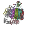 9bryC 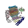 9brzC M: map data used to model this data C: citing same article ( |
|---|---|
| Similar structure data |


-Links
 PDBj
PDBj


- Assembly
| Deposited unit | 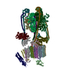
|
|---|---|
| 1 |
|
-Components
-V-type proton ATPase ... , 14 types, 30 molecules 89QRTV01234567UXabdghijklmnoce
| #1: Protein | Mass: 26196.449 Da / Num. of mol.: 3 / Source method: isolated from a natural source / Details: VATE1_MOUSE V-type proton ATPase subunit E 1 / Source: (natural) #2: Protein | Mass: 13674.476 Da / Num. of mol.: 3 / Source method: isolated from a natural source / Details: VATG2_MOUSE V-type proton ATPase subunit G 2 / Source: (natural) #3: Protein | Mass: 68402.875 Da / Num. of mol.: 3 / Source method: isolated from a natural source Details: VATA_MOUSE V-type proton ATPase catalytic subunit A Source: (natural) References: UniProt: P50516, H+-transporting two-sector ATPase #4: Protein | Mass: 56611.570 Da / Num. of mol.: 3 / Source method: isolated from a natural source / Details: VATB2_MOUSE V-type proton ATPase subunit B / Source: (natural) #5: Protein | | Mass: 43945.449 Da / Num. of mol.: 1 / Source method: isolated from a natural source / Details: VATC1_MOUSE V-type proton ATPase subunit C 1 / Source: (natural) #6: Protein | | Mass: 28419.117 Da / Num. of mol.: 1 / Source method: isolated from a natural source / Details: VATD_MOUSE V-type proton ATPase subunit D / Source: (natural) #7: Protein | | Mass: 55922.859 Da / Num. of mol.: 1 / Source method: isolated from a natural source / Details: VATH_MOUSE V-type proton ATPase subunit H / Source: (natural) #8: Protein | | Mass: 13389.262 Da / Num. of mol.: 1 / Source method: isolated from a natural source / Details: VATF_MOUSE V-type proton ATPase subunit F / Source: (natural) #9: Protein | | Mass: 96442.500 Da / Num. of mol.: 1 / Source method: isolated from a natural source Details: VPP1_MOUSE V-type proton ATPase 116 kDa subunit a isoform 1 Source: (natural) #10: Protein | | Mass: 21618.553 Da / Num. of mol.: 1 / Source method: isolated from a natural source Details: VATO_MOUSE V-type proton ATPase 21 kDa proteolipid subunit Source: (natural) #11: Protein | | Mass: 40341.934 Da / Num. of mol.: 1 / Source method: isolated from a natural source / Details: VA0D1_MOUSE V-type proton ATPase subunit d 1 / Source: (natural) #12: Protein | Mass: 15815.833 Da / Num. of mol.: 9 / Source method: isolated from a natural source Details: VATL_MOUSE V-type proton ATPase 16 kDa proteolipid subunit Source: (natural) #14: Protein | | Mass: 51046.215 Da / Num. of mol.: 1 / Source method: isolated from a natural source / Details: VAS1_MOUSE V-type proton ATPase subunit S1 / Source: (natural) #17: Protein | | Mass: 9203.020 Da / Num. of mol.: 1 / Source method: isolated from a natural source / Details: VA0E2_MOUSE V-type proton ATPase subunit e 2 / Source: (natural) |
|---|
-Protein , 3 types, 3 molecules pfs
| #13: Protein | Mass: 39128.789 Da / Num. of mol.: 1 / Source method: isolated from a natural source / Details: RENR_MOUSE Renin receptor / Source: (natural) |
|---|---|
| #15: Protein | Mass: 11000.004 Da / Num. of mol.: 1 / Source method: isolated from a natural source / Details: RNK_MOUSE Ribonuclease kappa / Source: (natural) References: UniProt: Q8K3C0, Hydrolases; Acting on ester bonds |
| #16: Protein | Mass: 34045.406 Da / Num. of mol.: 1 / Source method: isolated from a natural source / Details: SYPH_MOUSE Synaptophysin / Source: (natural) |
-Experimental details
-Experiment
| Experiment | Method: ELECTRON MICROSCOPY |
|---|---|
| EM experiment | Aggregation state: CELL / 3D reconstruction method: single particle reconstruction |
- Sample preparation
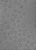
| Component | Name: Mouse brain isolated glutamatergic synaptic vesicles / Type: ORGANELLE OR CELLULAR COMPONENT / Entity ID: all / Source: NATURAL |
|---|---|
| Molecular weight | Experimental value: NO |
| Source (natural) | Organism: |
| Buffer solution | pH: 7.4 |
| Specimen | Embedding applied: NO / Shadowing applied: NO / Staining applied: NO / Vitrification applied: YES Details: The specimen state should be an intact subcellular component. |
| Vitrification | Cryogen name: ETHANE |
- Electron microscopy imaging
| Experimental equipment |  Model: Titan Krios / Image courtesy: FEI Company |
|---|---|
| Microscopy | Model: TFS KRIOS |
| Electron gun | Electron source:  FIELD EMISSION GUN / Accelerating voltage: 300 kV / Illumination mode: FLOOD BEAM FIELD EMISSION GUN / Accelerating voltage: 300 kV / Illumination mode: FLOOD BEAM |
| Electron lens | Mode: BRIGHT FIELD / Nominal defocus max: 4000 nm / Nominal defocus min: 1000 nm |
| Image recording | Electron dose: 50 e/Å2 / Film or detector model: GATAN K3 BIOQUANTUM (6k x 4k) |
- Processing
| Software | Name: PHENIX / Version: 1.21_5207: / Classification: refinement | |||||||||
|---|---|---|---|---|---|---|---|---|---|---|
| EM software |
| |||||||||
| CTF correction | Type: PHASE FLIPPING AND AMPLITUDE CORRECTION | |||||||||
| 3D reconstruction | Resolution: 4.3 Å / Resolution method: FSC 0.143 CUT-OFF / Num. of particles: 25667 / Symmetry type: POINT | |||||||||
| Atomic model building | Protocol: FLEXIBLE FIT |
