 Movie
Movie Controller

[English] 日本語
 Yorodumi
- EMDB-4134: Structure of the mammalian rescue complex with Pelota and Hbs1l a... -
+ Open data

- Basic information
| Entry | Database: EMDB / ID: EMD-4134 | ||||||||||||
|---|---|---|---|---|---|---|---|---|---|---|---|---|---|
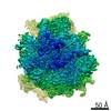
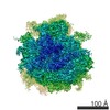
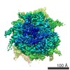
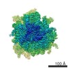
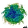
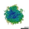
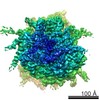
| Title | Structure of the mammalian rescue complex with Pelota and Hbs1l assembled on a truncated mRNA. | ||||||||||||
 Map data Map data | Postprocessed, sharpened map. | ||||||||||||
 Sample Sample |
| ||||||||||||
 Keywords Keywords | Translation / Elongation / Ribosome | ||||||||||||
| Function / homology |  Function and homology information Function and homology informationstalled ribosome sensor activity / Dom34-Hbs1 complex / RNA surveillance / nuclear-transcribed mRNA catabolic process, no-go decay / mRNA decay by 3' to 5' exoribonuclease / nuclear-transcribed mRNA catabolic process, non-stop decay / mesenchymal to epithelial transition / endoderm development / positive regulation of BMP signaling pathway / ribosome disassembly ...stalled ribosome sensor activity / Dom34-Hbs1 complex / RNA surveillance / nuclear-transcribed mRNA catabolic process, no-go decay / mRNA decay by 3' to 5' exoribonuclease / nuclear-transcribed mRNA catabolic process, non-stop decay / mesenchymal to epithelial transition / endoderm development / positive regulation of BMP signaling pathway / ribosome disassembly / inner cell mass cell proliferation / nonfunctional rRNA decay / stem cell population maintenance / chromosome organization / ubiquitin ligase inhibitor activity / 90S preribosome / translation elongation factor activity / positive regulation of signal transduction by p53 class mediator / phagocytic cup / translation regulator activity / rough endoplasmic reticulum / ribosomal small subunit export from nucleus / gastrulation / MDM2/MDM4 family protein binding / rescue of stalled cytosolic ribosome / class I DNA-(apurinic or apyrimidinic site) endonuclease activity / cytosolic ribosome / DNA-(apurinic or apyrimidinic site) lyase / ribosomal large subunit biogenesis / maturation of LSU-rRNA from tricistronic rRNA transcript (SSU-rRNA, 5.8S rRNA, LSU-rRNA) / maturation of SSU-rRNA from tricistronic rRNA transcript (SSU-rRNA, 5.8S rRNA, LSU-rRNA) / positive regulation of apoptotic signaling pathway / maturation of SSU-rRNA / small-subunit processome / spindle / rRNA processing / rhythmic process / positive regulation of canonical Wnt signaling pathway / regulation of translation / antimicrobial humoral immune response mediated by antimicrobial peptide / large ribosomal subunit / ribosomal small subunit assembly / ribosome binding / ribosomal small subunit biogenesis / 5S rRNA binding / ribosomal large subunit assembly / small ribosomal subunit / small ribosomal subunit rRNA binding / large ribosomal subunit rRNA binding / cytosolic small ribosomal subunit / killing of cells of another organism / cytosolic large ribosomal subunit / defense response to Gram-negative bacterium / Hydrolases; Acting on acid anhydrides; Acting on GTP to facilitate cellular and subcellular movement / perikaryon / cell differentiation / cytoplasmic translation / tRNA binding / mitochondrial inner membrane / postsynaptic density / rRNA binding / structural constituent of ribosome / ribosome / translation / ribonucleoprotein complex / cell division / DNA repair / mRNA binding / GTPase activity / apoptotic process / centrosome / synapse / dendrite / GTP binding / nucleolus / perinuclear region of cytoplasm / Golgi apparatus / signal transduction / endoplasmic reticulum / DNA binding / RNA binding / extracellular exosome / zinc ion binding / membrane / metal ion binding / nucleus / cytoplasm / cytosol Similarity search - Function | ||||||||||||
| Biological species |   Homo sapiens (human) Homo sapiens (human) | ||||||||||||
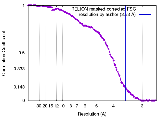
| Method | single particle reconstruction / cryo EM / Resolution: 3.53 Å | ||||||||||||
 Authors Authors | Shao S / Murray J | ||||||||||||
| Funding support |  United Kingdom, 3 items United Kingdom, 3 items
| ||||||||||||
 Citation Citation | Journal: Cell / Year: 2016 Title: Decoding Mammalian Ribosome-mRNA States by Translational GTPase Complexes. Authors: Sichen Shao / Jason Murray / Alan Brown / Jack Taunton / V Ramakrishnan / Ramanujan S Hegde /  Abstract: In eukaryotes, accurate protein synthesis relies on a family of translational GTPases that pair with specific decoding factors to decipher the mRNA code on ribosomes. We present structures of the ...In eukaryotes, accurate protein synthesis relies on a family of translational GTPases that pair with specific decoding factors to decipher the mRNA code on ribosomes. We present structures of the mammalian ribosome engaged with decoding factor⋅GTPase complexes representing intermediates of translation elongation (aminoacyl-tRNA⋅eEF1A), termination (eRF1⋅eRF3), and ribosome rescue (Pelota⋅Hbs1l). Comparative analyses reveal that each decoding factor exploits the plasticity of the ribosomal decoding center to differentially remodel ribosomal proteins and rRNA. This leads to varying degrees of large-scale ribosome movements and implies distinct mechanisms for communicating information from the decoding center to each GTPase. Additional structural snapshots of the translation termination pathway reveal the conformational changes that choreograph the accommodation of decoding factors into the peptidyl transferase center. Our results provide a structural framework for how different states of the mammalian ribosome are selectively recognized by the appropriate decoding factor⋅GTPase complex to ensure translational fidelity. | ||||||||||||
| History |
|
- Structure visualization
| Movie |
Movie viewer |
|---|---|
| Structure viewer | EM map: SurfViewMolmilJmol/JSmol |
| Supplemental images |

- Downloads & links
-EMDB archive
| Map data | emd_4134.map.gz | 13.9 MB | EMDB map data format | |
|---|---|---|---|---|
| Header (meta data) | emd-4134-v30.xmlemd-4134.xml | 115.2 KB 115.2 KB | Display Display | EMDB header |
| FSC (resolution estimation) | emd_4134_fsc.xml | 14.7 KB | Display | FSC data file |
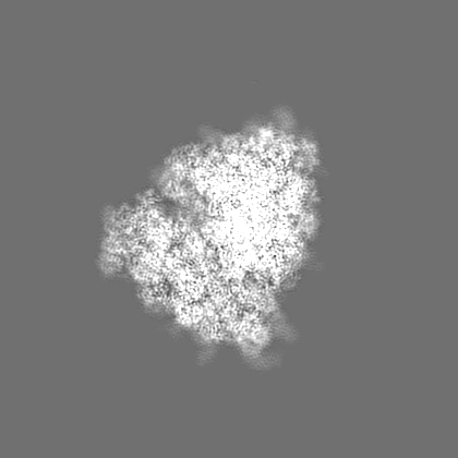
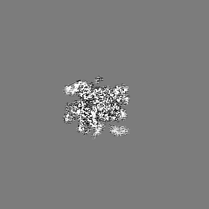
| Images | 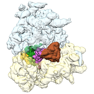 emd_4134.png emd_4134.png | 189 KB | ||
| Filedesc metadata | emd-4134.cif.gz | 22.8 KB | ||
| Others | emd_4134_half_map_1.map.gzemd_4134_half_map_2.map.gz | 247 MB 245.9 MB | ||
| Archive directory |  http://ftp.pdbj.org/pub/emdb/structures/EMD-4134ftp://ftp.pdbj.org/pub/emdb/structures/EMD-4134 http://ftp.pdbj.org/pub/emdb/structures/EMD-4134ftp://ftp.pdbj.org/pub/emdb/structures/EMD-4134 | HTTPS FTP |
-Related structure data
| Related structure data |  5lzwMC 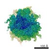 4129C 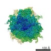 4130C 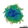 4131C 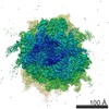 4132C 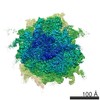 4133C 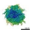 4135C 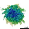 4136C 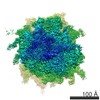 4137C  5lzsC 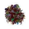 5lztC 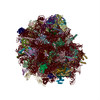 5lzuC 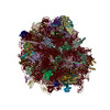 5lzvC  5lzxC  5lzyC  5lzzC M: atomic model generated by this map C: citing same article ( |
|---|---|
| Similar structure data |

-Links
| EMDB pages | EMDB (EBI/PDBe) / EMDataResource |
|---|---|
| Related items in Molecule of the Month |


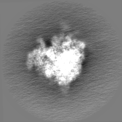
-Map
| File | Download / File: emd_4134.map.gz / Format: CCP4 / Size: 282.6 MB / Type: IMAGE STORED AS FLOATING POINT NUMBER (4 BYTES) | ||||||||||||||||||||||||||||||||||||||||||||||||||||||||||||||||||||
|---|---|---|---|---|---|---|---|---|---|---|---|---|---|---|---|---|---|---|---|---|---|---|---|---|---|---|---|---|---|---|---|---|---|---|---|---|---|---|---|---|---|---|---|---|---|---|---|---|---|---|---|---|---|---|---|---|---|---|---|---|---|---|---|---|---|---|---|---|---|
| Annotation | Postprocessed, sharpened map. | ||||||||||||||||||||||||||||||||||||||||||||||||||||||||||||||||||||
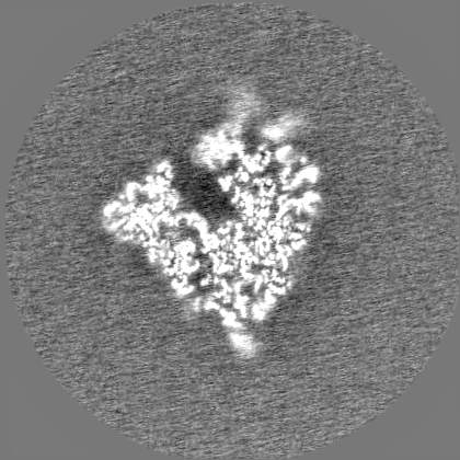
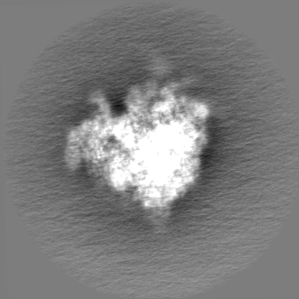
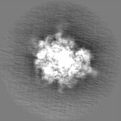
| Projections & slices | Image control
Images are generated by Spider. | ||||||||||||||||||||||||||||||||||||||||||||||||||||||||||||||||||||
| Voxel size | X=Y=Z: 1.34 Å | ||||||||||||||||||||||||||||||||||||||||||||||||||||||||||||||||||||
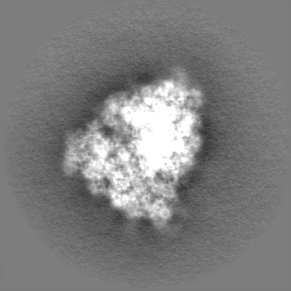
| Density |
| ||||||||||||||||||||||||||||||||||||||||||||||||||||||||||||||||||||
| Symmetry | Space group: 1 | ||||||||||||||||||||||||||||||||||||||||||||||||||||||||||||||||||||
| Details | EMDB XML:
CCP4 map header:
| ||||||||||||||||||||||||||||||||||||||||||||||||||||||||||||||||||||
 Z (Sec.)
 Y (Row.)
 X (Col.)


-Supplemental data
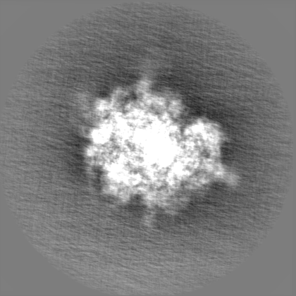
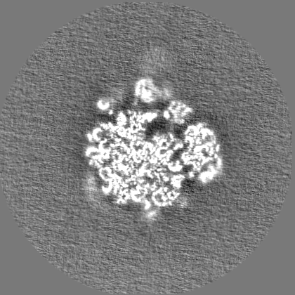
-Half map: Half map 1.
| File | emd_4134_half_map_1.map | ||||||||||||
|---|---|---|---|---|---|---|---|---|---|---|---|---|---|
| Annotation | Half map 1. | ||||||||||||
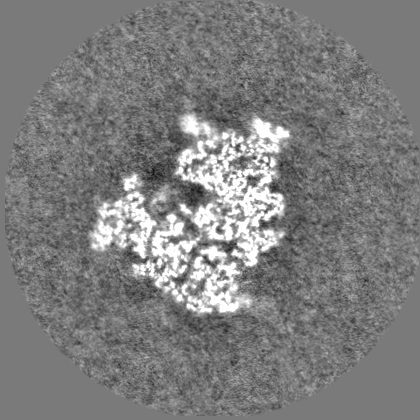
| Projections & Slices |
| ||||||||||||
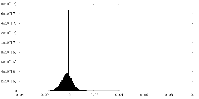
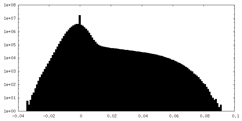
| Density Histograms |

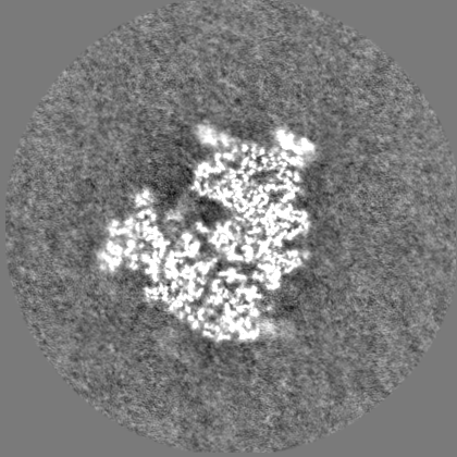

-Half map: Half map 2.
| File | emd_4134_half_map_2.map | ||||||||||||
|---|---|---|---|---|---|---|---|---|---|---|---|---|---|
| Annotation | Half map 2. | ||||||||||||
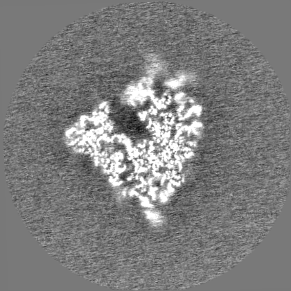
| Projections & Slices |
| ||||||||||||
| Density Histograms |


- Sample components
+Entire : Affinity-purified 80S ribosome-nascent chain complex reconstitute...
+Supramolecule #1: Affinity-purified 80S ribosome-nascent chain complex reconstitute...
+Macromolecule #1: uL2
+Macromolecule #2: uL3
+Macromolecule #3: uL4
+Macromolecule #4: uL18
+Macromolecule #5: eL6
+Macromolecule #6: uL30
+Macromolecule #7: eL8
+Macromolecule #8: uL6
+Macromolecule #9: uL16
+Macromolecule #10: uL5
+Macromolecule #11: eL13
+Macromolecule #12: eL14
+Macromolecule #13: eL15
+Macromolecule #14: uL13
+Macromolecule #15: uL22
+Macromolecule #16: eL18
+Macromolecule #17: eL19
+Macromolecule #18: eL20
+Macromolecule #19: eL21
+Macromolecule #20: eL22
+Macromolecule #21: uL14
+Macromolecule #22: eL24
+Macromolecule #23: uL23
+Macromolecule #24: uL24
+Macromolecule #25: eL27
+Macromolecule #26: uL15
+Macromolecule #27: eL29
+Macromolecule #28: eL30
+Macromolecule #29: eL31
+Macromolecule #30: eL32
+Macromolecule #31: eL33
+Macromolecule #32: eL34
+Macromolecule #33: uL29
+Macromolecule #34: eL36
+Macromolecule #35: eL37
+Macromolecule #36: eL38
+Macromolecule #37: eL39
+Macromolecule #38: eL40
+Macromolecule #39: eL41
+Macromolecule #40: eL42
+Macromolecule #41: eL43
+Macromolecule #42: eL28
+Macromolecule #43: uL10
+Macromolecule #44: uL11
+Macromolecule #45: Nascent chain
+Macromolecule #52: uS2
+Macromolecule #53: eS1
+Macromolecule #54: uS5
+Macromolecule #55: uS3
+Macromolecule #56: eS4
+Macromolecule #57: uS7
+Macromolecule #58: eS6
+Macromolecule #59: eS7
+Macromolecule #60: eS8
+Macromolecule #61: uS4
+Macromolecule #62: eS10
+Macromolecule #63: uS17
+Macromolecule #64: eS12
+Macromolecule #65: uS15
+Macromolecule #66: uS11
+Macromolecule #67: uS19
+Macromolecule #68: uS9
+Macromolecule #69: eS17
+Macromolecule #70: uS13
+Macromolecule #71: eS19
+Macromolecule #72: uS10
+Macromolecule #73: eS21
+Macromolecule #74: uS8
+Macromolecule #75: uS12
+Macromolecule #76: eS24
+Macromolecule #77: eS25
+Macromolecule #78: eS26
+Macromolecule #79: eS27
+Macromolecule #80: eS28
+Macromolecule #81: uS14
+Macromolecule #82: eS30
+Macromolecule #83: eS31
+Macromolecule #84: RACK1
+Macromolecule #86: Pelota

+Macromolecule #87: Hbs1l
+Macromolecule #46: P-site tRNA
+Macromolecule #47: E-site tRNA
+Macromolecule #48: 28S ribosomal RNA
+Macromolecule #49: 5S ribosomal RNA
+Macromolecule #50: 5.8S ribosomal RNA
+Macromolecule #51: 18S ribosomal RNA
+Macromolecule #85: mRNA (truncated)
+Macromolecule #88: MAGNESIUM ION
+Macromolecule #89: ZINC ION
+Macromolecule #90: PHOSPHOMETHYLPHOSPHONIC ACID GUANYLATE ESTER

-Experimental details
-Structure determination
| Method | cryo EM |
|---|---|
 Processing Processing | single particle reconstruction |
| Aggregation state | particle |
-Sample preparation
| Concentration | 0.2 mg/mL | |||||||||||||||
|---|---|---|---|---|---|---|---|---|---|---|---|---|---|---|---|---|
| Buffer | pH: 7.4 Component:
| |||||||||||||||
| Grid | Model: Quantifoil R2/2 / Material: COPPER / Mesh: 400 / Support film - Material: CARBON / Support film - topology: CONTINUOUS / Support film - Film thickness: 5 / Pretreatment - Type: GLOW DISCHARGE | |||||||||||||||
| Vitrification | Cryogen name: ETHANE / Chamber humidity: 100 % / Chamber temperature: 277 K / Instrument: FEI VITROBOT MARK III Details: 3 ul aliquots were applied to the grid and incubated for 30 s, before blotting for 3s to remove excess solution.. |
- Electron microscopy
| Microscope | FEI TITAN KRIOS |
|---|---|
| Image recording | Film or detector model: FEI FALCON II (4k x 4k) / Detector mode: INTEGRATING / Number real images: 1815 / Average exposure time: 1.0 sec. / Average electron dose: 30.0 e/Å2 |
| Electron beam | Acceleration voltage: 300 kV / Electron source:  FIELD EMISSION GUN FIELD EMISSION GUN |
| Electron optics | C2 aperture diameter: 70.0 µm / Calibrated magnification: 104478 / Illumination mode: FLOOD BEAM / Imaging mode: BRIGHT FIELD / Cs: 2.7 mm / Nominal magnification: 59000 |
| Sample stage | Specimen holder model: FEI TITAN KRIOS AUTOGRID HOLDER / Cooling holder cryogen: NITROGEN |
| Experimental equipment |  Model: Titan Krios / Image courtesy: FEI Company |
+Image processing
-Atomic model buiding 1
| Refinement | Space: RECIPROCAL / Protocol: OTHER / Overall B value: 84.1 / Target criteria: FSCaverage |
|---|---|
| Output model | PDB-5lzw: |
