Movie
Movie Controller

[English] 日本語
 Yorodumi
- EMDB-25404: Activated filamentous SgrAI endonuclease with Ca2+ and intact pri... -
+ Open data

- Basic information
| Entry | Database: EMDB / ID: EMD-25404 | |||||||||
|---|---|---|---|---|---|---|---|---|---|---|
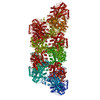
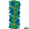
| Title | Activated filamentous SgrAI endonuclease with Ca2+ and intact primary site DNA | |||||||||
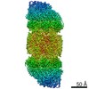
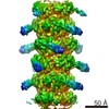
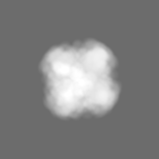
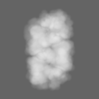
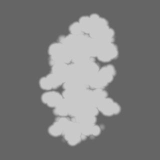
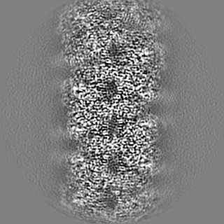
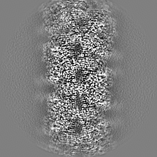
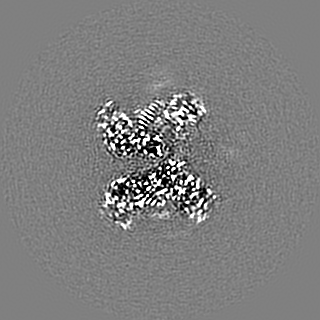
 Map data Map data | Full reconstruction of filamentous SgrAI bound to Ca2 and intact primary site DNA | |||||||||
 Sample Sample |
| |||||||||
 Keywords Keywords | restriction endonuclease / DNAse / allostery / bacterial innate immunity / filament / hyper-activation / substrate specificity / HYDROLASE-DNA complex | |||||||||
| Function / homology | Restriction endonuclease, type II, Cfr10I/Bse634I / Cfr10I/Bse634I restriction endonuclease / Restriction endonuclease type II-like / metal ion binding / identical protein binding / SgraIR restriction enzyme Function and homology information Function and homology information | |||||||||
| Biological species |  Streptomyces griseus (bacteria) Streptomyces griseus (bacteria) | |||||||||
| Method | helical reconstruction / cryo EM / Resolution: 2.7 Å | |||||||||
 Authors Authors | Shan Z / Lyumkis D | |||||||||
| Funding support |  United States, 2 items United States, 2 items
| |||||||||
 Citation Citation | Journal: J Biol Chem / Year: 2022 Title: Pretransition state and apo structures of the filament-forming enzyme SgrAI elucidate mechanisms of activation and substrate specificity. Authors: Zelin Shan / Niloofar Ghadirian / Dmitry Lyumkis / Nancy C Horton / Abstract: Enzyme filamentation is a widespread phenomenon that mediates enzyme regulation and function. For the filament-forming sequence-specific DNA endonuclease SgrAI, the process of filamentation both ...Enzyme filamentation is a widespread phenomenon that mediates enzyme regulation and function. For the filament-forming sequence-specific DNA endonuclease SgrAI, the process of filamentation both accelerates its DNA cleavage activity and expands its DNA sequence specificity, thus allowing for many additional DNA sequences to be rapidly cleaved. Both outcomes-the acceleration of DNA cleavage and the expansion of sequence specificity-are proposed to regulate critical processes in bacterial innate immunity. However, the mechanistic bases underlying these events remain unclear. Herein, we describe two new structures of the SgrAI enzyme that shed light on its catalytic function. First, we present the cryo-EM structure of filamentous SgrAI bound to intact primary site DNA and Ca resolved to ∼2.5 Å within the catalytic center, which represents the trapped enzyme-DNA complex prior to the DNA cleavage reaction. This structure reveals important conformational changes that contribute to the catalytic mechanism and the binding of a second divalent cation in the enzyme active site, which is expected to contribute to increased DNA cleavage activity of SgrAI in the filamentous state. Second, we present an X-ray crystal structure of DNA-free (apo) SgrAI resolved to 2.0 Å resolution, which reveals a disordered loop involved in DNA recognition. Collectively, these multiple new observations clarify the mechanism of expansion of DNA sequence specificity of SgrAI, including the indirect readout of sequence-dependent DNA structure, changes in protein-DNA interactions, and the disorder-to-order transition of a crucial DNA recognition element. | |||||||||
| History |
|
- Structure visualization
| Movie |
Movie viewer |
|---|---|
| Structure viewer | EM map: SurfViewMolmilJmol/JSmol |
| Supplemental images |

- Downloads & links
-EMDB archive
| Map data | emd_25404.map.gz | 98.6 MB | EMDB map data format | |
|---|---|---|---|---|
| Header (meta data) | emd-25404-v30.xmlemd-25404.xml | 22.7 KB 22.7 KB | Display Display | EMDB header |
| FSC (resolution estimation) | emd_25404_fsc.xml | 11.4 KB | Display | FSC data file |
| Images | 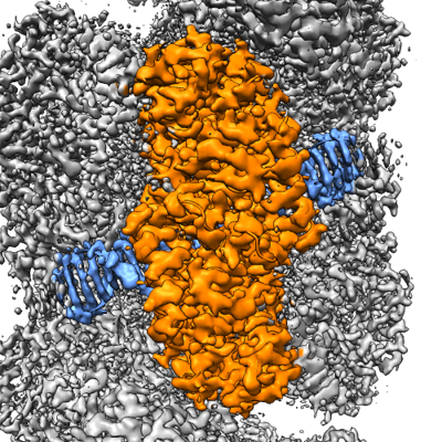 emd_25404.png emd_25404.png | 289.1 KB | ||
| Masks | emd_25404_msk_1.map | 125 MB | Mask map | |
| Filedesc metadata | emd-25404.cif.gz | 6.9 KB | ||
| Others | emd_25404_additional_1.map.gzemd_25404_half_map_1.map.gzemd_25404_half_map_2.map.gz | 117.1 MB 98.6 MB 98.7 MB | ||
| Archive directory |  http://ftp.pdbj.org/pub/emdb/structures/EMD-25404ftp://ftp.pdbj.org/pub/emdb/structures/EMD-25404 http://ftp.pdbj.org/pub/emdb/structures/EMD-25404ftp://ftp.pdbj.org/pub/emdb/structures/EMD-25404 | HTTPS FTP |
-Related structure data
| Related structure data |  7ss5MC  7s8dC M: atomic model generated by this map C: citing same article ( |
|---|---|
| Similar structure data |

-Links
| EMDB pages | EMDB (EBI/PDBe) / EMDataResource |
|---|---|
| Related items in Molecule of the Month |


-Map
| File | Download / File: emd_25404.map.gz / Format: CCP4 / Size: 125 MB / Type: IMAGE STORED AS FLOATING POINT NUMBER (4 BYTES) | ||||||||||||||||||||||||||||||||||||||||||||||||||||||||||||||||||||
|---|---|---|---|---|---|---|---|---|---|---|---|---|---|---|---|---|---|---|---|---|---|---|---|---|---|---|---|---|---|---|---|---|---|---|---|---|---|---|---|---|---|---|---|---|---|---|---|---|---|---|---|---|---|---|---|---|---|---|---|---|---|---|---|---|---|---|---|---|---|
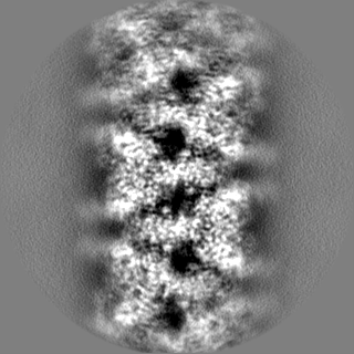
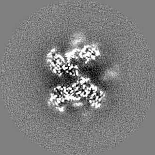
| Annotation | Full reconstruction of filamentous SgrAI bound to Ca2 and intact primary site DNA | ||||||||||||||||||||||||||||||||||||||||||||||||||||||||||||||||||||
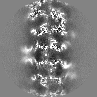
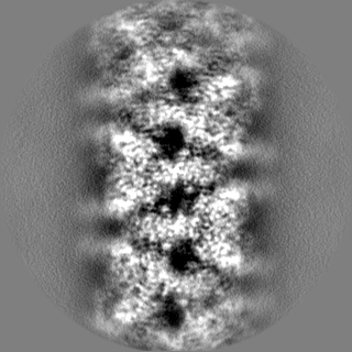
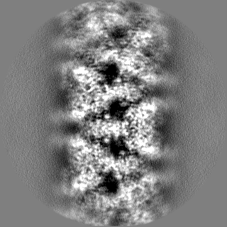
| Projections & slices | Image control
Images are generated by Spider. | ||||||||||||||||||||||||||||||||||||||||||||||||||||||||||||||||||||
| Voxel size | X=Y=Z: 0.83 Å | ||||||||||||||||||||||||||||||||||||||||||||||||||||||||||||||||||||
| Density |
| ||||||||||||||||||||||||||||||||||||||||||||||||||||||||||||||||||||
| Symmetry | Space group: 1 | ||||||||||||||||||||||||||||||||||||||||||||||||||||||||||||||||||||
| Details | EMDB XML:
CCP4 map header:
| ||||||||||||||||||||||||||||||||||||||||||||||||||||||||||||||||||||
 Z (Sec.)
 Y (Row.)
 X (Col.)


-Supplemental data
-Mask #1
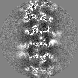
| File | emd_25404_msk_1.map | ||||||||||||
|---|---|---|---|---|---|---|---|---|---|---|---|---|---|
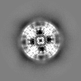
| Projections & Slices |
| ||||||||||||
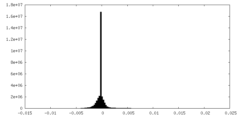
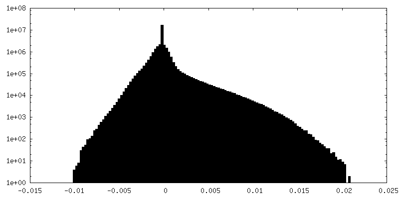
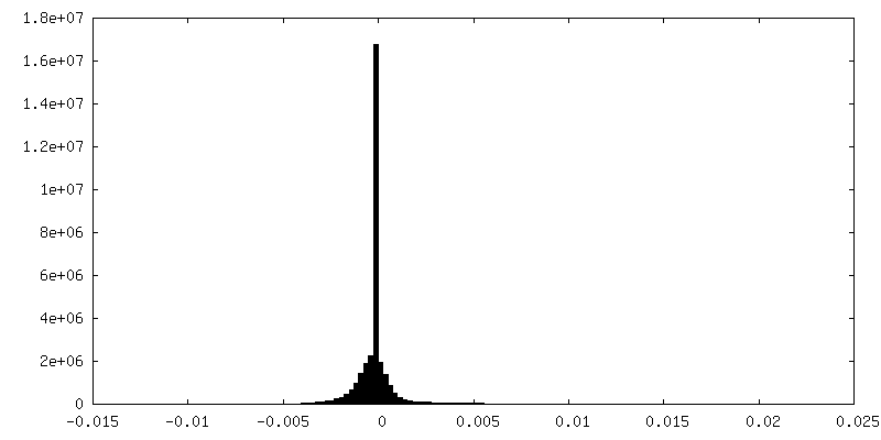
| Density Histograms |


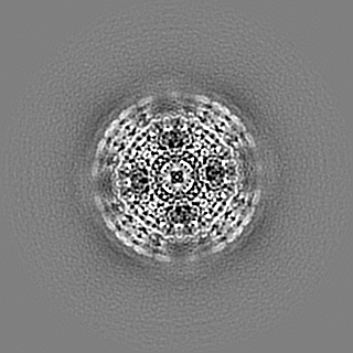
-Additional map: Reconstruction of filamentous SgrAI bound to Ca2 and...
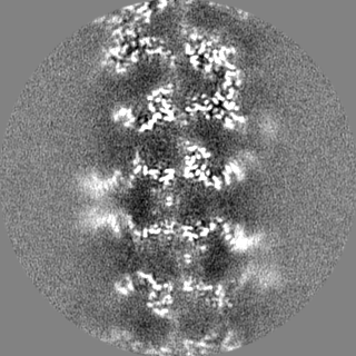
| File | emd_25404_additional_1.map | ||||||||||||
|---|---|---|---|---|---|---|---|---|---|---|---|---|---|
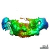
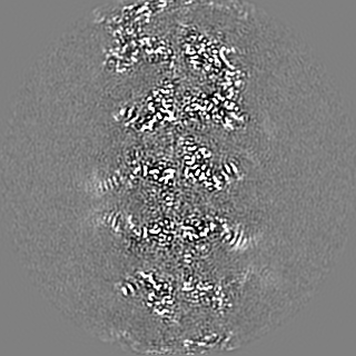
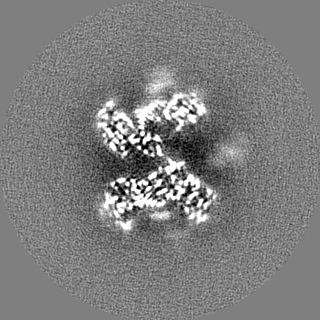
| Annotation | Reconstruction of filamentous SgrAI bound to Ca2 and intact primary site DNA, density-modified, 2-fold symmetrized, and averaged along Z-axis | ||||||||||||
| Projections & Slices |
| ||||||||||||
| Density Histograms |


-Half map: half map #1 of filamentous SgrAI bound to...
| File | emd_25404_half_map_1.map | ||||||||||||
|---|---|---|---|---|---|---|---|---|---|---|---|---|---|
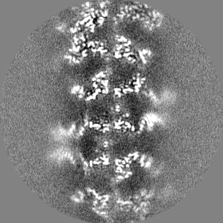
| Annotation | half map #1 of filamentous SgrAI bound to Ca2 and intact primary site DNA | ||||||||||||
| Projections & Slices |
| ||||||||||||
| Density Histograms |


-Half map: half map #2 of filamentous SgrAI bound to...
| File | emd_25404_half_map_2.map | ||||||||||||
|---|---|---|---|---|---|---|---|---|---|---|---|---|---|
| Annotation | half map #2 of filamentous SgrAI bound to Ca2 and intact primary site DNA | ||||||||||||
| Projections & Slices |
| ||||||||||||
| Density Histograms |

- Sample components
-Entire : Activated filamentous SgrAI endonuclease with Ca2+ and intact pri...
| Entire | Name: Activated filamentous SgrAI endonuclease with Ca2+ and intact primary site DNA |
|---|---|
| Components |
|
-Supramolecule #1: Activated filamentous SgrAI endonuclease with Ca2+ and intact pri...
| Supramolecule | Name: Activated filamentous SgrAI endonuclease with Ca2+ and intact primary site DNA type: complex / ID: 1 / Parent: 0 / Macromolecule list: #1-#2 |
|---|---|
| Source (natural) | Organism: Streptomyces griseus (bacteria) |
| Molecular weight | Theoretical: 74 kDa/nm |
-Macromolecule #1: SgraIR restriction enzyme
| Macromolecule | Name: SgraIR restriction enzyme / type: protein_or_peptide / ID: 1 / Number of copies: 2 / Enantiomer: LEVO |
|---|---|
| Source (natural) | Organism: Streptomyces griseus (bacteria) |
| Molecular weight | Theoretical: 39.783895 KDa |
| Recombinant expression | Organism: |
| Sequence | String: MPFTYSIEAT RNLATTERCI QDIRNAPVRN RSTQFQLAQQ NMLAYTFGEV IPGFASAGIN GMDYRDVIGR PVENAVTEGT HFFRDDFRV DSNAKAKVAG DIFEIVSSAV MWNCAARWNS LMVGEGWRSQ PRYSRPTLSP SPRRQVAVLN LPRSFDWVSL L VPESQEVI ...String: MPFTYSIEAT RNLATTERCI QDIRNAPVRN RSTQFQLAQQ NMLAYTFGEV IPGFASAGIN GMDYRDVIGR PVENAVTEGT HFFRDDFRV DSNAKAKVAG DIFEIVSSAV MWNCAARWNS LMVGEGWRSQ PRYSRPTLSP SPRRQVAVLN LPRSFDWVSL L VPESQEVI EEFRAGLRKD GLGLPTSTPD LAVVVLPEEF QNDEMWREEI AGLTRPNQIL LSGAYQRLQG RVQPGEISLA VA FKRSLRS DRLYQPLYEA NVMQLLLEGK LGAPKVEFEV HTLAPEGTNA FVTYEAASLY GLAEGRSAVH RAIRELYVPP TAA DLARRF FAFLNERMEL VNGENLYFQS HHHHHH UniProtKB: SgraIR restriction enzyme |
-Macromolecule #2: DNA (5'-D(P*GP*GP*TP*CP*TP*TP*CP*AP*CP*AP*CP*CP*GP*GP*TP*GP*TP*GP...
| Macromolecule | Name: DNA (5'-D(P*GP*GP*TP*CP*TP*TP*CP*AP*CP*AP*CP*CP*GP*GP*TP*GP*TP*GP*AP*AP*GP*AP*CP*C)-3') type: dna / ID: 2 / Number of copies: 2 / Classification: DNA |
|---|---|
| Source (natural) | Organism: Streptomyces griseus (bacteria) |
| Molecular weight | Theoretical: 12.314889 KDa |
| Sequence | String: (DG)(DA)(DT)(DG)(DC)(DG)(DT)(DG)(DG)(DG) (DT)(DC)(DT)(DT)(DC)(DA)(DC)(DA)(DC)(DC) (DG)(DG)(DT)(DG)(DT)(DG)(DA)(DA)(DG) (DA)(DC)(DC)(DC)(DA)(DC)(DG)(DC)(DA)(DT) (DC) |
-Macromolecule #3: CALCIUM ION
| Macromolecule | Name: CALCIUM ION / type: ligand / ID: 3 / Number of copies: 6 / Formula: CA |
|---|---|
| Molecular weight | Theoretical: 40.078 Da |
-Macromolecule #4: water
| Macromolecule | Name: water / type: ligand / ID: 4 / Number of copies: 593 / Formula: HOH |
|---|---|
| Molecular weight | Theoretical: 18.015 Da |
| Chemical component information |  ChemComp-HOH: |
-Experimental details
-Structure determination
| Method | cryo EM |
|---|---|
 Processing Processing | helical reconstruction |
| Aggregation state | filament |
-Sample preparation
| Buffer | pH: 8 Component:
| |||||||||||||||
|---|---|---|---|---|---|---|---|---|---|---|---|---|---|---|---|---|
| Grid | Model: UltrAuFoil R1.2/1.3 / Material: GOLD / Support film - Material: GOLD / Support film - topology: HOLEY / Pretreatment - Type: PLASMA CLEANING / Pretreatment - Time: 7 sec. / Pretreatment - Atmosphere: AIR | |||||||||||||||
| Vitrification | Cryogen name: ETHANE / Instrument: HOMEMADE PLUNGER Details: Cryo-EM grids were prepared by freezing using a manual plunger in cold room at 4C. |
- Electron microscopy
| Microscope | FEI TITAN KRIOS |
|---|---|
| Specialist optics | Energy filter - Name: GIF Bioquantum / Energy filter - Slit width: 30 eV |
| Image recording | Film or detector model: GATAN K2 SUMMIT (4k x 4k) / Detector mode: COUNTING / Digitization - Dimensions - Width: 3838 pixel / Digitization - Dimensions - Height: 3710 pixel / Digitization - Frames/image: 1-60 / Number real images: 216 / Average electron dose: 55.0 e/Å2 |
| Electron beam | Acceleration voltage: 300 kV / Electron source:  FIELD EMISSION GUN FIELD EMISSION GUN |
| Electron optics | C2 aperture diameter: 70.0 µm / Calibrated magnification: 60240 / Illumination mode: FLOOD BEAM / Imaging mode: BRIGHT FIELD / Cs: 2.7 mm / Nominal defocus max: 2.0 µm / Nominal defocus min: 0.8 µm / Nominal magnification: 165000 |
| Sample stage | Specimen holder model: FEI TITAN KRIOS AUTOGRID HOLDER / Cooling holder cryogen: NITROGEN |
| Experimental equipment |  Model: Titan Krios / Image courtesy: FEI Company |
-Image processing
| Final reconstruction | Applied symmetry - Helical parameters - Δz: 21.2 Å Applied symmetry - Helical parameters - Δ&Phi: -85.8 ° Applied symmetry - Helical parameters - Axial symmetry: C1 (asymmetric) Algorithm: FOURIER SPACE / Resolution.type: BY AUTHOR / Resolution: 2.7 Å / Resolution method: FSC 0.143 CUT-OFF / Software - Name: RELION (ver. 3.1) / Number images used: 220067 |
|---|---|
| Startup model | Type of model: EMDB MAP EMDB ID: |
| Final angle assignment | Type: PROJECTION MATCHING / Software - Name: RELION (ver. 3.1) |
| FSC plot (resolution estimation) |  |
-Atomic model buiding 1
| Initial model | PDB ID: Chain - Source name: PDB / Chain - Initial model type: experimental model |
|---|---|
| Refinement | Space: REAL / Protocol: FLEXIBLE FIT / Overall B value: 107.7 / Target criteria: Correlation coefficient |
| Output model | PDB-7ss5: |