Movie
Movie Controller

[English] 日本語
 Yorodumi
- PDB-7ss5: Activated SgrAI endonuclease DNA-bound dimer with Ca2+ and intact... -
+ Open data

- Basic information
| Entry | Database: PDB / ID: 7ss5 | |||||||||
|---|---|---|---|---|---|---|---|---|---|---|
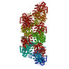
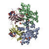
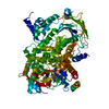
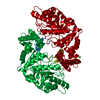
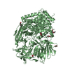
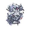
| Title | Activated SgrAI endonuclease DNA-bound dimer with Ca2+ and intact primary site DNA | |||||||||
 Components Components |
| |||||||||
 Keywords Keywords | HYDROLASE/DNA / restriction endonuclease / DNAse / allostery / bacterial innate immunity / filament / hyper-activation / substrate specificity / HYDROLASE-DNA complex | |||||||||
| Function / homology | Restriction endonuclease, type II, Cfr10I/Bse634I / Cfr10I/Bse634I restriction endonuclease / Restriction endonuclease type II-like / metal ion binding / identical protein binding / DNA / DNA (> 10) / SgraIR restriction enzyme Function and homology information Function and homology information | |||||||||
| Biological species |  Streptomyces griseus (bacteria) Streptomyces griseus (bacteria) | |||||||||
| Method | ELECTRON MICROSCOPY / helical reconstruction / cryo EM / Resolution: 2.7 Å | |||||||||
 Authors Authors | Shan, Z. / Lyumkis, D. / Horton, N.C. | |||||||||
| Funding support |  United States, 2items United States, 2items
| |||||||||
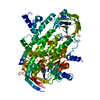
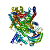
 Citation Citation | Journal: J Biol Chem / Year: 2022 Title: Pretransition state and apo structures of the filament-forming enzyme SgrAI elucidate mechanisms of activation and substrate specificity. Authors: Zelin Shan / Niloofar Ghadirian / Dmitry Lyumkis / Nancy C Horton / Abstract: Enzyme filamentation is a widespread phenomenon that mediates enzyme regulation and function. For the filament-forming sequence-specific DNA endonuclease SgrAI, the process of filamentation both ...Enzyme filamentation is a widespread phenomenon that mediates enzyme regulation and function. For the filament-forming sequence-specific DNA endonuclease SgrAI, the process of filamentation both accelerates its DNA cleavage activity and expands its DNA sequence specificity, thus allowing for many additional DNA sequences to be rapidly cleaved. Both outcomes-the acceleration of DNA cleavage and the expansion of sequence specificity-are proposed to regulate critical processes in bacterial innate immunity. However, the mechanistic bases underlying these events remain unclear. Herein, we describe two new structures of the SgrAI enzyme that shed light on its catalytic function. First, we present the cryo-EM structure of filamentous SgrAI bound to intact primary site DNA and Ca resolved to ∼2.5 Å within the catalytic center, which represents the trapped enzyme-DNA complex prior to the DNA cleavage reaction. This structure reveals important conformational changes that contribute to the catalytic mechanism and the binding of a second divalent cation in the enzyme active site, which is expected to contribute to increased DNA cleavage activity of SgrAI in the filamentous state. Second, we present an X-ray crystal structure of DNA-free (apo) SgrAI resolved to 2.0 Å resolution, which reveals a disordered loop involved in DNA recognition. Collectively, these multiple new observations clarify the mechanism of expansion of DNA sequence specificity of SgrAI, including the indirect readout of sequence-dependent DNA structure, changes in protein-DNA interactions, and the disorder-to-order transition of a crucial DNA recognition element. | |||||||||
| History |
|
- Structure visualization
| Movie |
Movie viewer |
|---|---|
| Structure viewer | Molecule: MolmilJmol/JSmol |
- Downloads & links
-Download
| PDBx/mmCIF format | 7ss5.cif.gz | 170.6 KB | Display | PDBx/mmCIF format |
|---|---|---|---|---|
| PDB format | pdb7ss5.ent.gz | 127.8 KB | Display | PDB format |
| PDBx/mmJSON format | 7ss5.json.gz | Tree view | PDBx/mmJSON format | |
| Others |  Other downloads Other downloads |
-Validation report
| Arichive directory | https://data.pdbj.org/pub/pdb/validation_reports/ss/7ss5ftp://data.pdbj.org/pub/pdb/validation_reports/ss/7ss5 | HTTPS FTP |
|---|
-Related structure data
| Related structure data | 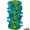 25404MC  7s8dC M: map data used to model this data C: citing same article ( |
|---|---|
| Similar structure data |

-Links
 PDBj
PDBj


- Assembly
| Deposited unit | 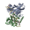
|
|---|---|
| 1 |
|
-Components
| #1: Protein | Mass: 39783.895 Da / Num. of mol.: 2 Source method: isolated from a genetically manipulated source Source: (gene. exp.) Streptomyces griseus (bacteria) / Gene: sgraIR / Production host: #2: DNA chain | Mass: 12314.889 Da / Num. of mol.: 2 / Source method: obtained synthetically / Source: (synth.) Streptomyces griseus (bacteria)#3: Chemical | ChemComp-CA /   Mass: 40.078 Da / Num. of mol.: 6 / Source method: obtained synthetically / Formula: Ca / Feature type: SUBJECT OF INVESTIGATION Mass: 40.078 Da / Num. of mol.: 6 / Source method: obtained synthetically / Formula: Ca / Feature type: SUBJECT OF INVESTIGATION#4: Water | ChemComp-HOH / |  Mass: 18.015 Da / Num. of mol.: 593 / Source method: isolated from a natural source / Formula: H2O Mass: 18.015 Da / Num. of mol.: 593 / Source method: isolated from a natural source / Formula: H2OHas ligand of interest | Y | |
|---|
-Experimental details
-Experiment
| Experiment | Method: ELECTRON MICROSCOPY |
|---|---|
| EM experiment | Aggregation state: FILAMENT / 3D reconstruction method: helical reconstruction |
- Sample preparation
| Component | Name: Activated filamentous SgrAI endonuclease with Ca2+ and intact primary site DNA Type: COMPLEX / Entity ID: #1-#2 / Source: MULTIPLE SOURCES | |||||||||||||||||||||||||
|---|---|---|---|---|---|---|---|---|---|---|---|---|---|---|---|---|---|---|---|---|---|---|---|---|---|---|
| Molecular weight | Value: 74 kDa/nm / Experimental value: NO | |||||||||||||||||||||||||
| Source (natural) | Organism: Streptomyces griseus (bacteria) | |||||||||||||||||||||||||
| Source (recombinant) | Organism: | |||||||||||||||||||||||||
| Buffer solution | pH: 8 | |||||||||||||||||||||||||
| Buffer component |
| |||||||||||||||||||||||||
| Specimen | Embedding applied: NO / Shadowing applied: NO / Staining applied: NO / Vitrification applied: YES | |||||||||||||||||||||||||
| Specimen support | Grid material: GOLD / Grid type: UltrAuFoil R1.2/1.3 | |||||||||||||||||||||||||
| Vitrification | Instrument: HOMEMADE PLUNGER / Cryogen name: ETHANE Details: Cryo-EM grids were prepared by freezing using a manual plunger in cold room at 4C |
- Electron microscopy imaging
| Experimental equipment |  Model: Titan Krios / Image courtesy: FEI Company |
|---|---|
| Microscopy | Model: FEI TITAN KRIOS |
| Electron gun | Electron source:  FIELD EMISSION GUN / Accelerating voltage: 300 kV / Illumination mode: FLOOD BEAM FIELD EMISSION GUN / Accelerating voltage: 300 kV / Illumination mode: FLOOD BEAM |
| Electron lens | Mode: BRIGHT FIELD / Nominal magnification: 165000 X / Calibrated magnification: 60240 X / Nominal defocus max: 2000 nm / Nominal defocus min: 800 nm / Cs: 2.7 mm / C2 aperture diameter: 70 µm / Alignment procedure: COMA FREE |
| Specimen holder | Cryogen: NITROGEN / Specimen holder model: FEI TITAN KRIOS AUTOGRID HOLDER |
| Image recording | Electron dose: 55 e/Å2 / Detector mode: COUNTING / Film or detector model: GATAN K2 SUMMIT (4k x 4k) / Num. of real images: 216 |
| EM imaging optics | Energyfilter name: GIF Bioquantum / Energyfilter slit width: 30 eV |
| Image scans | Width: 3838 / Height: 3710 / Movie frames/image: 60 / Used frames/image: 1-60 |
- Processing
| Software | Name: PHENIX / Version: 1.19.2_4158: / Classification: refinement | ||||||||||||||||||||||||||||||||
|---|---|---|---|---|---|---|---|---|---|---|---|---|---|---|---|---|---|---|---|---|---|---|---|---|---|---|---|---|---|---|---|---|---|
| EM software |
| ||||||||||||||||||||||||||||||||
| CTF correction | Type: PHASE FLIPPING AND AMPLITUDE CORRECTION | ||||||||||||||||||||||||||||||||
| Helical symmerty | Angular rotation/subunit: -85.8 ° / Axial rise/subunit: 21.2 Å / Axial symmetry: C1 | ||||||||||||||||||||||||||||||||
| 3D reconstruction | Resolution: 2.7 Å / Resolution method: FSC 0.143 CUT-OFF / Num. of particles: 220067 / Algorithm: FOURIER SPACE / Symmetry type: HELICAL | ||||||||||||||||||||||||||||||||
| Atomic model building | B value: 107.7 / Protocol: FLEXIBLE FIT / Space: REAL / Target criteria: Correlation coefficient | ||||||||||||||||||||||||||||||||
| Atomic model building | PDB-ID: 6OBJ Accession code: 6OBJ / Source name: PDB / Type: experimental model | ||||||||||||||||||||||||||||||||
| Refinement | Highest resolution: 2.7 Å | ||||||||||||||||||||||||||||||||
| Refine LS restraints |
|