[English] 日本語
Yorodumi
- PDB-7s8d: Structure of DNA-free SgrAI -

+
Open data


ID or keywords:

Loading...

-
Basic information

Entry
Database: PDB / ID: 7s8d
TitleStructure of DNA-free SgrAI
ComponentsSgraIR restriction enzyme
KeywordsDNA BINDING PROTEIN / type II restriction endonuclease / nuclease / DNA cleavage / anti-viral / bacterial innate immunity / filament forming
Function / homologyRestriction endonuclease, type II, Cfr10I/Bse634I / Cfr10I/Bse634I restriction endonuclease / Restriction endonuclease type II-like / identical protein binding / metal ion binding / SgraIR restriction enzyme
Function and homology information
Biological speciesStreptomyces griseus (bacteria)
MethodX-RAY DIFFRACTION / SYNCHROTRON / MOLECULAR REPLACEMENT / Resolution: 2.02 Å
AuthorsHorton, N.C.
Funding support United States, 2items
OrganizationGrant numberCountry
National Science Foundation (NSF, United States)MCB-1934291 United States
National Science Foundation (NSF, United States)1410355 United States
Citation
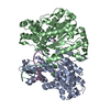
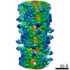
Journal: J Biol Chem / Year: 2022
Title: Pretransition state and apo structures of the filament-forming enzyme SgrAI elucidate mechanisms of activation and substrate specificity.
Authors: Zelin Shan / Niloofar Ghadirian / Dmitry Lyumkis / Nancy C Horton /
Abstract: Enzyme filamentation is a widespread phenomenon that mediates enzyme regulation and function. For the filament-forming sequence-specific DNA endonuclease SgrAI, the process of filamentation both ...Enzyme filamentation is a widespread phenomenon that mediates enzyme regulation and function. For the filament-forming sequence-specific DNA endonuclease SgrAI, the process of filamentation both accelerates its DNA cleavage activity and expands its DNA sequence specificity, thus allowing for many additional DNA sequences to be rapidly cleaved. Both outcomes-the acceleration of DNA cleavage and the expansion of sequence specificity-are proposed to regulate critical processes in bacterial innate immunity. However, the mechanistic bases underlying these events remain unclear. Herein, we describe two new structures of the SgrAI enzyme that shed light on its catalytic function. First, we present the cryo-EM structure of filamentous SgrAI bound to intact primary site DNA and Ca resolved to ∼2.5 Å within the catalytic center, which represents the trapped enzyme-DNA complex prior to the DNA cleavage reaction. This structure reveals important conformational changes that contribute to the catalytic mechanism and the binding of a second divalent cation in the enzyme active site, which is expected to contribute to increased DNA cleavage activity of SgrAI in the filamentous state. Second, we present an X-ray crystal structure of DNA-free (apo) SgrAI resolved to 2.0 Å resolution, which reveals a disordered loop involved in DNA recognition. Collectively, these multiple new observations clarify the mechanism of expansion of DNA sequence specificity of SgrAI, including the indirect readout of sequence-dependent DNA structure, changes in protein-DNA interactions, and the disorder-to-order transition of a crucial DNA recognition element.
History
DepositionSep 17, 2021Deposition site: RCSB / Processing site: RCSB
Revision 1.0Aug 24, 2022Provider: repository / Type: Initial release
Revision 1.1Oct 18, 2023Group: Data collection / Refinement description
Category: chem_comp_atom / chem_comp_bond / pdbx_initial_refinement_model

-
Structure visualization

Structure viewerMolecule:
MolmilJmol/JSmol

Downloads & links

-
Assembly

Deposited unit
A: SgraIR restriction enzyme
B: SgraIR restriction enzyme
hetero molecules


Theoretical massNumber of molelcules
Total (without water)79,6484
Polymers79,5682
Non-polymers802
Water10,106561
1


  • Idetical with deposited unit
  • defined by author&software
  • Evidence: equilibrium centrifugation
TypeNameSymmetry operationNumber
identity operation1_555x,y,z1
Buried area2380 Å2
ΔGint-46 kcal/mol
Surface area26700 Å2
MethodPISA
Unit cell
Length a, b, c (Å)70.110, 72.379, 78.640
Angle α, β, γ (deg.)90.000, 107.949, 90.000
Int Tables number4
Space group name H-MP1211
Space group name HallP2yb
Symmetry operation#1: x,y,z
#2: -x,y+1/2,-z

-
Components

#1: Protein SgraIR restriction enzyme


Mass: 39783.895 Da / Num. of mol.: 2
Source method: isolated from a genetically manipulated source
Source: (gene. exp.) Streptomyces griseus (bacteria) / Gene: sgraIR / Production host: Escherichia coli (E. coli) / References: UniProt: Q9F6L0
#2: Chemical ChemComp-CA / CALCIUM ION


Mass: 40.078 Da / Num. of mol.: 2 / Source method: obtained synthetically / Formula: Ca
#3: Water ChemComp-HOH / water


Mass: 18.015 Da / Num. of mol.: 561 / Source method: isolated from a natural source / Formula: H2O
Has ligand of interestN

-
Experimental details

-
Experiment

ExperimentMethod: X-RAY DIFFRACTION / Number of used crystals: 1

-
Sample preparation

CrystalDensity Matthews: 2.39 Å3/Da / Density % sol: 48.44 %
Crystal growTemperature: 290 K / Method: vapor diffusion, hanging drop / pH: 6.5
Details: 5% PEG 4000 0.1 M imidazole pH 6.5 0.1 M NaCl 5 mM Ca(0Ac)2

-
Data collection

DiffractionMean temperature: 100 K / Serial crystal experiment: N
Diffraction sourceSource: SYNCHROTRON / Site: SSRL / Beamline: BL9-2 / Wavelength: 1 Å
DetectorType: DECTRIS PILATUS3 6M / Detector: PIXEL / Date: Mar 23, 2014
RadiationProtocol: SINGLE WAVELENGTH / Monochromatic (M) / Laue (L): M / Scattering type: x-ray
Radiation wavelengthWavelength: 1 Å / Relative weight: 1
ReflectionResolution: 2.02→38 Å / Num. obs: 47684 / % possible obs: 97.5 % / Redundancy: 2 % / Biso Wilson estimate: 22.11 Å2 / CC1/2: 0.993 / CC star: 0.998 / Rmerge(I) obs: 0.07552 / Rpim(I) all: 0.07552 / Rrim(I) all: 0.1068 / Net I/av σ(I): 8.92 / Net I/σ(I): 8.92
Reflection shellResolution: 2.02→2.097 Å / Redundancy: 2 % / Rmerge(I) obs: 0.3917 / Mean I/σ(I) obs: 2.16 / Num. unique obs: 4428 / CC1/2: 0.522 / CC star: 0.828 / Rpim(I) all: 0.3917 / Rrim(I) all: 0.5539 / % possible all: 91.34

-
Processing

Software
NameVersionClassification
PHENIX1.19_4080refinement
Blu-Icedata collection
XDSdata scaling
PHASERphasing
Cootmodel building
XDSdata reduction
RefinementMethod to determine structure: MOLECULAR REPLACEMENT
Starting model: 3DVO chain A
Resolution: 2.02→38 Å / SU ML: 0.2361 / Cross valid method: FREE R-VALUE / σ(F): 2.25 / Phase error: 24.5963
Stereochemistry target values: GeoStd + Monomer Library + CDL v1.2
RfactorNum. reflection% reflection
Rfree0.2458 1999 4.19 %
Rwork0.2015 45676 -
obs0.2034 47675 97.52 %
Solvent computationShrinkage radii: 0.9 Å / VDW probe radii: 1.11 Å / Solvent model: FLAT BULK SOLVENT MODEL
Displacement parametersBiso mean: 25.61 Å2
Refinement stepCycle: LAST / Resolution: 2.02→38 Å
ProteinNucleic acidLigandSolventTotal
Num. atoms4908 0 2 561 5471
Refine LS restraints
Refine-IDTypeDev idealNumber
X-RAY DIFFRACTIONf_bond_d0.00145019
X-RAY DIFFRACTIONf_angle_d0.43176834
X-RAY DIFFRACTIONf_chiral_restr0.0394772
X-RAY DIFFRACTIONf_plane_restr0.0035892
X-RAY DIFFRACTIONf_dihedral_angle_d11.55371783
LS refinement shell
Resolution (Å)Rfactor RfreeNum. reflection RfreeRfactor RworkNum. reflection RworkRefine-ID% reflection obs (%)
2.02-2.080.33971280.28442921X-RAY DIFFRACTION88.27
2.08-2.130.28671450.2623323X-RAY DIFFRACTION98.89
2.13-2.190.30941450.25423294X-RAY DIFFRACTION98.4
2.19-2.270.2891410.23863220X-RAY DIFFRACTION97.79
2.27-2.350.26741390.23453198X-RAY DIFFRACTION96.22
2.35-2.440.25791440.22673262X-RAY DIFFRACTION98.35
2.44-2.550.26471440.2183298X-RAY DIFFRACTION98.96
2.55-2.690.28061450.20853301X-RAY DIFFRACTION98.82
2.69-2.850.28111430.2093290X-RAY DIFFRACTION98.91
2.85-3.070.23971420.20473218X-RAY DIFFRACTION96.03
3.07-3.380.23421460.18883334X-RAY DIFFRACTION99.6
3.38-3.870.24941450.16633352X-RAY DIFFRACTION99.57
3.87-4.880.17881450.1563296X-RAY DIFFRACTION97.42
4.88-380.21321470.19553369X-RAY DIFFRACTION97.97

+
About Yorodumi

-
News

-
Feb 9, 2022. New format data for meta-information of EMDB entries

New format data for meta-information of EMDB entries

  • Version 3 of the EMDB header file is now the official format.
  • The previous official version 1.9 will be removed from the archive.

Related info.:EMDB header

External links:wwPDB to switch to version 3 of the EMDB data model

-
Aug 12, 2020. Covid-19 info

Covid-19 info

URL: https://pdbjlvh1.pdbj.org/emnavi/covid19.php

New page: Covid-19 featured information page in EM Navigator.

Related info.:Covid-19 info / Mar 5, 2020. Novel coronavirus structure data

+
Mar 5, 2020. Novel coronavirus structure data

Novel coronavirus structure data

Related info.:Yorodumi Speices / Aug 12, 2020. Covid-19 info

External links:COVID-19 featured content - PDBj / Molecule of the Month (242):Coronavirus Proteases

+
Jan 31, 2019. EMDB accession codes are about to change! (news from PDBe EMDB page)

EMDB accession codes are about to change! (news from PDBe EMDB page)

  • The allocation of 4 digits for EMDB accession codes will soon come to an end. Whilst these codes will remain in use, new EMDB accession codes will include an additional digit and will expand incrementally as the available range of codes is exhausted. The current 4-digit format prefixed with “EMD-” (i.e. EMD-XXXX) will advance to a 5-digit format (i.e. EMD-XXXXX), and so on. It is currently estimated that the 4-digit codes will be depleted around Spring 2019, at which point the 5-digit format will come into force.
  • The EM Navigator/Yorodumi systems omit the EMD- prefix.

Related info.:Q: What is EMD? / ID/Accession-code notation in Yorodumi/EM Navigator

External links:EMDB Accession Codes are Changing Soon! / Contact to PDBj

+
Jul 12, 2017. Major update of PDB

Major update of PDB

  • wwPDB released updated PDB data conforming to the new PDBx/mmCIF dictionary.
  • This is a major update changing the version number from 4 to 5, and with Remediation, in which all the entries are updated.
  • In this update, many items about electron microscopy experimental information are reorganized (e.g. em_software).
  • Now, EM Navigator and Yorodumi are based on the updated data.

External links:wwPDB Remediation / Enriched Model Files Conforming to OneDep Data Standards Now Available in the PDB FTP Archive

-
Yorodumi

Thousand views of thousand structures

  • Yorodumi is a browser for structure data from EMDB, PDB, SASBDB, etc.
  • This page is also the successor to EM Navigator detail page, and also detail information page/front-end page for Omokage search.
  • The word "yorodu" (or yorozu) is an old Japanese word meaning "ten thousand". "mi" (miru) is to see.

Related info.:EMDB / PDB / SASBDB / Comparison of 3 databanks / Yorodumi Search / Aug 31, 2016. New EM Navigator & Yorodumi / Yorodumi Papers / Jmol/JSmol / Function and homology information / Changes in new EM Navigator and Yorodumi

Read more