 Movie
Movie Controller

[English] 日本語
 Yorodumi
- EMDB-20015: Structure of filamentous SgrAI endonuclease in its activated form -
+ Open data

- Basic information
| Entry | Database: EMDB / ID: EMD-20015 | |||||||||
|---|---|---|---|---|---|---|---|---|---|---|
| Title | Structure of filamentous SgrAI endonuclease in its activated form | |||||||||
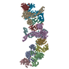
 Map data Map data | Cryo-EM reconstruction of filamentous SgrAI in its activated form | |||||||||
 Sample Sample |
| |||||||||
 Keywords Keywords | restriction endonuclease / DNAse / allostery / bacterial innate immunity / filament / hyper-activation / substrate specificity / HYDROLASE-DNA complex | |||||||||
| Function / homology | Restriction endonuclease, type II, Cfr10I/Bse634I / Cfr10I/Bse634I restriction endonuclease / Restriction endonuclease type II-like / metal ion binding / identical protein binding / SgraIR restriction enzyme Function and homology information Function and homology information | |||||||||
| Biological species |  Streptomyces griseus (bacteria) Streptomyces griseus (bacteria) | |||||||||
| Method | helical reconstruction / cryo EM / Resolution: 3.5 Å | |||||||||
 Authors Authors | Polley S / Lyumkis D | |||||||||
| Funding support |  United States, 2 items United States, 2 items
| |||||||||
 Citation Citation | Journal: Structure / Year: 2019 Title: Mechanism of Filamentation-Induced Allosteric Activation of the SgrAI Endonuclease. Authors: Smarajit Polley / Dmitry Lyumkis / Nancy C Horton /  Abstract: Filament formation by enzymes is increasingly recognized as an important phenomenon with potentially unique regulatory properties and biological roles. SgrAI is an allosterically regulated type II ...Filament formation by enzymes is increasingly recognized as an important phenomenon with potentially unique regulatory properties and biological roles. SgrAI is an allosterically regulated type II restriction endonuclease that forms filaments with enhanced DNA cleavage activity and altered sequence specificity. Here, we present the cryoelectron microscopy (cryo-EM) structure of the filament of SgrAI in its activated configuration. The structural data illuminate the mechanistic origin of hyperaccelerated DNA cleavage activity and suggests how indirect DNA sequence readout within filamentous SgrAI may enable recognition of substantially more nucleotide sequences than its low-activity form, thereby altering and partially relaxing its DNA sequence specificity. Together, substrate DNA binding, indirect readout, and filamentation simultaneously enhance SgrAI's catalytic activity and modulate substrate preference. This unusual enzyme mechanism may have evolved to perform the specialized functions of bacterial innate immunity in rapid defense against invading phage DNA without causing damage to the host DNA. | |||||||||
| History |
|
- Structure visualization
| Movie |
Movie viewer |
|---|---|
| Structure viewer | EM map: SurfViewMolmilJmol/JSmol |
| Supplemental images |

- Downloads & links
-EMDB archive
| Map data | emd_20015.map.gz | 58.3 MB | EMDB map data format | |
|---|---|---|---|---|
| Header (meta data) | emd-20015-v30.xmlemd-20015.xml | 19 KB 19 KB | Display Display | EMDB header |
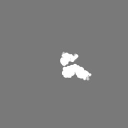
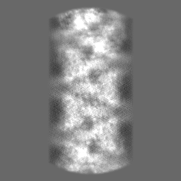
| Images | 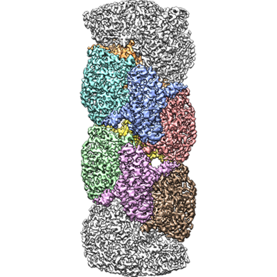 emd_20015.png emd_20015.png | 195 KB | ||
| Masks | emd_20015_msk_1.map | 64 MB | Mask map | |
| Filedesc metadata | emd-20015.cif.gz | 6.6 KB | ||
| Others | emd_20015_half_map_1.map.gzemd_20015_half_map_2.map.gz | 9.9 MB 9.9 MB | ||
| Archive directory |  http://ftp.pdbj.org/pub/emdb/structures/EMD-20015ftp://ftp.pdbj.org/pub/emdb/structures/EMD-20015 http://ftp.pdbj.org/pub/emdb/structures/EMD-20015ftp://ftp.pdbj.org/pub/emdb/structures/EMD-20015 | HTTPS FTP |
-Related structure data
| Related structure data | 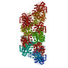 6objMC M: atomic model generated by this map C: citing same article ( |
|---|---|
| Similar structure data |

-Links
| EMDB pages | EMDB (EBI/PDBe) / EMDataResource |
|---|---|
| Related items in Molecule of the Month |


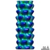
-Map
| File | Download / File: emd_20015.map.gz / Format: CCP4 / Size: 64 MB / Type: IMAGE STORED AS FLOATING POINT NUMBER (4 BYTES) | ||||||||||||||||||||||||||||||||||||||||||||||||||||||||||||||||||||
|---|---|---|---|---|---|---|---|---|---|---|---|---|---|---|---|---|---|---|---|---|---|---|---|---|---|---|---|---|---|---|---|---|---|---|---|---|---|---|---|---|---|---|---|---|---|---|---|---|---|---|---|---|---|---|---|---|---|---|---|---|---|---|---|---|---|---|---|---|---|
| Annotation | Cryo-EM reconstruction of filamentous SgrAI in its activated form | ||||||||||||||||||||||||||||||||||||||||||||||||||||||||||||||||||||
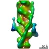
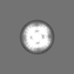
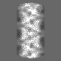
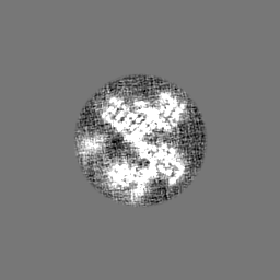
| Projections & slices | Image control
Images are generated by Spider. | ||||||||||||||||||||||||||||||||||||||||||||||||||||||||||||||||||||
| Voxel size | X=Y=Z: 1.31 Å | ||||||||||||||||||||||||||||||||||||||||||||||||||||||||||||||||||||
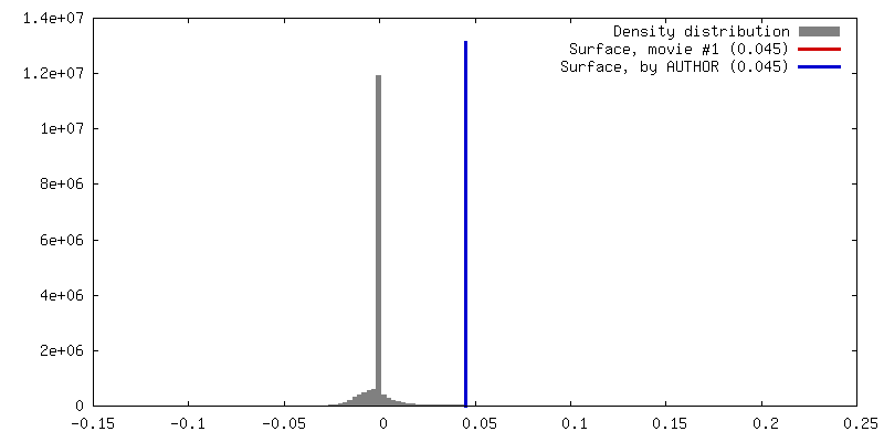
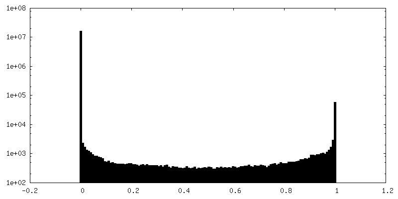
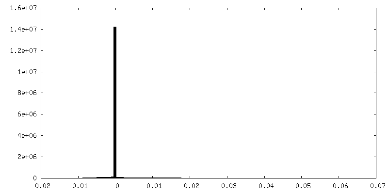
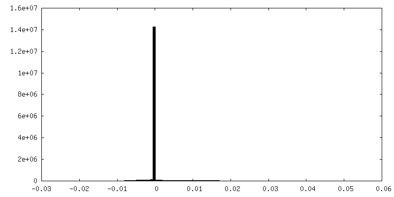
| Density |
| ||||||||||||||||||||||||||||||||||||||||||||||||||||||||||||||||||||
| Symmetry | Space group: 1 | ||||||||||||||||||||||||||||||||||||||||||||||||||||||||||||||||||||
| Details | EMDB XML:
CCP4 map header:
| ||||||||||||||||||||||||||||||||||||||||||||||||||||||||||||||||||||
 Z (Sec.)
 Y (Row.)
 X (Col.)


-Supplemental data
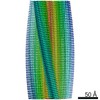
-Mask #1
| File | emd_20015_msk_1.map | ||||||||||||
|---|---|---|---|---|---|---|---|---|---|---|---|---|---|
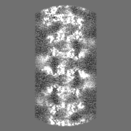
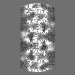
| Projections & Slices |
| ||||||||||||
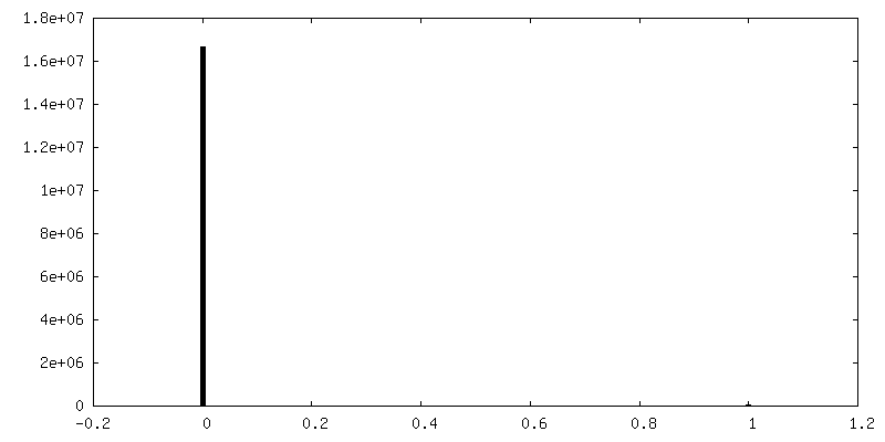
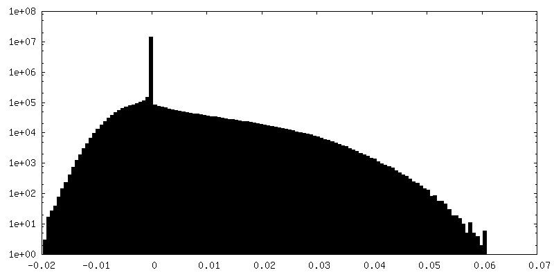
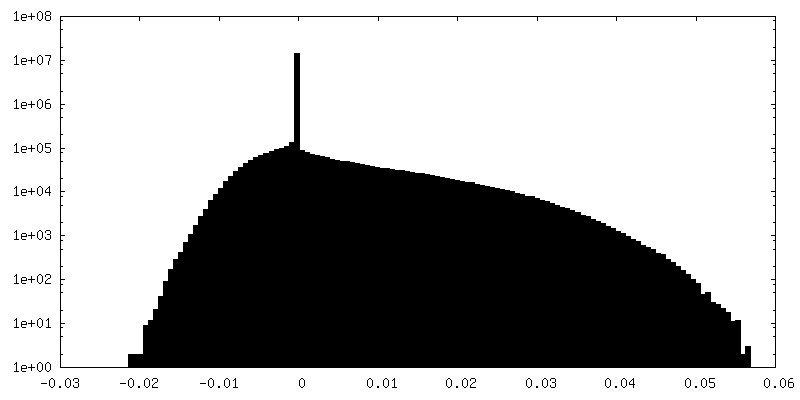
| Density Histograms |


-Half map: half-map 1 for the Cryo-EM reconstruction of filamentous...
| File | emd_20015_half_map_1.map | ||||||||||||
|---|---|---|---|---|---|---|---|---|---|---|---|---|---|
| Annotation | half-map 1 for the Cryo-EM reconstruction of filamentous SgrAI in its activated form | ||||||||||||
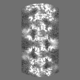
| Projections & Slices |
| ||||||||||||
| Density Histograms |

-Half map: half-map 2 for the Cryo-EM reconstruction of filamentous...
| File | emd_20015_half_map_2.map | ||||||||||||
|---|---|---|---|---|---|---|---|---|---|---|---|---|---|
| Annotation | half-map 2 for the Cryo-EM reconstruction of filamentous SgrAI in its activated form | ||||||||||||
| Projections & Slices |
| ||||||||||||
| Density Histograms |


- Sample components
-Entire : Filamentous assembly of SgrAI protein bound to pre-cleaved DNA
| Entire | Name: Filamentous assembly of SgrAI protein bound to pre-cleaved DNA |
|---|---|
| Components |
|
-Supramolecule #1: Filamentous assembly of SgrAI protein bound to pre-cleaved DNA
| Supramolecule | Name: Filamentous assembly of SgrAI protein bound to pre-cleaved DNA type: complex / ID: 1 / Parent: 0 / Macromolecule list: #1-#2 |
|---|---|
| Source (natural) | Organism: Streptomyces griseus (bacteria) |
| Molecular weight | Theoretical: 74 kDa/nm |
-Macromolecule #1: SgraIR restriction enzyme
| Macromolecule | Name: SgraIR restriction enzyme / type: protein_or_peptide / ID: 1 / Number of copies: 2 / Enantiomer: LEVO EC number: Hydrolases; Acting on ester bonds; Endodeoxyribonucleases producing 5'-phosphomonoesters |
|---|---|
| Source (natural) | Organism: Streptomyces griseus (bacteria) |
| Molecular weight | Theoretical: 38.073102 KDa |
| Recombinant expression | Organism: |
| Sequence | String: MPFTYSIEAT RNLATTERCI QDIRNAPVRN RSTQFQLAQQ NMLAYTFGEV IPGFASAGIN GMDYRDVIGR PVENAVTEGT HFFRDDFRV DSNAKAKVAG DIFEIVSSAV MWNCAARWNS LMVGEGWRSQ PRYSRPTLSP SPRRQVAVLN LPRSFDWVSL L VPESQEVI ...String: MPFTYSIEAT RNLATTERCI QDIRNAPVRN RSTQFQLAQQ NMLAYTFGEV IPGFASAGIN GMDYRDVIGR PVENAVTEGT HFFRDDFRV DSNAKAKVAG DIFEIVSSAV MWNCAARWNS LMVGEGWRSQ PRYSRPTLSP SPRRQVAVLN LPRSFDWVSL L VPESQEVI EEFRAGLRKD GLGLPTSTPD LAVVVLPEEF QNDEMWREEI AGLTRPNQIL LSGAYQRLQG RVQPGEISLA VA FKRSLRS DRLYQPLYEA NVMQLLLEGK LGAPKVEFEV HTLAPEGTNA FVTYEAASLY GLAEGRSAVH RAIRELYVPP TAA DLARRF FAFLNERMEL VNG UniProtKB: SgraIR restriction enzyme |
-Macromolecule #2: DNA (26-MER)
| Macromolecule | Name: DNA (26-MER) / type: dna / ID: 2 / Details: assembled from pre-cleaved DNAs / Number of copies: 2 / Classification: DNA |
|---|---|
| Source (natural) | Organism: Streptomyces griseus (bacteria) |
| Molecular weight | Theoretical: 12.314889 KDa |
| Sequence | String: (DG)(DA)(DT)(DG)(DC)(DG)(DT)(DG)(DG)(DG) (DT)(DC)(DT)(DT)(DC)(DA)(DC)(DA)(DC)(DC) (DG)(DG)(DT)(DG)(DT)(DG)(DA)(DA)(DG) (DA)(DC)(DC)(DC)(DA)(DC)(DG)(DC)(DA)(DT) (DC) |
-Macromolecule #3: MAGNESIUM ION
| Macromolecule | Name: MAGNESIUM ION / type: ligand / ID: 3 / Number of copies: 2 / Formula: MG |
|---|---|
| Molecular weight | Theoretical: 24.305 Da |
-Experimental details
-Structure determination
| Method | cryo EM |
|---|---|
 Processing Processing | helical reconstruction |
| Aggregation state | filament |
-Sample preparation
| Concentration | 1 mg/mL | |||||||||||||||
|---|---|---|---|---|---|---|---|---|---|---|---|---|---|---|---|---|
| Buffer | pH: 8 Component:
| |||||||||||||||
| Grid | Model: UltrAuFoil / Material: GOLD / Mesh: 400 / Pretreatment - Type: PLASMA CLEANING / Pretreatment - Time: 6 sec. / Pretreatment - Atmosphere: OTHER | |||||||||||||||
| Vitrification | Cryogen name: ETHANE / Chamber humidity: 80 % / Instrument: HOMEMADE PLUNGER |
- Electron microscopy
| Microscope | FEI TITAN KRIOS |
|---|---|
| Image recording | Film or detector model: GATAN K2 SUMMIT (4k x 4k) / Detector mode: COUNTING / Digitization - Dimensions - Width: 3838 pixel / Digitization - Dimensions - Height: 3710 pixel / Digitization - Frames/image: 1-60 / Number real images: 216 / Average electron dose: 55.0 e/Å2 |
| Electron beam | Acceleration voltage: 300 kV / Electron source:  FIELD EMISSION GUN FIELD EMISSION GUN |
| Electron optics | C2 aperture diameter: 70.0 µm / Calibrated magnification: 38167 / Illumination mode: FLOOD BEAM / Imaging mode: BRIGHT FIELD / Cs: 2.7 mm / Nominal defocus max: 2.6 µm / Nominal defocus min: 0.6 µm / Nominal magnification: 22500 |
| Sample stage | Specimen holder model: FEI TITAN KRIOS AUTOGRID HOLDER / Cooling holder cryogen: NITROGEN |
| Experimental equipment |  Model: Titan Krios / Image courtesy: FEI Company |
-Image processing
| Final reconstruction | Applied symmetry - Helical parameters - Δz: 21.6 Å Applied symmetry - Helical parameters - Δ&Phi: -86.2 ° Applied symmetry - Helical parameters - Axial symmetry: C1 (asymmetric) Algorithm: FOURIER SPACE / Resolution.type: BY AUTHOR / Resolution: 3.5 Å / Resolution method: FSC 0.143 CUT-OFF / Software - Name: RELION (ver. 3.0) / Number images used: 7 |
|---|---|
| Startup model | Type of model: NONE |
| Final angle assignment | Type: PROJECTION MATCHING / Software - Name: RELION (ver. 3.0) |
-Atomic model buiding 1
| Refinement | Space: REAL / Protocol: AB INITIO MODEL / Target criteria: Correlation coefficient |
|---|---|
| Output model | PDB-6obj: |
