Movie
Movie Controller


+ Open data

- Basic information
| Entry | Database: EMDB / ID: EMD-2430 | |||||||||
|---|---|---|---|---|---|---|---|---|---|---|
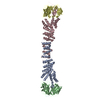
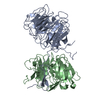
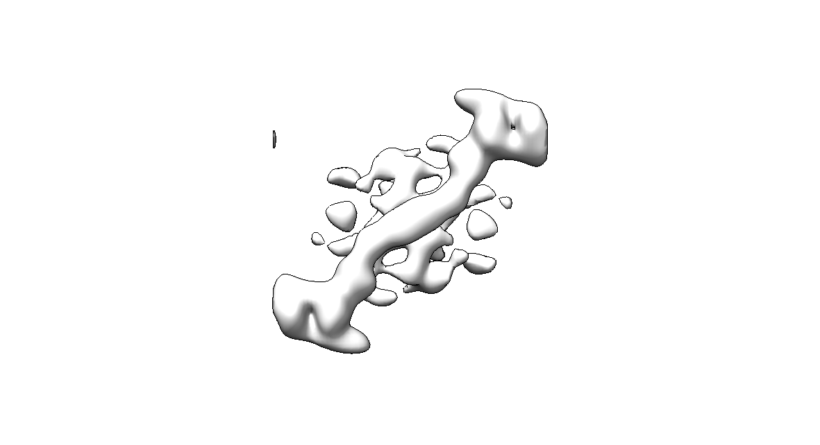
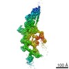
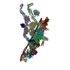
| Title | The structure of the COPII coat assembled on membranes | |||||||||
 Map data Map data | sec13/31 outer coat edge left-handed direction | |||||||||
 Sample Sample |
| |||||||||
 Keywords Keywords | COPII / coat / secretion / trafficking / Sec13 / Sec31 | |||||||||
| Function / homology |  Function and homology information Function and homology informationpositive regulation of ER to Golgi vesicle-mediated transport / Seh1-associated complex / COPII-coated vesicle budding / nuclear pore localization / protein exit from endoplasmic reticulum / COPII-mediated vesicle transport / regulation of TORC1 signaling / nuclear pore outer ring / COPII-coated vesicle cargo loading / Regulation of Glucokinase by Glucokinase Regulatory Protein ...positive regulation of ER to Golgi vesicle-mediated transport / Seh1-associated complex / COPII-coated vesicle budding / nuclear pore localization / protein exit from endoplasmic reticulum / COPII-mediated vesicle transport / regulation of TORC1 signaling / nuclear pore outer ring / COPII-coated vesicle cargo loading / Regulation of Glucokinase by Glucokinase Regulatory Protein / : / positive regulation of protein exit from endoplasmic reticulum / Regulation of HSF1-mediated heat shock response / COPII vesicle coat / SUMOylation of SUMOylation proteins / mating projection tip / endoplasmic reticulum organization / SUMOylation of RNA binding proteins / SUMOylation of chromatin organization proteins / vacuolar membrane / nucleocytoplasmic transport / endoplasmic reticulum exit site / positive regulation of TOR signaling / nuclear pore / mRNA transport / ERAD pathway / positive regulation of TORC1 signaling / cell periphery / protein import into nucleus / nuclear envelope / protein transport / endoplasmic reticulum membrane / positive regulation of DNA-templated transcription / structural molecule activity / endoplasmic reticulum Similarity search - Function | |||||||||
| Biological species |  | |||||||||
| Method | subtomogram averaging / cryo EM / negative staining / Resolution: 40.0 Å | |||||||||
 Authors Authors | Zanetti G / Prinz S / Daum S / Meister A / Schekman R / Bacia K / Briggs JAG | |||||||||
 Citation Citation | Journal: Elife / Year: 2013 Title: The structure of the COPII transport-vesicle coat assembled on membranes. Authors: Giulia Zanetti / Simone Prinz / Sebastian Daum / Annette Meister / Randy Schekman / Kirsten Bacia / John A G Briggs /  Abstract: Coat protein complex II (COPII) mediates formation of the membrane vesicles that export newly synthesised proteins from the endoplasmic reticulum. The inner COPII proteins bind to cargo and membrane, ...Coat protein complex II (COPII) mediates formation of the membrane vesicles that export newly synthesised proteins from the endoplasmic reticulum. The inner COPII proteins bind to cargo and membrane, linking them to the outer COPII components that form a cage around the vesicle. Regulated flexibility in coat architecture is essential for transport of a variety of differently sized cargoes, but structural data on the assembled coat has not been available. We have used cryo-electron tomography and subtomogram averaging to determine the structure of the complete, membrane-assembled COPII coat. We describe a novel arrangement of the outer coat and find that the inner coat can assemble into regular lattices. The data reveal how coat subunits interact with one another and with the membrane, suggesting how coordinated assembly of inner and outer coats can mediate and regulate packaging of vesicles ranging from small spheres to large tubular carriers. DOI:http://dx.doi.org/10.7554/eLife.00951.001. | |||||||||
| History |
|
- Structure visualization
| Movie |
Movie viewer |
|---|---|
| Structure viewer | EM map: SurfViewMolmilJmol/JSmol |
| Supplemental images |
- Downloads & links
-EMDB archive
| Map data | emd_2430.map.gz | 1.3 MB | EMDB map data format | |
|---|---|---|---|---|
| Header (meta data) | emd-2430-v30.xmlemd-2430.xml | 14.3 KB 14.3 KB | Display Display | EMDB header |
| Images |  emd_2430.png emd_2430.png | 46.3 KB | ||
| Archive directory |  http://ftp.pdbj.org/pub/emdb/structures/EMD-2430ftp://ftp.pdbj.org/pub/emdb/structures/EMD-2430 http://ftp.pdbj.org/pub/emdb/structures/EMD-2430ftp://ftp.pdbj.org/pub/emdb/structures/EMD-2430 | HTTPS FTP |
-Related structure data
| Related structure data | 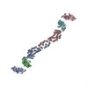 4bzjMC 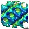 2428C 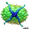 2429C 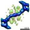 2431C  2432C 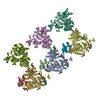 4bziC 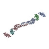 4bzkC C: citing same article ( M: atomic model generated by this map |
|---|---|
| Similar structure data |

-Links
| EMDB pages | EMDB (EBI/PDBe) / EMDataResource |
|---|---|
| Related items in Molecule of the Month |


-Map
| File | Download / File: emd_2430.map.gz / Format: CCP4 / Size: 1.4 MB / Type: IMAGE STORED AS FLOATING POINT NUMBER (4 BYTES) | ||||||||||||||||||||||||||||||||||||||||||||||||||||||||||||
|---|---|---|---|---|---|---|---|---|---|---|---|---|---|---|---|---|---|---|---|---|---|---|---|---|---|---|---|---|---|---|---|---|---|---|---|---|---|---|---|---|---|---|---|---|---|---|---|---|---|---|---|---|---|---|---|---|---|---|---|---|---|
| Annotation | sec13/31 outer coat edge left-handed direction | ||||||||||||||||||||||||||||||||||||||||||||||||||||||||||||
| Projections & slices | Image control
Images are generated by Spider. | ||||||||||||||||||||||||||||||||||||||||||||||||||||||||||||
| Voxel size | X=Y=Z: 4.3 Å | ||||||||||||||||||||||||||||||||||||||||||||||||||||||||||||
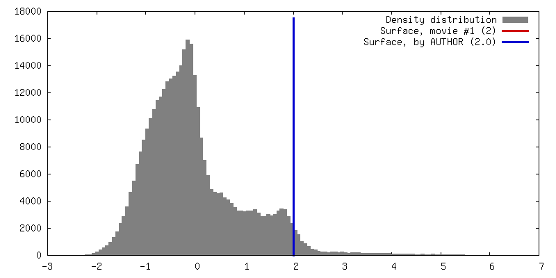
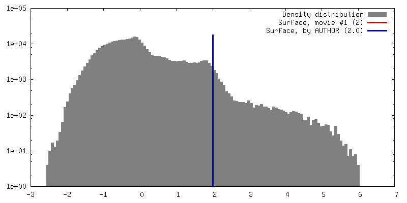
| Density |
| ||||||||||||||||||||||||||||||||||||||||||||||||||||||||||||
| Symmetry | Space group: 1 | ||||||||||||||||||||||||||||||||||||||||||||||||||||||||||||
| Details | EMDB XML:
CCP4 map header:
| ||||||||||||||||||||||||||||||||||||||||||||||||||||||||||||
 Z (Sec.)
 Y (Row.)
 X (Col.)


-Supplemental data
- Sample components
-Entire : Sec13/31 complex (as part of complete COPII assembled on membrane...
| Entire | Name: Sec13/31 complex (as part of complete COPII assembled on membrane) edge in left-handed direction |
|---|---|
| Components |
|
-Supramolecule #1000: Sec13/31 complex (as part of complete COPII assembled on membrane...
| Supramolecule | Name: Sec13/31 complex (as part of complete COPII assembled on membrane) edge in left-handed direction type: sample / ID: 1000 / Oligomeric state: heterotetramers of sec13 and sec31 / Number unique components: 2 |
|---|---|
| Molecular weight | Experimental: 319.236 KDa / Theoretical: 319.236 KDa |
-Macromolecule #1: Sec31
| Macromolecule | Name: Sec31 / type: protein_or_peptide / ID: 1 / Number of copies: 4 / Oligomeric state: heterotetramer / Recombinant expression: Yes |
|---|---|
| Source (natural) | Organism: |
| Molecular weight | Experimental: 138.824 KDa / Theoretical: 138.824 KDa |
| Recombinant expression | Organism: |
| Sequence | UniProtKB: UNIPROTKB: E7Q1I6 |
-Macromolecule #2: Sec13
| Macromolecule | Name: Sec13 / type: protein_or_peptide / ID: 2 / Number of copies: 4 / Oligomeric state: heterotetramer / Recombinant expression: Yes |
|---|---|
| Source (natural) | Organism: |
| Molecular weight | Experimental: 20.79 KDa / Theoretical: 27.9 KDa |
| Recombinant expression | Organism: |
| Sequence | UniProtKB: UNIPROTKB: E7Q6Z3 |
-Experimental details
-Structure determination
| Method | negative staining, cryo EM |
|---|---|
 Processing Processing | subtomogram averaging |
| Aggregation state | helical array |
-Sample preparation
| Concentration | 0.03 mg/mL |
|---|---|
| Buffer | pH: 6.8 / Details: HEPES, 50 mM KOAc, 1.2 mM MgCl2 |
| Staining | Type: NEGATIVE / Details: plunge frozen |
| Grid | Details: C-flat grids |
| Vitrification | Cryogen name: ETHANE / Instrument: HOMEMADE PLUNGER |
- Electron microscopy #1
| Microscopy ID | 1 |
|---|---|
| Microscope | FEI TITAN KRIOS |
| Specialist optics | Energy filter - Name: GATAN GIF 2002 |
| Date | Sep 18, 2012 |
| Image recording | Category: CCD / Film or detector model: GATAN MULTISCAN / Number real images: 26 / Average electron dose: 80 e/Å2 / Bits/pixel: 16 |
| Electron beam | Acceleration voltage: 200 kV / Electron source:  FIELD EMISSION GUN FIELD EMISSION GUN |
| Electron optics | Illumination mode: FLOOD BEAM / Imaging mode: BRIGHT FIELD / Cs: 2.7 mm / Nominal defocus max: 3.2 µm / Nominal defocus min: 2.0 µm / Nominal magnification: 19500 |
| Sample stage | Specimen holder model: FEI TITAN KRIOS AUTOGRID HOLDER / Tilt series - Axis1 - Min angle: -60 ° / Tilt series - Axis1 - Max angle: 60 ° |
| Experimental equipment |  Model: Titan Krios / Image courtesy: FEI Company |
-Electron microscopy #2
| Microscopy ID | 2 |
|---|---|
| Microscope | FEI TITAN KRIOS |
| Specialist optics | Energy filter - Name: GATAN GIF 2002 |
| Date | Jun 19, 2012 |
| Image recording | Category: CCD / Film or detector model: GATAN MULTISCAN / Number real images: 26 / Average electron dose: 80 e/Å2 / Bits/pixel: 16 |
| Electron beam | Acceleration voltage: 200 kV / Electron source: FIELD EMISSION GUN |
| Electron optics | Illumination mode: FLOOD BEAM / Imaging mode: BRIGHT FIELD / Cs: 2.7 mm / Nominal defocus max: 3.2 µm / Nominal defocus min: 2.0 µm / Nominal magnification: 19500 |
| Sample stage | Specimen holder model: FEI TITAN KRIOS AUTOGRID HOLDER / Tilt series - Axis1 - Min angle: -60 ° / Tilt series - Axis1 - Max angle: 60 ° |
| Experimental equipment | Model: Titan Krios / Image courtesy: FEI Company |
-Image processing
| Details | see materials and methods in relevant publication |
|---|---|
| Final reconstruction | Applied symmetry - Point group: C2 (2 fold cyclic) / Algorithm: OTHER / Resolution.type: BY AUTHOR / Resolution: 40.0 Å / Resolution method: FSC 0.5 CUT-OFF / Software - Name: TOM/AV3, Matlab / Number subtomograms used: 182 |
| CTF correction | Details: each tilted image within tomogram |
| Final angle assignment | Details: 0 0 0 in zyz convention |
-Atomic model buiding 1
| Initial model | PDB ID: |
|---|---|
| Software | Name: Chimera |
| Refinement | Space: REAL / Protocol: RIGID BODY FIT / Target criteria: Cross-correlation |
| Output model | PDB-4bzj: |