- PDB-2pm6: Crystal Structure of yeast Sec13/31 edge element of the COPII ves... -
+
Open data
ID or keywords:
Loading...
-
Basic information
Entry
Database: PDB / ID: 2pm6
Title
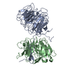
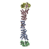
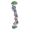
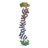
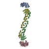
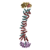
Crystal Structure of yeast Sec13/31 edge element of the COPII vesicular coat, native version
Components
Protein transport protein SEC13
Protein transport protein SEC31
Keywords
PROTEIN TRANSPORT / beta propeller / alpha solenoid
Function / homology
Function and homology information
positive regulation of ER to Golgi vesicle-mediated transport / Seh1-associated complex / COPII-coated vesicle budding / nuclear pore localization / protein exit from endoplasmic reticulum / COPII-mediated vesicle transport / regulation of TORC1 signaling / nuclear pore outer ring / Regulation of Glucokinase by Glucokinase Regulatory Protein / COPII-coated vesicle cargo loading ...positive regulation of ER to Golgi vesicle-mediated transport / Seh1-associated complex / COPII-coated vesicle budding / nuclear pore localization / protein exit from endoplasmic reticulum / COPII-mediated vesicle transport / regulation of TORC1 signaling / nuclear pore outer ring / Regulation of Glucokinase by Glucokinase Regulatory Protein / COPII-coated vesicle cargo loading / : / positive regulation of protein exit from endoplasmic reticulum / Regulation of HSF1-mediated heat shock response / COPII vesicle coat / SUMOylation of SUMOylation proteins / mating projection tip / endoplasmic reticulum organization / SUMOylation of RNA binding proteins / SUMOylation of chromatin organization proteins / vacuolar membrane / nucleocytoplasmic transport / endoplasmic reticulum exit site / positive regulation of TOR signaling / nuclear pore / mRNA transport / ERAD pathway / positive regulation of TORC1 signaling / cell periphery / protein import into nucleus / nuclear envelope / protein transport / endoplasmic reticulum membrane / positive regulation of DNA-templated transcription / structural molecule activity / endoplasmic reticulum Similarity search - Function
Serine Threonine Protein Phosphatase 5, Tetratricopeptide repeat - #1030 / Protein transport protein SEC31 / Protein transport protein SEC31 / Protein transport protein SEC31-like / SRA1/Sec31 / Steroid receptor RNA activator (SRA1) / Ancestral coatomer element 1, Sec16/Sec31 / Sec16 Sec23-binding domain / Sec13/Seh1 family / YVTN repeat-like/Quinoprotein amine dehydrogenase ...Serine Threonine Protein Phosphatase 5, Tetratricopeptide repeat - #1030 / Protein transport protein SEC31 / Protein transport protein SEC31 / Protein transport protein SEC31-like / SRA1/Sec31 / Steroid receptor RNA activator (SRA1) / Ancestral coatomer element 1, Sec16/Sec31 / Sec16 Sec23-binding domain / Sec13/Seh1 family / YVTN repeat-like/Quinoprotein amine dehydrogenase / 7 Propeller / Methylamine Dehydrogenase; Chain H / Serine Threonine Protein Phosphatase 5, Tetratricopeptide repeat / Alpha Horseshoe / WD domain, G-beta repeat / WD40 repeat, conserved site / Trp-Asp (WD) repeats signature. / Trp-Asp (WD) repeats profile. / Trp-Asp (WD) repeats circular profile. / WD40 repeats / WD40 repeat / WD40-repeat-containing domain superfamily / WD40/YVTN repeat-like-containing domain superfamily / Mainly Beta / Mainly Alpha Similarity search - Domain/homology
In the structure databanks used in Yorodumi, some data are registered as the other names, "COVID-19 virus" and "2019-nCoV". Here are the details of the virus and the list of structure data.
Jan 31, 2019. EMDB accession codes are about to change! (news from PDBe EMDB page)
EMDB accession codes are about to change! (news from PDBe EMDB page)
The allocation of 4 digits for EMDB accession codes will soon come to an end. Whilst these codes will remain in use, new EMDB accession codes will include an additional digit and will expand incrementally as the available range of codes is exhausted. The current 4-digit format prefixed with “EMD-” (i.e. EMD-XXXX) will advance to a 5-digit format (i.e. EMD-XXXXX), and so on. It is currently estimated that the 4-digit codes will be depleted around Spring 2019, at which point the 5-digit format will come into force.
The EM Navigator/Yorodumi systems omit the EMD- prefix.
Related info.:Q: What is EMD? / ID/Accession-code notation in Yorodumi/EM Navigator
Yorodumi is a browser for structure data from EMDB, PDB, SASBDB, etc.
This page is also the successor to EM Navigator detail page, and also detail information page/front-end page for Omokage search.
The word "yorodu" (or yorozu) is an old Japanese word meaning "ten thousand". "mi" (miru) is to see.
Related info.:EMDB / PDB / SASBDB / Comparison of 3 databanks / Yorodumi Search / Aug 31, 2016. New EM Navigator & Yorodumi / Yorodumi Papers / Jmol/JSmol / Function and homology information / Changes in new EM Navigator and Yorodumi
 Movie
Movie Controller

 Yorodumi
 Open data
 Basic information
 Components
 Keywords
 Function and homology information
 X-RAY DIFFRACTION /
  Authors
 Citation
 Structure visualization
 Downloads & links
 Other downloads

 PDBj
PDBj

 Assembly
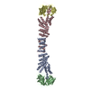
 Mass: 18.015 Da / Num. of mol.: 261 / Source method: isolated from a natural source / Formula: H2O
 Sample preparation
 Processing