- EMDB-2316: Structural Basis of Signal Sequence Surveillance and Selection by... -
+
Open data
ID or keywords:
Loading...
-
Basic information
Entry
Database: EMDB / ID: EMD-2316
Title
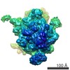
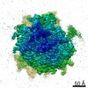
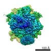
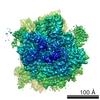
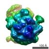
Structural Basis of Signal Sequence Surveillance and Selection by the SRP-SR Complex
Map data
RNCEspP-SRP-FtsY
Sample
Sample: Ribosome-SRP-FtsY complex with EspP nascent chain
Complex: 70S ribosome
Protein or peptide: SIGNAL RECOGNITION PARTICLE PROTEIN
Protein or peptide: SIGNAL RECOGNITION PARTICLE RECEPTOR FTSY
Protein or peptide: 4.5 S RNA
Protein or peptide: SIGNAL RECOGNITION PARTICLE 54 KDA PROTEIN
Protein or peptide: DIPEPTIDYL AMINOPEPTIDASE B
Keywords
Ribosome / SRP / signal recognition particle / SR / FtsY / cryo-EM
Function / homology
Function and homology information
Synthesis, secretion, and inactivation of Glucagon-like Peptide-1 (GLP-1) / Hydrolases; Acting on peptide bonds (peptidases); Dipeptidyl-peptidases and tripeptidyl-peptidases / signal recognition particle binding / signal recognition particle / signal-recognition-particle GTPase / 7S RNA binding / SRP-dependent cotranslational protein targeting to membrane / dipeptidyl-peptidase activity / fungal-type vacuole membrane / stringent response ...Synthesis, secretion, and inactivation of Glucagon-like Peptide-1 (GLP-1) / Hydrolases; Acting on peptide bonds (peptidases); Dipeptidyl-peptidases and tripeptidyl-peptidases / signal recognition particle binding / signal recognition particle / signal-recognition-particle GTPase / 7S RNA binding / SRP-dependent cotranslational protein targeting to membrane / dipeptidyl-peptidase activity / fungal-type vacuole membrane / stringent response / protein targeting / aminopeptidase activity / protein processing / cytoplasmic side of plasma membrane / serine-type endopeptidase activity / GTPase activity / GTP binding / protein homodimerization activity / ATP hydrolysis activity / proteolysis / plasma membrane / cytosol Similarity search - Function
Signal-recognition particle receptor FtsY / Signal recognition particle protein / SRP/SRP receptor, N-terminal / Signal recognition particle, SRP54 subunit / Signal recognition particle, SRP54 subunit, M-domain / Signal recognition particle, SRP54 subunit, M-domain superfamily / Signal peptide binding domain / SRP54-type proteins GTP-binding domain signature. / Signal recognition particle SRP54, helical bundle / Signal recognition particle SRP54, N-terminal domain superfamily ...Signal-recognition particle receptor FtsY / Signal recognition particle protein / SRP/SRP receptor, N-terminal / Signal recognition particle, SRP54 subunit / Signal recognition particle, SRP54 subunit, M-domain / Signal recognition particle, SRP54 subunit, M-domain superfamily / Signal peptide binding domain / SRP54-type proteins GTP-binding domain signature. / Signal recognition particle SRP54, helical bundle / Signal recognition particle SRP54, N-terminal domain superfamily / SRP54-type protein, helical bundle domain / SRP54-type protein, helical bundle domain / Signal recognition particle, SRP54 subunit, GTPase domain / SRP54-type protein, GTPase domain / SRP54-type protein, GTPase domain / : / Dipeptidylpeptidase IV, N-terminal domain / Dipeptidyl peptidase IV (DPP IV) N-terminal region / Peptidase S9, serine active site / Prolyl endopeptidase family serine active site. / Peptidase S9, prolyl oligopeptidase, catalytic domain / Prolyl oligopeptidase family / Alpha/Beta hydrolase fold / ATPases associated with a variety of cellular activities / AAA+ ATPase domain / P-loop containing nucleoside triphosphate hydrolase Similarity search - Domain/homology
Signal recognition particle protein / Signal recognition particle receptor FtsY / Dipeptidyl aminopeptidase B / Signal recognition particle 54 kDa protein Similarity search - Component
Journal: Nat Struct Mol Biol / Year: 2013 Title: Structural basis of signal sequence surveillance and selection by the SRP-FtsY complex. Authors: Ottilie von Loeffelholz / Kèvin Knoops / Aileen Ariosa / Xin Zhang / Manikandan Karuppasamy / Karine Huard / Guy Schoehn / Imre Berger / Shu-ou Shan / Christiane Schaffitzel / Abstract: Signal-recognition particle (SRP)-dependent targeting of translating ribosomes to membranes is a multistep quality-control process. Ribosomes that are translating weakly hydrophobic signal sequences ...Signal-recognition particle (SRP)-dependent targeting of translating ribosomes to membranes is a multistep quality-control process. Ribosomes that are translating weakly hydrophobic signal sequences can be rejected from the targeting reaction even after they are bound to the SRP. Here we show that the early complex, formed by Escherichia coli SRP and its receptor FtsY with ribosomes translating the incorrect cargo EspP, is unstable and rearranges inefficiently into subsequent conformational states, such that FtsY dissociation is favored over successful targeting. The N-terminal extension of EspP is responsible for these defects in the early targeting complex. The cryo-electron microscopy structure of this 'false' early complex with EspP revealed an ordered M domain of SRP protein Ffh making two ribosomal contacts, and the NG domains of Ffh and FtsY forming a distorted, flexible heterodimer. Our results provide a structural basis for SRP-mediated signal-sequence selection during recruitment of the SRP receptor.
History
Deposition
Feb 13, 2013
-
Header (metadata) release
Feb 27, 2013
-
Map release
Feb 26, 2014
-
Update
Feb 26, 2014
-
Current status
Feb 26, 2014
Processing site: PDBe / Status: Released
-
Structure visualization
Movie
Surface view with section colored by density value
Organism: Escherichia coli BL21 (bacteria) / Recombinant strain: star
-
Experimental details
-
Structure determination
Method
cryo EM
Processing
single particle reconstruction
Aggregation state
particle
-
Sample preparation
Buffer
pH: 7.5 Details: 50 mM Hepes-KOH, 100 mM KOAc, 8 mM Mg(OAc)2, pH 7.5
Vitrification
Cryogen name: NITROGEN / Chamber humidity: 100 % / Chamber temperature: 77 K / Instrument: FEI VITROBOT MARK IV Method: grids were glow discharged on both sides for 30 s, blottime 1s, blotforce 1
-
Electron microscopy
Microscope
FEI POLARA 300
Temperature
Min: 77 K / Max: 80 K / Average: 78 K
Alignment procedure
Legacy - Astigmatism: correction based on power spectrum from images taken at 100000 x magnification
Details
low dose
Date
Jan 11, 2011
Image recording
Category: CCD / Film or detector model: GENERIC GATAN (4k x 4k) / Digitization - Sampling interval: 15 µm / Number real images: 1974 / Average electron dose: 15 e/Å2 / Camera length: 61.44 / Details: Images recorded on CCD camera / Bits/pixel: 32
Tilt angle min
0
Electron beam
Acceleration voltage: 300 kV / Electron source: FIELD EMISSION GUN
In the structure databanks used in Yorodumi, some data are registered as the other names, "COVID-19 virus" and "2019-nCoV". Here are the details of the virus and the list of structure data.
Jan 31, 2019. EMDB accession codes are about to change! (news from PDBe EMDB page)
EMDB accession codes are about to change! (news from PDBe EMDB page)
The allocation of 4 digits for EMDB accession codes will soon come to an end. Whilst these codes will remain in use, new EMDB accession codes will include an additional digit and will expand incrementally as the available range of codes is exhausted. The current 4-digit format prefixed with “EMD-” (i.e. EMD-XXXX) will advance to a 5-digit format (i.e. EMD-XXXXX), and so on. It is currently estimated that the 4-digit codes will be depleted around Spring 2019, at which point the 5-digit format will come into force.
The EM Navigator/Yorodumi systems omit the EMD- prefix.
Related info.:Q: What is EMD? / ID/Accession-code notation in Yorodumi/EM Navigator
Yorodumi is a browser for structure data from EMDB, PDB, SASBDB, etc.
This page is also the successor to EM Navigator detail page, and also detail information page/front-end page for Omokage search.
The word "yorodu" (or yorozu) is an old Japanese word meaning "ten thousand". "mi" (miru) is to see.
Related info.:EMDB / PDB / SASBDB / Comparison of 3 databanks / Yorodumi Search / Aug 31, 2016. New EM Navigator & Yorodumi / Yorodumi Papers / Jmol/JSmol / Function and homology information / Changes in new EM Navigator and Yorodumi
 Movie
Movie Controller

 Yorodumi
 Open data
 Basic information
 Map data
 Sample
 Keywords
 Function and homology information

 Sulfolobus solfataricus (archaea) /
 Authors
 Citation
 Structure visualization
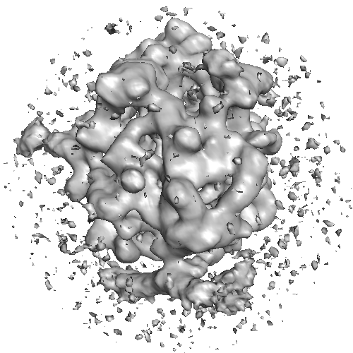
 Downloads & links
 2316-view_2.png
 http://ftp.pdbj.org/pub/emdb/structures/EMD-2316


 Z (Sec.)
 Y (Row.)
 X (Col.)


 Sample components
 Processing
 Electron microscopy
 FIELD EMISSION GUN
