Movie
Movie Controller

[English] 日本語
 Yorodumi
- PDB-3zn8: Structural Basis of Signal Sequence Surveillance and Selection by... -
+ Open data

- Basic information
| Entry | Database: PDB / ID: 3zn8 | |||||||||
|---|---|---|---|---|---|---|---|---|---|---|
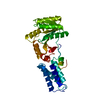
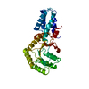
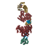
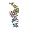
| Title | Structural Basis of Signal Sequence Surveillance and Selection by the SRP-SR Complex | |||||||||
 Components Components |
| |||||||||
 Keywords Keywords | PROTEIN TRANSPORT / HYDROLASE | |||||||||
| Function / homology |  Function and homology information Function and homology informationSynthesis, secretion, and inactivation of Glucagon-like Peptide-1 (GLP-1) / Hydrolases; Acting on peptide bonds (peptidases); Dipeptidyl-peptidases and tripeptidyl-peptidases / signal recognition particle / signal recognition particle binding / signal-recognition-particle GTPase / 7S RNA binding / SRP-dependent cotranslational protein targeting to membrane / dipeptidyl-peptidase activity / fungal-type vacuole membrane / stringent response ...Synthesis, secretion, and inactivation of Glucagon-like Peptide-1 (GLP-1) / Hydrolases; Acting on peptide bonds (peptidases); Dipeptidyl-peptidases and tripeptidyl-peptidases / signal recognition particle / signal recognition particle binding / signal-recognition-particle GTPase / 7S RNA binding / SRP-dependent cotranslational protein targeting to membrane / dipeptidyl-peptidase activity / fungal-type vacuole membrane / stringent response / aminopeptidase activity / protein targeting / protein processing / cytoplasmic side of plasma membrane / serine-type endopeptidase activity / GTPase activity / GTP binding / protein homodimerization activity / proteolysis / plasma membrane / cytosol Similarity search - Function | |||||||||
| Biological species |    SULFOLOBUS SOLFATARICUS (archaea) SULFOLOBUS SOLFATARICUS (archaea) | |||||||||
| Method | ELECTRON MICROSCOPY / single particle reconstruction / cryo EM / Resolution: 12 Å | |||||||||
 Authors Authors | von Loeffelholz, O. / Knoops, K. / Ariosa, A. / Zhang, X. / Karuppasamy, M. / Huard, K. / Schoehn, G. / Berger, I. / Shan, S.O. / Schaffitzel, C. | |||||||||
 Citation Citation | Journal: Nat Struct Mol Biol / Year: 2013 Title: Structural basis of signal sequence surveillance and selection by the SRP-FtsY complex. Authors: Ottilie von Loeffelholz / Kèvin Knoops / Aileen Ariosa / Xin Zhang / Manikandan Karuppasamy / Karine Huard / Guy Schoehn / Imre Berger / Shu-ou Shan / Christiane Schaffitzel /  Abstract: Signal-recognition particle (SRP)-dependent targeting of translating ribosomes to membranes is a multistep quality-control process. Ribosomes that are translating weakly hydrophobic signal sequences ...Signal-recognition particle (SRP)-dependent targeting of translating ribosomes to membranes is a multistep quality-control process. Ribosomes that are translating weakly hydrophobic signal sequences can be rejected from the targeting reaction even after they are bound to the SRP. Here we show that the early complex, formed by Escherichia coli SRP and its receptor FtsY with ribosomes translating the incorrect cargo EspP, is unstable and rearranges inefficiently into subsequent conformational states, such that FtsY dissociation is favored over successful targeting. The N-terminal extension of EspP is responsible for these defects in the early targeting complex. The cryo-electron microscopy structure of this 'false' early complex with EspP revealed an ordered M domain of SRP protein Ffh making two ribosomal contacts, and the NG domains of Ffh and FtsY forming a distorted, flexible heterodimer. Our results provide a structural basis for SRP-mediated signal-sequence selection during recruitment of the SRP receptor. | |||||||||
| History |
|
- Structure visualization
| Movie |
Movie viewer |
|---|---|
| Structure viewer | Molecule: MolmilJmol/JSmol |
- Downloads & links
-Download
| PDBx/mmCIF format | 3zn8.cif.gz | 199.5 KB | Display | PDBx/mmCIF format |
|---|---|---|---|---|
| PDB format | pdb3zn8.ent.gz | 152.7 KB | Display | PDB format |
| PDBx/mmJSON format | 3zn8.json.gz | Tree view | PDBx/mmJSON format | |
| Others |  Other downloads Other downloads |
-Validation report
| Arichive directory | https://data.pdbj.org/pub/pdb/validation_reports/zn/3zn8ftp://data.pdbj.org/pub/pdb/validation_reports/zn/3zn8 | HTTPS FTP |
|---|
-Related structure data
| Related structure data | 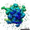 2316MC M: map data used to model this data C: citing same article ( |
|---|---|
| Similar structure data |


-Links
 PDBj
PDBj


- Assembly
| Deposited unit | 
|
|---|---|
| 1 |
|
-Components
-SIGNAL RECOGNITION PARTICLE ... , 3 types, 3 molecules ADM
| #1: Protein | Mass: 32256.250 Da / Num. of mol.: 1 / Fragment: NG, RESIDUES 2-295 Source method: isolated from a genetically manipulated source Source: (gene. exp.) References: UniProt: O07347, signal-recognition-particle GTPase |
|---|---|
| #2: Protein | Mass: 32182.012 Da / Num. of mol.: 1 / Fragment: NG, RESIDUES 201-495 Source method: isolated from a genetically manipulated source Source: (gene. exp.) References: UniProt: P10121, signal-recognition-particle GTPase |
| #4: Protein | Mass: 14591.145 Da / Num. of mol.: 1 / Fragment: M, RESIDUES 327-431 Source method: isolated from a genetically manipulated source Source: (gene. exp.) SULFOLOBUS SOLFATARICUS (archaea) / Production host: References: UniProt: Q97ZE7, signal-recognition-particle GTPase |
-RNA chain / Protein/peptide , 2 types, 2 molecules GS
| #3: RNA chain | Mass: 28505.969 Da / Num. of mol.: 1 Source method: isolated from a genetically manipulated source Source: (gene. exp.) |
|---|---|
| #5: Protein/peptide | Mass: 1522.956 Da / Num. of mol.: 1 / Fragment: RESIDUES 31-44 Source method: isolated from a genetically manipulated source Source: (gene. exp.) Production host: References: UniProt: P18962, Hydrolases; Acting on peptide bonds (peptidases); Dipeptidyl-peptidases and tripeptidyl-peptidases |
-Non-polymers , 2 types, 122 molecules 

| #6: Chemical | ChemComp-MG /  Mass: 24.305 Da / Num. of mol.: 1 / Source method: obtained synthetically / Formula: Mg Mass: 24.305 Da / Num. of mol.: 1 / Source method: obtained synthetically / Formula: Mg |
|---|---|
| #7: Water | ChemComp-HOH / Mass: 18.015 Da / Num. of mol.: 121 / Source method: isolated from a natural source / Formula: H2O |
-Experimental details
-Experiment
| Experiment | Method: ELECTRON MICROSCOPY |
|---|---|
| EM experiment | Aggregation state: PARTICLE / 3D reconstruction method: single particle reconstruction |
- Sample preparation
| Component | Name: RNCESPP-SRP-FTSY / Type: RIBOSOME / Details: MICROGRAPHS SELECTED BASED ON CTF |
|---|---|
| Buffer solution | Name: 50 MM HEPES-KOH, 100 MM KOAC, 8 MM MG(OAC)2, PH 7.5 / pH: 7.5 Details: 50 MM HEPES-KOH, 100 MM KOAC, 8 MM MG(OAC)2, PH 7.5 |
| Specimen | Embedding applied: NO / Shadowing applied: NO / Staining applied: NO / Vitrification applied: YES |
| Specimen support | Details: OTHER |
| Vitrification | Instrument: FEI VITROBOT MARK IV / Cryogen name: NITROGEN Details: THE GRIDS WERE PLUNGE FROZEN IN LIQUID ETHANE USING A FEI MARK IV VITRIFICATION ROBOT AFTER BLOTTING FOR 1 S AT RT AND 100 PER CENT RELATIVE HUMIDITY |
- Electron microscopy imaging
| Experimental equipment |  Model: Tecnai Polara / Image courtesy: FEI Company |
|---|---|
| Microscopy | Model: FEI POLARA 300 / Date: Jan 11, 2011 |
| Electron gun | Electron source:  FIELD EMISSION GUN / Accelerating voltage: 300 kV / Illumination mode: FLOOD BEAM FIELD EMISSION GUN / Accelerating voltage: 300 kV / Illumination mode: FLOOD BEAM |
| Electron lens | Mode: BRIGHT FIELD / Nominal magnification: 59000 X / Calibrated magnification: 76000 X / Nominal defocus max: 5700 nm / Nominal defocus min: 700 nm / Cs: 2.3 mm |
| Specimen holder | Temperature: 77 K / Tilt angle min: 0 ° |
| Image recording | Electron dose: 15 e/Å2 / Film or detector model: GATAN ULTRASCAN 4000 (4k x 4k) |
| Image scans | Num. digital images: 1974 |
- Processing
| EM software | Name: SPIDER / Category: 3D reconstruction | |||||||||||||||||||||||||||||||||||
|---|---|---|---|---|---|---|---|---|---|---|---|---|---|---|---|---|---|---|---|---|---|---|---|---|---|---|---|---|---|---|---|---|---|---|---|---|
| CTF correction | Details: MICROGRAPH | |||||||||||||||||||||||||||||||||||
| Symmetry | Point symmetry: C1 (asymmetric) | |||||||||||||||||||||||||||||||||||
| 3D reconstruction | Method: SINGLE PARTICLE, REFERENCE BASED PROJECTION MATCHING Resolution: 12 Å / Num. of particles: 46945 / Nominal pixel size: 3.75 Å / Actual pixel size: 3.75 Å / Details: BP RP SUBPROGRAM IN SPIDER / Symmetry type: POINT | |||||||||||||||||||||||||||||||||||
| Atomic model building | Protocol: RIGID BODY FIT / Space: REAL / Target criteria: FSC / Details: METHOD--RIGID BODY REFINEMENT PROTOCOL--X-RAY | |||||||||||||||||||||||||||||||||||
| Atomic model building |
| |||||||||||||||||||||||||||||||||||
| Refinement | Highest resolution: 12 Å | |||||||||||||||||||||||||||||||||||
| Refinement step | Cycle: LAST / Highest resolution: 12 Å
|