ムービー
ムービー コントローラー


+ データを開く

- 基本情報
基本情報
| 登録情報 | データベース: EMDB / ID: EMD-22586 | |||||||||
|---|---|---|---|---|---|---|---|---|---|---|
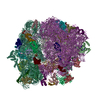
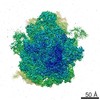
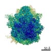
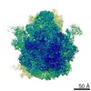
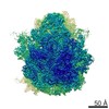
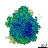
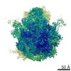
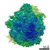
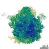
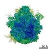
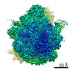
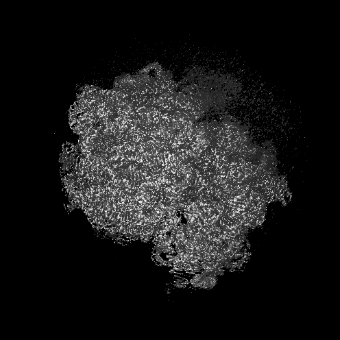
| タイトル | Structure of the Bacterial Ribosome at 2 Angstrom Resolution (composite structure) | |||||||||
 マップデータ マップデータ | 70S ribosome map (composite structure) | |||||||||
 試料 試料 |
| |||||||||
 キーワード キーワード | antibiotics / post-translational modifications / post-transcriptional modifications / RIBOSOME | |||||||||
| 機能・相同性 |  機能・相同性情報 機能・相同性情報positive regulation of ribosome biogenesis / DnaA-L2 complex / negative regulation of DNA-templated DNA replication initiation / assembly of large subunit precursor of preribosome / cytosolic ribosome assembly / regulation of cell growth / mRNA 5'-UTR binding / large ribosomal subunit / transferase activity / ribosome binding ...positive regulation of ribosome biogenesis / DnaA-L2 complex / negative regulation of DNA-templated DNA replication initiation / assembly of large subunit precursor of preribosome / cytosolic ribosome assembly / regulation of cell growth / mRNA 5'-UTR binding / large ribosomal subunit / transferase activity / ribosome binding / small ribosomal subunit / 5S rRNA binding / ribosomal large subunit assembly / cytosolic small ribosomal subunit / small ribosomal subunit rRNA binding / cytosolic large ribosomal subunit / cytoplasmic translation / tRNA binding / rRNA binding / structural constituent of ribosome / ribosome / translation / ribonucleoprotein complex / mRNA binding / RNA binding / zinc ion binding / metal ion binding / cytosol / cytoplasm 類似検索 - 分子機能 | |||||||||
| 生物種 |  | |||||||||
| 手法 | 単粒子再構成法 / クライオ電子顕微鏡法 / 解像度: 1.98 Å | |||||||||
 データ登録者 データ登録者 | Watson ZL / Ward FR | |||||||||
| 資金援助 |  米国, 2件 米国, 2件
| |||||||||
 引用 引用 | ジャーナル: Elife / 年: 2020 タイトル: Structure of the bacterial ribosome at 2 Å resolution. 著者: Zoe L Watson / Fred R Ward / Raphaël Méheust / Omer Ad / Alanna Schepartz / Jillian F Banfield / Jamie Hd Cate / 要旨: Using cryo-electron microscopy (cryo-EM), we determined the structure of the 70S ribosome with a global resolution of 2.0 Å. The maps reveal unambiguous positioning of protein and RNA residues, ...Using cryo-electron microscopy (cryo-EM), we determined the structure of the 70S ribosome with a global resolution of 2.0 Å. The maps reveal unambiguous positioning of protein and RNA residues, their detailed chemical interactions, and chemical modifications. Notable features include the first examples of isopeptide and thioamide backbone substitutions in ribosomal proteins, the former likely conserved in all domains of life. The maps also reveal extensive solvation of the small (30S) ribosomal subunit, and interactions with A-site and P-site tRNAs, mRNA, and the antibiotic paromomycin. The maps and models of the bacterial ribosome presented here now allow a deeper phylogenetic analysis of ribosomal components including structural conservation to the level of solvation. The high quality of the maps should enable future structural analyses of the chemical basis for translation and aid the development of robust tools for cryo-EM structure modeling and refinement. | |||||||||
| 履歴 |
|
- 構造の表示
構造の表示
| ムービー |
ムービービューア |
|---|---|
| 構造ビューア | EMマップ: SurfViewMolmilJmol/JSmol |
| 添付画像 |

- ダウンロードとリンク
-EMDBアーカイブ
| マップデータ | emd_22586.map.gz | 431.5 MB | EMDBマップデータ形式 | |
|---|---|---|---|---|
| ヘッダ (付随情報) | emd-22586-v30.xmlemd-22586.xml | 75.6 KB 75.6 KB | 表示 表示 | EMDBヘッダ |
| 画像 |  emd_22586.png emd_22586.png | 275.1 KB | ||
| Filedesc metadata | emd-22586.cif.gz | 15.3 KB | ||
| アーカイブディレクトリ |  http://ftp.pdbj.org/pub/emdb/structures/EMD-22586ftp://ftp.pdbj.org/pub/emdb/structures/EMD-22586 http://ftp.pdbj.org/pub/emdb/structures/EMD-22586ftp://ftp.pdbj.org/pub/emdb/structures/EMD-22586 | HTTPS FTP |
-検証レポート
| 文書・要旨 | emd_22586_validation.pdf.gz | 670.3 KB | 表示 | EMDB検証レポート |
|---|---|---|---|---|
| 文書・詳細版 | emd_22586_full_validation.pdf.gz | 669.8 KB | 表示 | |
| XML形式データ | emd_22586_validation.xml.gz | 7.6 KB | 表示 | |
| CIF形式データ | emd_22586_validation.cif.gz | 8.9 KB | 表示 | |
| アーカイブディレクトリ | https://ftp.pdbj.org/pub/emdb/validation_reports/EMD-22586ftp://ftp.pdbj.org/pub/emdb/validation_reports/EMD-22586 | HTTPS FTP |
-関連構造データ
| 関連構造データ |  7k00MC M: このマップから作成された原子モデル C: 同じ文献を引用 ( |
|---|---|
| 類似構造データ | |
| 電子顕微鏡画像生データ | EMPIAR-10509 (タイトル: Structure of the Bacterial Ribosome at 2 Å Resolution Data size: 2.1 TB Data #1: Unaligned movies of 70S ribosome complex 1 [micrographs - multiframe] Data #2: Unaligned movies of 70S ribosome complex 2 [micrographs - multiframe]) |


-リンク
| EMDBのページ | EMDB (EBI/PDBe) / EMDataResource |
|---|---|
| 「今月の分子」の関連する項目 |


-マップ
| ファイル | ダウンロード / ファイル: emd_22586.map.gz / 形式: CCP4 / 大きさ: 465.5 MB / タイプ: IMAGE STORED AS FLOATING POINT NUMBER (4 BYTES) | ||||||||||||||||||||||||||||||||||||||||||||||||||||||||||||||||||||
|---|---|---|---|---|---|---|---|---|---|---|---|---|---|---|---|---|---|---|---|---|---|---|---|---|---|---|---|---|---|---|---|---|---|---|---|---|---|---|---|---|---|---|---|---|---|---|---|---|---|---|---|---|---|---|---|---|---|---|---|---|---|---|---|---|---|---|---|---|---|
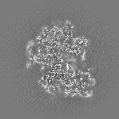
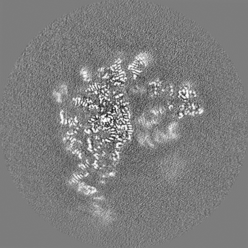
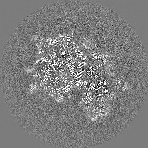
| 注釈 | 70S ribosome map (composite structure) | ||||||||||||||||||||||||||||||||||||||||||||||||||||||||||||||||||||
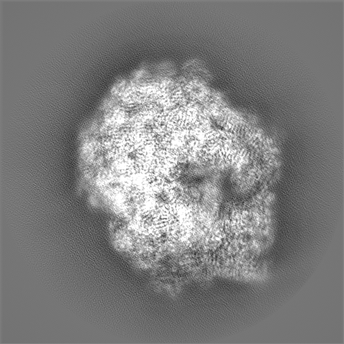
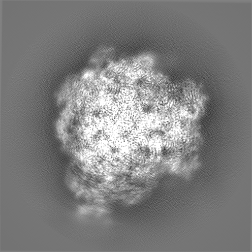
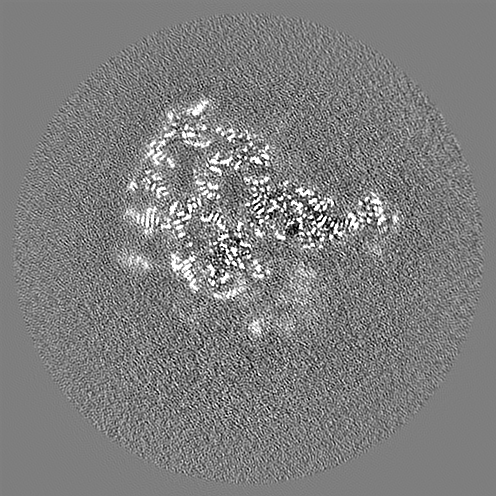
| 投影像・断面図 | 画像のコントロール
画像は Spider により作成 | ||||||||||||||||||||||||||||||||||||||||||||||||||||||||||||||||||||
| ボクセルのサイズ | X=Y=Z: 0.7118 Å | ||||||||||||||||||||||||||||||||||||||||||||||||||||||||||||||||||||
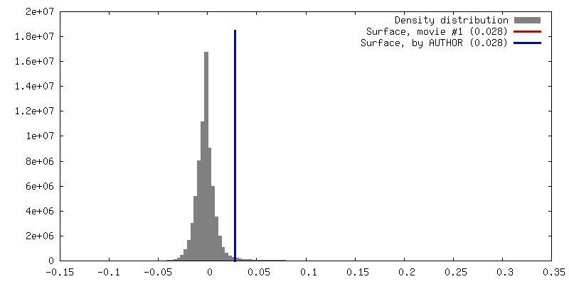
| 密度 |
| ||||||||||||||||||||||||||||||||||||||||||||||||||||||||||||||||||||
| 対称性 | 空間群: 1 | ||||||||||||||||||||||||||||||||||||||||||||||||||||||||||||||||||||
| 詳細 | EMDB XML:
CCP4マップ ヘッダ情報:
| ||||||||||||||||||||||||||||||||||||||||||||||||||||||||||||||||||||
 Z (Sec.)
 Y (Row.)
 X (Col.)


-添付データ
- 試料の構成要素
+全体 : E. coli 70S ribosome
+超分子 #1: E. coli 70S ribosome
+分子 #1: 16S rRNA
+分子 #22: 23S rRNA
+分子 #23: 5S rRNA
+分子 #53: mRNA
+分子 #54: A-site tRNA-val
+分子 #55: P-site tRNA-fMet
+分子 #56: E-site tRNA
+分子 #2: 30S ribosomal protein S2
+分子 #3: 30S ribosomal protein S3
+分子 #4: 30S ribosomal protein S4
+分子 #5: 30S ribosomal protein S5
+分子 #6: 30S ribosomal protein S6
+分子 #7: 30S ribosomal protein S7
+分子 #8: 30S ribosomal protein S8
+分子 #9: 30S ribosomal protein S9
+分子 #10: 30S ribosomal protein S10
+分子 #11: 30S ribosomal protein S11
+分子 #12: 30S ribosomal protein S12
+分子 #13: 30S ribosomal protein S13
+分子 #14: 30S ribosomal protein S14
+分子 #15: 30S ribosomal protein S15
+分子 #16: 30S ribosomal protein S16
+分子 #17: 30S ribosomal protein S17
+分子 #18: 30S ribosomal protein S18
+分子 #19: 30S ribosomal protein S19
+分子 #20: 30S ribosomal protein S20
+分子 #21: 30S ribosomal protein S21
+分子 #24: 50S ribosomal protein L2
+分子 #25: 50S ribosomal protein L3
+分子 #26: 50S ribosomal protein L4
+分子 #27: 50S ribosomal protein L5
+分子 #28: 50S ribosomal protein L6
+分子 #29: 50S ribosomal protein L9
+分子 #30: 50S ribosomal protein L13
+分子 #31: 50S ribosomal protein L14
+分子 #32: 50S ribosomal protein L15
+分子 #33: 50S ribosomal protein L16
+分子 #34: 50S ribosomal protein L17
+分子 #35: 50S ribosomal protein L18
+分子 #36: 50S ribosomal protein L19
+分子 #37: 50S ribosomal protein L20
+分子 #38: 50S ribosomal protein L21
+分子 #39: 50S ribosomal protein L22
+分子 #40: 50S ribosomal protein L23
+分子 #41: 50S ribosomal protein L24
+分子 #42: 50S ribosomal protein L25
+分子 #43: 50S ribosomal protein L27
+分子 #44: 50S ribosomal protein L28
+分子 #45: 50S ribosomal protein L29
+分子 #46: 50S ribosomal protein L30
+分子 #47: 50S ribosomal protein L32
+分子 #48: 50S ribosomal protein L33
+分子 #49: 50S ribosomal protein L34
+分子 #50: 50S ribosomal protein L35
+分子 #51: 50S ribosomal protein L36
+分子 #52: 50S ribosomal protein L31
+分子 #57: PAROMOMYCIN
+分子 #58: MAGNESIUM ION
+分子 #59: SPERMIDINE

+分子 #60: SPERMINE

+分子 #61: ZINC ION
+分子 #62: water

-実験情報
-構造解析
| 手法 | クライオ電子顕微鏡法 |
|---|---|
 解析 解析 | 単粒子再構成法 |
| 試料の集合状態 | particle |
-試料調製
| 濃度 | 0.27 mg/mL |
|---|---|
| 緩衝液 | pH: 7.5 |
| グリッド | モデル: UltrAuFoil / 材質: GOLD / メッシュ: 300 / 支持フィルム - 材質: CARBON / 前処理 - タイプ: GLOW DISCHARGE |
| 凍結 | 凍結剤: ETHANE / チャンバー内湿度: 100 % / チャンバー内温度: 277 K / 装置: FEI VITROBOT MARK III |
- 電子顕微鏡法
| 顕微鏡 | FEI TITAN KRIOS |
|---|---|
| 撮影 | フィルム・検出器のモデル: GATAN K3 (6k x 4k) / 撮影したグリッド数: 2 / 平均電子線量: 40.0 e/Å2 |
| 電子線 | 加速電圧: 300 kV / 電子線源:  FIELD EMISSION GUN FIELD EMISSION GUN |
| 電子光学系 | 照射モード: OTHER / 撮影モード: BRIGHT FIELD / Cs: 2.7 mm |
| 実験機器 |  モデル: Titan Krios / 画像提供: FEI Company |
-画像解析
| 初期モデル | モデルのタイプ: PDB ENTRY PDBモデル - PDB ID: |
|---|---|
| 最終 再構成 | 解像度のタイプ: BY AUTHOR / 解像度: 1.98 Å / 解像度の算出法: FSC 0.143 CUT-OFF / 詳細: Ewald sphere corrected in RELION / 使用した粒子像数: 307495 |
| 初期 角度割当 | タイプ: MAXIMUM LIKELIHOOD |
| 最終 角度割当 | タイプ: MAXIMUM LIKELIHOOD / ソフトウェア - 名称: RELION |