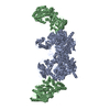
Complex: Complex consisting of actin-filament nucleator, Arp2/3 complex associated with nucleation promoting factor Dip1 and first four actin subunits from the pointed end of the nucleated actin-filament
Protein or peptide: x 10 types
Ligand: x 3 types
Keywords
Arp2/3 / actin / Dip1 / cytoskeletal protein / actin regulator / STRUCTURAL PROTEIN
Function / homology
Function and homology information
protein localization to actin cortical patch / Regulation of actin dynamics for phagocytic cup formation / RHO GTPases Activate WASPs and WAVEs / Clathrin-mediated endocytosis / actin cortical patch organization / medial cortex / Neutrophil degranulation / actin filament branching / cell cortex of cell tip / actin cortical patch localization ...protein localization to actin cortical patch / Regulation of actin dynamics for phagocytic cup formation / RHO GTPases Activate WASPs and WAVEs / Clathrin-mediated endocytosis / actin cortical patch organization / medial cortex / Neutrophil degranulation / actin filament branching / cell cortex of cell tip / actin cortical patch localization / actin cortical patch assembly / Arp2/3 protein complex / Arp2/3 complex-mediated actin nucleation / Arp2/3 complex binding / actin cortical patch / cell tip / regulation of actin filament polymerization / mating projection tip / cortical actin cytoskeleton organization / cytoskeletal motor activator activity / cell division site / myosin heavy chain binding / tropomyosin binding / actin filament bundle / troponin I binding / filamentous actin / mesenchyme migration / cortical cytoskeleton / establishment or maintenance of cell polarity / skeletal muscle myofibril / actin filament bundle assembly / striated muscle thin filament / skeletal muscle thin filament assembly / actin monomer binding / skeletal muscle fiber development / stress fiber / titin binding / actin filament polymerization / filopodium / actin filament / structural constituent of cytoskeleton / Hydrolases; Acting on acid anhydrides; Acting on acid anhydrides to facilitate cellular and subcellular movement / endocytosis / calcium-dependent protein binding / actin filament binding / lamellipodium / toxin activity / cell body / cell cortex / protein-macromolecule adaptor activity / protein domain specific binding / hydrolase activity / calcium ion binding / positive regulation of gene expression / magnesium ion binding / ATP binding / identical protein binding / nucleus / cytoplasm / cytosol Similarity search - Function
National Institutes of Health/National Institute of General Medical Sciences (NIH/NIGMS)
R01GM127440, R01GM092917, S10OD012272
United States
Citation
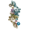
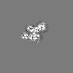
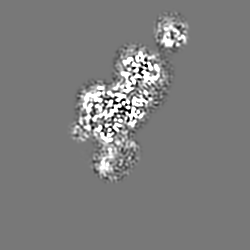
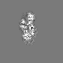
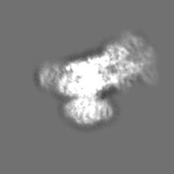
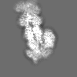
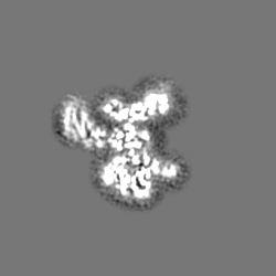
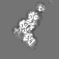
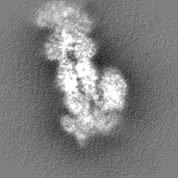
Journal: Nat Struct Mol Biol / Year: 2020 Title: Cryo-EM reveals the transition of Arp2/3 complex from inactive to nucleation-competent state. Authors: Mohammed Shaaban / Saikat Chowdhury / Brad J Nolen / Abstract: Arp2/3 complex, a crucial actin filament nucleator, undergoes structural rearrangements during activation by nucleation-promoting factors (NPFs). However, the conformational pathway leading to the ...Arp2/3 complex, a crucial actin filament nucleator, undergoes structural rearrangements during activation by nucleation-promoting factors (NPFs). However, the conformational pathway leading to the nucleation-competent state is unclear due to lack of high-resolution structures of the activated state. Here we report a ~3.9 Å resolution cryo-EM structure of activated Schizosaccharomyces pombe Arp2/3 complex bound to the S. pombe NPF Dip1 and attached to the end of the nucleated actin filament. The structure reveals global and local conformational changes that allow the two actin-related proteins in Arp2/3 complex to mimic a filamentous actin dimer and template nucleation. Activation occurs through a clamp-twisting mechanism, in which Dip1 forces two core subunits in Arp2/3 complex to pivot around one another, shifting half of the complex into a new activated position. By showing how Dip1 stimulates activation, the structure reveals how NPFs can activate Arp2/3 complex in diverse cellular processes.
History
Deposition
Mar 3, 2020
-
Header (metadata) release
Apr 1, 2020
-
Map release
Aug 12, 2020
-
Update
Apr 2, 2025
-
Current status
Apr 2, 2025
Processing site: RCSB / Status: Released
-
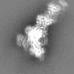
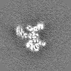
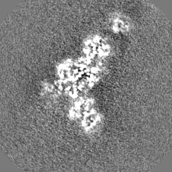
Structure visualization
Movie
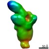
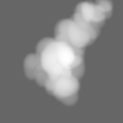
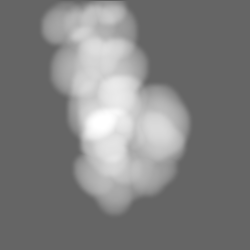
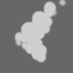
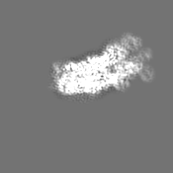
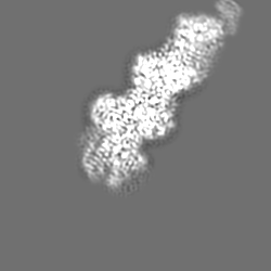
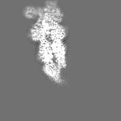
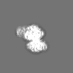
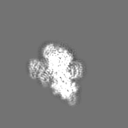
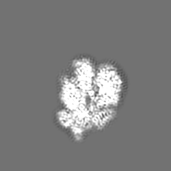
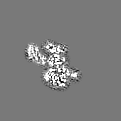
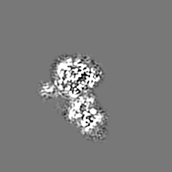
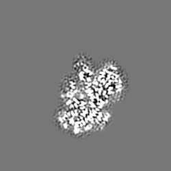
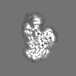
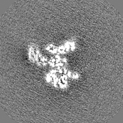
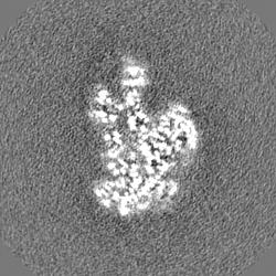
Surface view with section colored by density value
Entire : Complex consisting of actin-filament nucleator, Arp2/3 complex as...
Entire
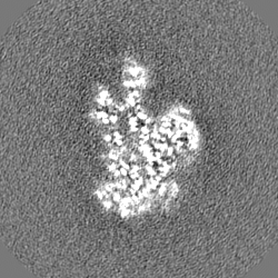
Name: Complex consisting of actin-filament nucleator, Arp2/3 complex associated with nucleation promoting factor Dip1 and first four actin subunits from the pointed end of the nucleated actin-filament
Components
Complex: Complex consisting of actin-filament nucleator, Arp2/3 complex associated with nucleation promoting factor Dip1 and first four actin subunits from the pointed end of the nucleated actin-filament
Protein or peptide: Actin-related protein 3
Protein or peptide: Actin-related protein 2
Protein or peptide: Actin-related protein 2/3 complex subunit 1
Protein or peptide: Actin-related protein 2/3 complex subunit 2
Protein or peptide: Actin-related protein 2/3 complex subunit 3
Protein or peptide: Actin-related protein 2/3 complex subunit 4
Protein or peptide: Actin-related protein 2/3 complex subunit 5
Protein or peptide: Protein dip1
Protein or peptide: Actin, alpha skeletal muscle
Protein or peptide: Phalloidin
Ligand: MAGNESIUM ION
Ligand: ADENOSINE-5'-TRIPHOSPHATE
Ligand: ADENOSINE-5'-DIPHOSPHATE
+
Supramolecule #1: Complex consisting of actin-filament nucleator, Arp2/3 complex as...
Supramolecule
Name: Complex consisting of actin-filament nucleator, Arp2/3 complex associated with nucleation promoting factor Dip1 and first four actin subunits from the pointed end of the nucleated actin-filament type: complex / ID: 1 / Parent: 0 / Macromolecule list: #1-#10
Data were collected by stage shifting to targeted exposure positions.
Image recording
Film or detector model: FEI FALCON III (4k x 4k) / Detector mode: COUNTING / Digitization - Dimensions - Width: 4000 pixel / Digitization - Dimensions - Height: 4000 pixel / Number grids imaged: 2 / Number real images: 5109 / Average exposure time: 60.0 sec. / Average electron dose: 36.35 e/Å2 Details: Each micrograph was collected as dose-fractionated movies consisting of 45 fractions per movie.
Electron beam
Acceleration voltage: 200 kV / Electron source: FIELD EMISSION GUN
Model: Talos Arctica / Image courtesy: FEI Company
+
Image processing
Particle selection
Number selected: 3500000 Details: Particles were picked using Laplacian Gaussian auto-picking.
Startup model
#0 - Type of model: INSILICO MODEL #0 - In silico model: Ab-initio initial model was determined using CryoSPARCv2. #1 - Type of model: INSILICO MODEL #1 - In silico model: Ab-initio initial model was determined using CryoSPARCv2.
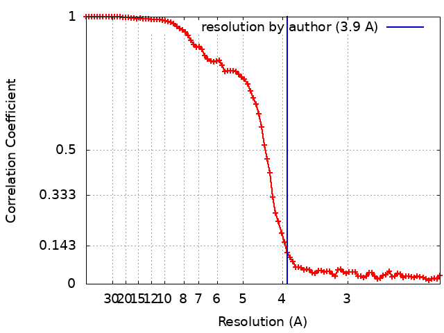
Final reconstruction
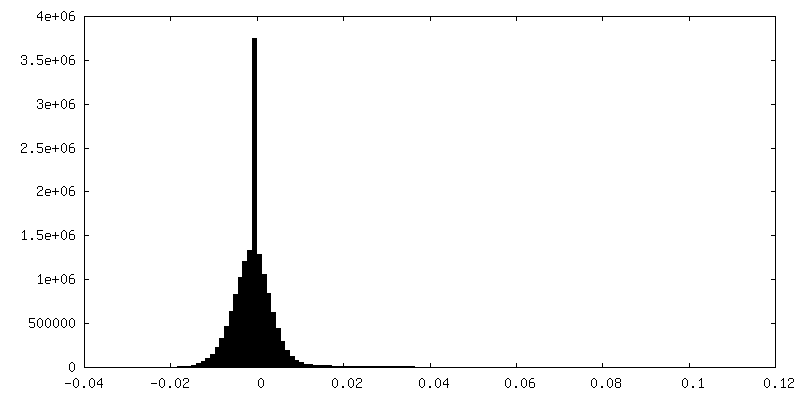
Number classes used: 1 / Applied symmetry - Point group: C1 (asymmetric) / Algorithm: BACK PROJECTION / Resolution.type: BY AUTHOR / Resolution: 3.9 Å / Resolution method: FSC 0.143 CUT-OFF / Software - Name: RELION (ver. 3.0.6) / Number images used: 110433
Initial angle assignment
Type: NOT APPLICABLE
Final angle assignment
Type: MAXIMUM LIKELIHOOD / Software - Name: RELION (ver. 3.0.6)
Final 3D classification
Number classes: 3 / Software - Name: RELION (ver. 3.0.6) Details: Classification was performed without image alignments
In the structure databanks used in Yorodumi, some data are registered as the other names, "COVID-19 virus" and "2019-nCoV". Here are the details of the virus and the list of structure data.
Jan 31, 2019. EMDB accession codes are about to change! (news from PDBe EMDB page)
EMDB accession codes are about to change! (news from PDBe EMDB page)
The allocation of 4 digits for EMDB accession codes will soon come to an end. Whilst these codes will remain in use, new EMDB accession codes will include an additional digit and will expand incrementally as the available range of codes is exhausted. The current 4-digit format prefixed with “EMD-” (i.e. EMD-XXXX) will advance to a 5-digit format (i.e. EMD-XXXXX), and so on. It is currently estimated that the 4-digit codes will be depleted around Spring 2019, at which point the 5-digit format will come into force.
The EM Navigator/Yorodumi systems omit the EMD- prefix.
Related info.:Q: What is EMD? / ID/Accession-code notation in Yorodumi/EM Navigator
Yorodumi is a browser for structure data from EMDB, PDB, SASBDB, etc.
This page is also the successor to EM Navigator detail page, and also detail information page/front-end page for Omokage search.
The word "yorodu" (or yorozu) is an old Japanese word meaning "ten thousand". "mi" (miru) is to see.
Related info.:EMDB / PDB / SASBDB / Comparison of 3 databanks / Yorodumi Search / Aug 31, 2016. New EM Navigator & Yorodumi / Yorodumi Papers / Jmol/JSmol / Function and homology information / Changes in new EM Navigator and Yorodumi
 Movie
Movie Controller

 Yorodumi
 Open data
 Basic information
 Map data
 Sample
 Keywords
 Function and homology information

 Amanita phalloides (death cap)
 Authors
 United States, 1 items
  Citation
 Structure visualization
 Downloads & links
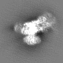
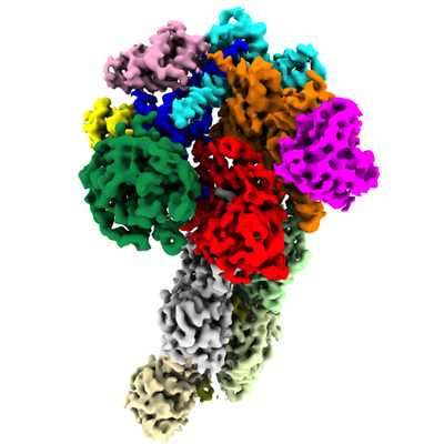 emd_21502.png
 http://ftp.pdbj.org/pub/emdb/structures/EMD-21502


 Z (Sec.)
 X (Row.)
 Y (Col.)


 Sample components


 Processing
 Electron microscopy
 FIELD EMISSION GUN