 Movie
Movie Controller
[English] 日本語
 Yorodumi
- SASDFS4: HP0242 from Helicobacter pylori, N-terminal domain of syntaxin-1A... -
+ Open data

- Basic information
| Entry | Database: SASBDB / ID: SASDFS4 |
|---|---|
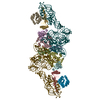
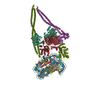
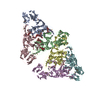
 Sample Sample | HP0242 from Helicobacter pylori, N-terminal domain of syntaxin-1A from Rattus norvegicus, de novo designed coiled-coil trimer domain (Domain A (PDB:3MLI)-Linker (PDB:1BR0)-Domain B (PDB:4DZL))
|
| Biological species |   Helicobacter pylori (bacteria) Helicobacter pylori (bacteria) de novo design (unknown) |
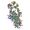
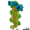
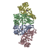
 Citation Citation | Journal: ACS Synth Biol / Year: 2019 Title: Construction of a Quadrangular Tetramer and a Cage-Like Hexamer from Three-Helix Bundle-Linked Fusion Proteins. Authors: Takaaki Miyamoto / Yugo Hayashi / Keito Yoshida / Hiroki Watanabe / Takayuki Uchihashi / Kento Yonezawa / Nobutaka Shimizu / Hironari Kamikubo / Shun Hirota /  Abstract: Self-assembled protein nanostructures have gained interest, owing to their potential applications in biomaterials; however, successful design and construction of protein nanostructures are limited. ...Self-assembled protein nanostructures have gained interest, owing to their potential applications in biomaterials; however, successful design and construction of protein nanostructures are limited. Herein, we constructed fusion protein 1 by linking the C-terminus of a dimerization domain and the N-terminus of another dimerization domain with a three-helix bundle protein, where it self-assembled mainly into tetramers. By replacing the C-terminal dimerization domain of 1 with a trimerization domain (fusion protein 2), hexamers were mainly obtained. According to ab initio structural models reconstructed from the small-angle X-ray scattering data, the tetramer of 1 and hexamer of 2 adopted quadrangle and cage-like structures, respectively, although they were combinations of different conformations. High-speed atomic force microscopy observations indicated that the tetramer and hexamer exhibit conformational dynamics. These results show that the present method utilizing three-helix bundle-linked fusion proteins is useful in the construction of protein nanostructures. |
 Contact author Contact author |
|
- Structure visualization
| Structure viewer | Molecule:  MolmilJmol/JSmol MolmilJmol/JSmol |
|---|
- Downloads & links
 SASDFS4
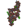


-Models
| Model #2930 |  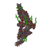 Type: mix / Radius of dummy atoms: 1.90 A / Chi-square value: 0.10 / P-value: 0.000088  Search similar-shape structures of this assembly by Omokage search (details) Search similar-shape structures of this assembly by Omokage search (details) |
|---|
-Sample
| Sample | Name: HP0242 from Helicobacter pylori, N-terminal domain of syntaxin-1A from Rattus norvegicus, de novo designed coiled-coil trimer domain (Domain A (PDB:3MLI)-Linker (PDB:1BR0)-Domain B (PDB:4DZL)) Specimen concentration: 1.90-6.80 |
|---|---|
| Buffer | Name: 20 mM Tris-HCl 150 mM NaCl / pH: 8 |
| Entity #1583 | Name: Fusion protein2 / Type: protein Description: HP0242 from Helicobacter pylori, N-terminal domain of syntaxin-1A from Rattus norvegicus, de novo designed coiled-coil trimer domain Formula weight: 29.855 / Num. of mol.: 6 Source: Helicobacter pylori, Rattus norvegicus, de novo design Sequence: MGSSHHHHHH SSGLVPRGSH MRDYSELEIF EGNPLDKWND IIFHASKKAS KKELERLLEL LALCETFIEK EDLEEKFESF AKALRIDEEL QQKIESRKTD IVIQSMANIL SALFMDEFFE QVEEIRGFID KIAENVEEVK RKHSAILASP NPDEKTKEEL EELMSDIKKT ...Sequence: MGSSHHHHHH SSGLVPRGSH MRDYSELEIF EGNPLDKWND IIFHASKKAS KKELERLLEL LALCETFIEK EDLEEKFESF AKALRIDEEL QQKIESRKTD IVIQSMANIL SALFMDEFFE QVEEIRGFID KIAENVEEVK RKHSAILASP NPDEKTKEEL EELMSDIKKT ANKVRSKLKS IEQSIEQEEG LNRSSADLRI RKTQHSTLSR KFVEVMSEYN ATQSDYGSGG EIAAIKQEIA AIKKEIAAIK WEIAAIKQGY G |
-Experimental information
| Beam | Instrument name: Nara Institute of Science and Technology Rigaku Nano-Viewer City: Ikoma / 国: Japan / Type of source: X-ray in house / Wavelength: 0.15418 Å / Dist. spec. to detc.: 0.75 mm | |||||||||||||||||||||||||||||||||||||||
|---|---|---|---|---|---|---|---|---|---|---|---|---|---|---|---|---|---|---|---|---|---|---|---|---|---|---|---|---|---|---|---|---|---|---|---|---|---|---|---|---|
| Detector | Name: Pilatus 200K / Type: Pilatus / Pixsize x: 172 mm | |||||||||||||||||||||||||||||||||||||||
| Scan |  Measurement date: Mar 29, 2017 / Storage temperature: 4 °C / Cell temperature: 20 °C / Exposure time: 60 sec. / Number of frames: 25 / Unit: 1/A /
| |||||||||||||||||||||||||||||||||||||||
| Distance distribution function P(R) | 
| |||||||||||||||||||||||||||||||||||||||
| Result | 
|
