 Movie
Movie Controller
[English] 日本語
 Yorodumi
- SASDFR4: HP2042 form from Helicobacter pylori, N-terminal domain of syntax... -
+ Open data

- Basic information
| Entry | Database: SASBDB / ID: SASDFR4 |
|---|---|
 Sample Sample | HP2042 form from Helicobacter pylori, N-terminal domain of syntaxin-1A from Rattus norvegicus, Trp repressor from Escherichia coli (Domain A (PDB:3MLI)-Linker (PDB:1BR0)-Domain B (PDB:1WRT))
|
| Biological species |   Helicobacter pylori (bacteria) Helicobacter pylori (bacteria) |
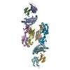
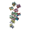
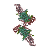
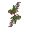
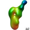
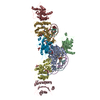
 Citation Citation | Journal: ACS Synth Biol / Year: 2019 Title: Construction of a Quadrangular Tetramer and a Cage-Like Hexamer from Three-Helix Bundle-Linked Fusion Proteins. Authors: Takaaki Miyamoto / Yugo Hayashi / Keito Yoshida / Hiroki Watanabe / Takayuki Uchihashi / Kento Yonezawa / Nobutaka Shimizu / Hironari Kamikubo / Shun Hirota /  Abstract: Self-assembled protein nanostructures have gained interest, owing to their potential applications in biomaterials; however, successful design and construction of protein nanostructures are limited. ...Self-assembled protein nanostructures have gained interest, owing to their potential applications in biomaterials; however, successful design and construction of protein nanostructures are limited. Herein, we constructed fusion protein 1 by linking the C-terminus of a dimerization domain and the N-terminus of another dimerization domain with a three-helix bundle protein, where it self-assembled mainly into tetramers. By replacing the C-terminal dimerization domain of 1 with a trimerization domain (fusion protein 2), hexamers were mainly obtained. According to ab initio structural models reconstructed from the small-angle X-ray scattering data, the tetramer of 1 and hexamer of 2 adopted quadrangle and cage-like structures, respectively, although they were combinations of different conformations. High-speed atomic force microscopy observations indicated that the tetramer and hexamer exhibit conformational dynamics. These results show that the present method utilizing three-helix bundle-linked fusion proteins is useful in the construction of protein nanostructures. |
 Contact author Contact author |
|
- Structure visualization
| Structure viewer | Molecule:  MolmilJmol/JSmol MolmilJmol/JSmol |
|---|
- Downloads & links
 SASDFR4
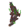


-Models
| Model #2929 |   Type: mix / Radius of dummy atoms: 1.90 A / Chi-square value: 0.10 / P-value: 0.046149  Search similar-shape structures of this assembly by Omokage search (details) Search similar-shape structures of this assembly by Omokage search (details) |
|---|
-Sample
| Sample | Name: HP2042 form from Helicobacter pylori, N-terminal domain of syntaxin-1A from Rattus norvegicus, Trp repressor from Escherichia coli (Domain A (PDB:3MLI)-Linker (PDB:1BR0)-Domain B (PDB:1WRT)) Specimen concentration: 2.30-8.30 |
|---|---|
| Buffer | Name: 20 mM Tris-HCl 150 mM NaCl / pH: 8 |
| Entity #1582 | Name: Fusion protein1 / Type: protein Description: HP2042 form from Helicobacter pylori, N-terminal domain of syntaxin-1A from Rattus norvegicus, Trp repressor from Escherichia coli Formula weight: 37.434 / Num. of mol.: 4 Source: Helicobacter pylori, Rattus norvegicus, Escherichia coli Sequence: MGSSHHHHHH SSGLVPRGSH MRDYSELEIF EGNPLDKWND IIFHASKKAS KKELERLLEL LALCETFIEK EDLEEKFESF AKALRIDEEL QQKIESRKTD IVIQSMANIL SALFMDEFFE QVEEIRGFID KIAENVEEVK RKHSAILASP NPDEKTKEEL EELMSDIKKT ...Sequence: MGSSHHHHHH SSGLVPRGSH MRDYSELEIF EGNPLDKWND IIFHASKKAS KKELERLLEL LALCETFIEK EDLEEKFESF AKALRIDEEL QQKIESRKTD IVIQSMANIL SALFMDEFFE QVEEIRGFID KIAENVEEVK RKHSAILASP NPDEKTKEEL EELMSDIKKT ANKVRSKLKS IEQSIEQEEG LNRSSADLRI RKTQHSTLSR KFVEVMSEYN ATQSDYGSGS GRHQEWLRFV DLLKNAYQND LHLPLLNLML TPDEREALGT RVRIVEELLR GEMSQRELKN ELGAGIATIT RGSNSLKAAP VELRQWLEEV LLKSD |
-Experimental information
| Beam | Instrument name: Nara Institute of Science and Technology Rigaku Nano-Viewer City: Ikoma / 国: Japan / Type of source: X-ray in house / Wavelength: 0.15418 Å / Dist. spec. to detc.: 0.75 mm | |||||||||||||||||||||||||||||||||||||||
|---|---|---|---|---|---|---|---|---|---|---|---|---|---|---|---|---|---|---|---|---|---|---|---|---|---|---|---|---|---|---|---|---|---|---|---|---|---|---|---|---|
| Detector | Name: Pilatus 200K / Type: Pilatus / Pixsize x: 172 mm | |||||||||||||||||||||||||||||||||||||||
| Scan |  Measurement date: Dec 8, 2016 / Storage temperature: 4 °C / Cell temperature: 20 °C / Exposure time: 60 sec. / Number of frames: 25 / Unit: 1/A /
| |||||||||||||||||||||||||||||||||||||||
| Distance distribution function P(R) | 
| |||||||||||||||||||||||||||||||||||||||
| Result | 
|
