National Institutes of Health/National Institute Of Allergy and Infectious Diseases (NIH/NIAID)
R01-AI11893
米国
引用
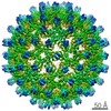
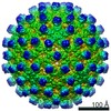
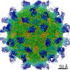
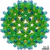
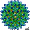
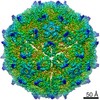
ジャーナル: ACS Chem Biol / 年: 2020 タイトル: The Integrity of the Intradimer Interface of the Hepatitis B Virus Capsid Protein Dimer Regulates Capsid Self-Assembly. 著者: Zhongchao Zhao / Joseph Che-Yen Wang / Carolina Pérez Segura / Jodi A Hadden-Perilla / Adam Zlotnick / 要旨: During the hepatitis B virus lifecycle, 120 copies of homodimeric capsid protein assemble around a copy of reverse transcriptase and viral RNA and go on to produce an infectious virion. Assembly ...During the hepatitis B virus lifecycle, 120 copies of homodimeric capsid protein assemble around a copy of reverse transcriptase and viral RNA and go on to produce an infectious virion. Assembly needs to be tightly regulated by protein conformational change to ensure symmetry, fidelity, and reproducibility. Here, we show that structures at the intradimer interface regulate conformational changes at the distal interdimer interface and so regulate assembly. A pair of interacting charged residues, D78 from each monomer, conspicuously located at the top of a four-helix bundle that forms the intradimer interface, were mutated to serine to disrupt communication between the two monomers. The mutation slowed assembly and destabilized the dimer to thermal and chemical denaturation. Mutant dimers showed evidence of transient partial unfolding based on the appearance of new proteolytically sensitive sites. Though the mutant dimer was less stable, the resulting capsids were as stable as the wildtype, based on assembly and thermal denaturation studies. Cryo-EM image reconstructions of capsid indicated that the subunits adopted an "open" state more usually associated with a free dimer and that the spike tips were either disordered or highly flexible. Molecular dynamics simulations provide mechanistic explanations for these results, suggesting that D78 stabilizes helix 4a, which forms part of the intradimer interface, by capping its N-terminus and hydrogen-bonding to nearby residues, whereas the D78S mutation disrupts these interactions, leading to partial unwinding of helix 4a. This in turn weakens the connection from helix 4 and the intradimer interface to helix 5, which forms the interdimer interface.
 ムービー
ムービー コントローラー


 データを開く
 基本情報
基本情報 マップデータ
 試料
試料 キーワード
キーワード 機能・相同性情報
 Hepatitis B virus genotype D subtype adw (isolate United Kingdom/adyw/1979) (ウイルス)
 データ登録者
 米国, 1件
  引用
引用 構造の表示
構造の表示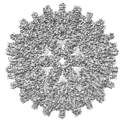
 ダウンロードとリンク
 emd_21497.png
 http://ftp.pdbj.org/pub/emdb/structures/EMD-21497


 Z (Sec.)
 Y (Row.)
 X (Col.)


 試料の構成要素
 解析
解析 電子顕微鏡法
 FIELD EMISSION GUN


