 Movie
Movie Controller

[English] 日本語
 Yorodumi
- EMDB-13957: Extended H/L (SLPH/SLPL) complex from C. difficile (CD630 strain)... -
+ Open data

- Basic information
| Entry | 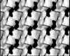 | |||||||||
|---|---|---|---|---|---|---|---|---|---|---|
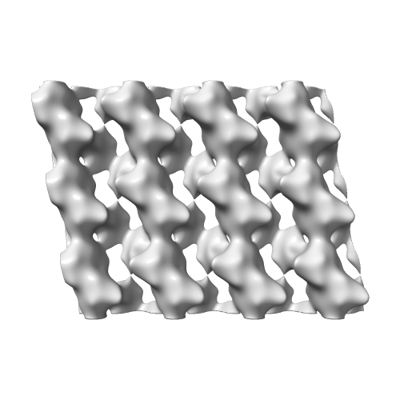
| Title | Extended H/L (SLPH/SLPL) complex from C. difficile (CD630 strain) fit into R20291 S-layer negative stain map | |||||||||
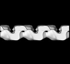
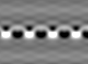
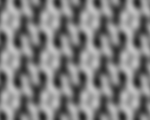
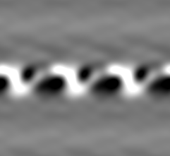
 Map data Map data | Electron crystallographic reconstruction of R20291 S-layer, extended to cover 12 molecules of SlpA for flexible fitting of X-ray structure to map. | |||||||||
 Sample Sample |
| |||||||||
 Keywords Keywords | Bacterial surface / S-layer / STRUCTURAL PROTEIN | |||||||||
| Function / homology | Low molecular weight S layer protein, N-terminal / Low molecular weight S layer protein, N-terminal, subdomain / Low molecular weight S layer protein N terminal / : / Putative cell wall binding repeat 2 / Cell wall binding domain 2 (CWB2) / identical protein binding / Precursor of the S-layer proteins Function and homology information Function and homology information | |||||||||
| Biological species |  Clostridioides difficile R20291 (bacteria) / Clostridioides difficile 630 (bacteria) Clostridioides difficile R20291 (bacteria) / Clostridioides difficile 630 (bacteria) | |||||||||
| Method | electron crystallography / negative staining | |||||||||
 Authors Authors | Banerji O / Wilson JS | |||||||||
| Funding support |  United Kingdom, 1 items United Kingdom, 1 items
| |||||||||
 Citation Citation | Journal: Nat Commun / Year: 2022 Title: Structure and assembly of the S-layer in C. difficile. Authors: Paola Lanzoni-Mangutchi / Oishik Banerji / Jason Wilson / Anna Barwinska-Sendra / Joseph A Kirk / Filipa Vaz / Shauna O'Beirne / Arnaud Baslé / Kamel El Omari / Armin Wagner / Neil F ...Authors: Paola Lanzoni-Mangutchi / Oishik Banerji / Jason Wilson / Anna Barwinska-Sendra / Joseph A Kirk / Filipa Vaz / Shauna O'Beirne / Arnaud Baslé / Kamel El Omari / Armin Wagner / Neil F Fairweather / Gillian R Douce / Per A Bullough / Robert P Fagan / Paula S Salgado /  Abstract: Many bacteria and archaea possess a two-dimensional protein array, or S-layer, that covers the cell surface and plays crucial roles in cell physiology. Here, we report the crystal structure of SlpA, ...Many bacteria and archaea possess a two-dimensional protein array, or S-layer, that covers the cell surface and plays crucial roles in cell physiology. Here, we report the crystal structure of SlpA, the main S-layer protein of the bacterial pathogen Clostridioides difficile, and use electron microscopy to study S-layer organisation and assembly. The SlpA crystal lattice mimics S-layer assembly in the cell, through tiling of triangular prisms above the cell wall, interlocked by distinct ridges facing the environment. Strikingly, the array is very compact, with pores of only ~10 Å in diameter, compared to other S-layers (30-100 Å). The surface-exposed flexible ridges are partially dispensable for overall structure and assembly, although a mutant lacking this region becomes susceptible to lysozyme, an important molecule in host defence. Thus, our work gives insights into S-layer organisation and provides a basis for development of C. difficile-specific therapeutics. | |||||||||
| History |
|
- Structure visualization
| Supplemental images |
|---|
- Downloads & links
-EMDB archive
| Map data | emd_13957.map.gz | 15.1 MB | EMDB map data format | |
|---|---|---|---|---|
| Header (meta data) | emd-13957-v30.xmlemd-13957.xml | 14.6 KB 14.6 KB | Display Display | EMDB header |
| Images |  emd_13957.png emd_13957.png | 59.5 KB | ||
| Filedesc metadata | emd-13957.cif.gz | 6 KB | ||
| Filedesc structureFactors | emd_13957_sf.cif.gz | 985 B | ||
| Archive directory |  http://ftp.pdbj.org/pub/emdb/structures/EMD-13957ftp://ftp.pdbj.org/pub/emdb/structures/EMD-13957 http://ftp.pdbj.org/pub/emdb/structures/EMD-13957ftp://ftp.pdbj.org/pub/emdb/structures/EMD-13957 | HTTPS FTP |
-Related structure data
| Related structure data |  7qgqMC  7acvC  7acwC 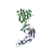 7acxC  7acyC  7aczC M: atomic model generated by this map C: citing same article ( |
|---|---|
| Similar structure data |
-Links
| EMDB pages | EMDB (EBI/PDBe) / EMDataResource |
|---|
-Map
| File | Download / File: emd_13957.map.gz / Format: CCP4 / Size: 70.6 MB / Type: IMAGE STORED AS FLOATING POINT NUMBER (4 BYTES) | ||||||||||||||||||||||||||||||||||||
|---|---|---|---|---|---|---|---|---|---|---|---|---|---|---|---|---|---|---|---|---|---|---|---|---|---|---|---|---|---|---|---|---|---|---|---|---|---|
| Annotation | Electron crystallographic reconstruction of R20291 S-layer, extended to cover 12 molecules of SlpA for flexible fitting of X-ray structure to map. | ||||||||||||||||||||||||||||||||||||
| Projections & slices | Image control
Images are generated by Spider. generated in cubic-lattice coordinate | ||||||||||||||||||||||||||||||||||||
| Voxel size | X: 1 Å / Y: 1 Å / Z: 1.5625 Å | ||||||||||||||||||||||||||||||||||||
| Density |
| ||||||||||||||||||||||||||||||||||||
| Symmetry | Space group: 1 | ||||||||||||||||||||||||||||||||||||
| Details | EMDB XML:
|
 Z (Sec.)
 Y (Row.)
 X (Col.)


-Supplemental data
- Sample components
-Entire : Electron crystallographic reconstruction of R20291 S-layer, exten...
| Entire | Name: Electron crystallographic reconstruction of R20291 S-layer, extended to cover 12 molecules of SlpA for flexible fitting of X-ray structure to map |
|---|---|
| Components |
|
-Supramolecule #1: Electron crystallographic reconstruction of R20291 S-layer, exten...
| Supramolecule | Name: Electron crystallographic reconstruction of R20291 S-layer, extended to cover 12 molecules of SlpA for flexible fitting of X-ray structure to map type: complex / ID: 1 / Parent: 0 / Macromolecule list: all |
|---|---|
| Source (natural) | Organism: Clostridioides difficile R20291 (bacteria) |
-Macromolecule #1: Precursor of the S-layer proteins
| Macromolecule | Name: Precursor of the S-layer proteins / type: protein_or_peptide / ID: 1 / Number of copies: 12 / Enantiomer: LEVO |
|---|---|
| Source (natural) | Organism: Clostridioides difficile 630 (bacteria) / Strain: 630 |
| Molecular weight | Theoretical: 39.495035 KDa |
| Sequence | String: NDTIASQDTP AKVVIKANKL KDLKDYVDDL KTYNNTYSNV VTVAGEDRIE TAIELSSKYY NSDDKNAITD KAVNDIVLVG STSIVDGLV ASPLASEKTA PLLLTSKDKL DSSVKSEIKR VMNLKSDTGI NTSKKVYLAG GVNSISKDVE NELKNMGLKV T RLSGEDRY ...String: NDTIASQDTP AKVVIKANKL KDLKDYVDDL KTYNNTYSNV VTVAGEDRIE TAIELSSKYY NSDDKNAITD KAVNDIVLVG STSIVDGLV ASPLASEKTA PLLLTSKDKL DSSVKSEIKR VMNLKSDTGI NTSKKVYLAG GVNSISKDVE NELKNMGLKV T RLSGEDRY ETSLAIADEI GLDNDKAFVV GGTGLADAMS IAPVASQLKD GDATPIVVVD GKAKEISDDA KSFLGTSDVD II GGKNSVS KEIEESIDSA TGKTPDRISG DDRQATNAEV LKEDDYFTDG EVVNYFVAKD GSTKEDQLVD ALAAAPIAGR FKE SPAPII LATDTLSSDQ NVAVSKAVPK DGGTNLVQVG KGIASSVINK MKDLLDM UniProtKB: Precursor of the S-layer proteins |
-Macromolecule #2: Precursor of the S-layer proteins
| Macromolecule | Name: Precursor of the S-layer proteins / type: protein_or_peptide / ID: 2 / Number of copies: 12 / Enantiomer: LEVO |
|---|---|
| Source (natural) | Organism: Clostridioides difficile 630 (bacteria) / Strain: 630 |
| Molecular weight | Theoretical: 33.949785 KDa |
| Sequence | String: ATTGTQGYTV VKNDWKKAVK QLQDGLKDNS IGKITVSFND GVVGEVAPKS ANKKADRDAA AEKLYNLVNT QLDKLGDGDY VDFSVDYNL ENKIITNQAD AEAIVTKLNS LNEKTLIDIA TKDTFGMVSK TQDSEGKNVA ATKALKVKDV ATFGLKSGGS E DTGYVVEM ...String: ATTGTQGYTV VKNDWKKAVK QLQDGLKDNS IGKITVSFND GVVGEVAPKS ANKKADRDAA AEKLYNLVNT QLDKLGDGDY VDFSVDYNL ENKIITNQAD AEAIVTKLNS LNEKTLIDIA TKDTFGMVSK TQDSEGKNVA ATKALKVKDV ATFGLKSGGS E DTGYVVEM KAGAVEDKYG KVGDSTAGIA INLPSTGLEY AGKGTTIDFN KTLKVDVTGG STPSAVAVSG FVTKDDTDLA KS GTINVRV INAKEESIDI DASSYTSAEN LAKRYVFDPD EISEAYKAIV ALQNDGIESN LVQLVNGKYQ VIFYPEGKRL E UniProtKB: Precursor of the S-layer proteins |
-Experimental details
-Structure determination
| Method | negative staining |
|---|---|
 Processing Processing | electron crystallography |
| Aggregation state | 2D array |
-Sample preparation
| Buffer | pH: 7.4 |
|---|---|
| Staining | Type: NEGATIVE / Material: Uranyl Formate Details: Negatively stained EM samples were prepared by depositing sample on continuous carbon layer and staining with 2 % Uranyl Formate with blotting |
| Grid | Model: Homemade / Material: COPPER / Mesh: 300 / Support film - Material: CARBON / Support film - topology: CONTINUOUS / Pretreatment - Type: GLOW DISCHARGE / Pretreatment - Time: 30 sec. / Pretreatment - Atmosphere: AIR |
- Electron microscopy
| Microscope | FEI/PHILIPS CM200FEG |
|---|---|
| Image recording | Film or detector model: GATAN ULTRASCAN 4000 (4k x 4k) / Digitization - Dimensions - Width: 2048 pixel / Digitization - Dimensions - Height: 2048 pixel / Number real images: 36 / Average electron dose: 0.1 e/Å2 / Details: Images binned by 2 before processing |
| Electron beam | Acceleration voltage: 200 kV / Electron source:  FIELD EMISSION GUN FIELD EMISSION GUN |
| Electron optics | Illumination mode: FLOOD BEAM / Imaging mode: BRIGHT FIELD / Nominal defocus max: 2.2 µm / Nominal defocus min: 0.8 µm / Camera length: 0.1 mm |
+Image processing
-Atomic model buiding 1
| Refinement | Space: REAL / Protocol: FLEXIBLE FIT |
|---|---|
| Output model | PDB-7qgq: |
