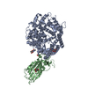
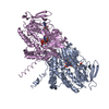
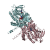
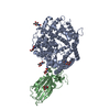
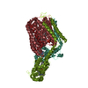
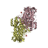
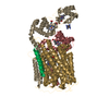
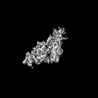
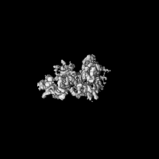
登録情報 データベース : EMDB / ID : EMD-13159タイトル Structure of the V. vulnificus ExoY-G-actin-profilin complex Main map, was used to build VvExoY and Actin 複合体 : Structure of the V. vulnificus ExoY-G-actin-profilin complexタンパク質・ペプチド : Maltose/maltodextrin-binding periplasmic protein,RTX-toxinタンパク質・ペプチド : Actin, cytoplasmic 1タンパク質・ペプチド : Profilin-1リガンド : CALCIUM IONリガンド : ADENOSINE-5'-TRIPHOSPHATE / / / 機能・相同性 分子機能 ドメイン・相同性 構成要素
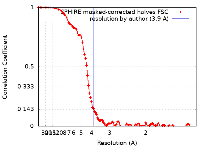
/ / / / / / / / / / / / / / / / / / / / / / / / / / / / / / / / / / / / / / / / / / / / / / / / / / / / / / / / / / / / / / / / / / / / / / / / / / / / / / / / / / / / / / / / / / / / / / / / / / / / / / / / / / / / / / / / / / / / / / / / / / / / / / / / / / / / / / / / / / / / / / / / / / / / / / / / / / / / 生物種 Vibrio vulnificus (バクテリア) / Homo sapiens (ヒト)手法 / / 解像度 : 3.9 Å Belyy A / Merino F 資金援助 Organization Grant number 国 Max Planck Society
ジャーナル : Nat Commun / 年 : 2021タイトル : Mechanism of actin-dependent activation of nucleotidyl cyclase toxins from bacterial human pathogens.著者 : Alexander Belyy / Felipe Merino / Undine Mechold / Stefan Raunser / 要旨 : Bacterial human pathogens secrete initially inactive nucleotidyl cyclases that become potent enzymes by binding to actin inside eukaryotic host cells. The underlying molecular mechanism of this ... Bacterial human pathogens secrete initially inactive nucleotidyl cyclases that become potent enzymes by binding to actin inside eukaryotic host cells. The underlying molecular mechanism of this activation is, however, unclear. Here, we report structures of ExoY from Pseudomonas aeruginosa and Vibrio vulnificus bound to their corresponding activators F-actin and profilin-G-actin. The structures reveal that in contrast to the apo-state, two flexible regions become ordered and interact strongly with actin. The specific stabilization of these regions results in an allosteric stabilization of the nucleotide binding pocket and thereby to an activation of the enzyme. Differences in the sequence and conformation of the actin-binding regions are responsible for the selective binding to either F- or G-actin. Other nucleotidyl cyclase toxins that bind to calmodulin rather than actin undergo a similar disordered-to-ordered transition during activation, suggesting that the allosteric activation-by-stabilization mechanism of ExoY is conserved in these enzymes, albeit the different activator. 履歴 登録 2021年7月1日 - ヘッダ(付随情報) 公開 2021年11月17日 - マップ公開 2021年11月17日 - 更新 2026年3月4日 - 現状 2026年3月4日 処理サイト : PDBe / 状態 : 公開
すべて表示 表示を減らす
 ムービー
ムービー コントローラー


 データを開く
 基本情報
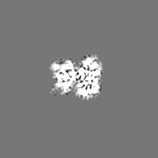
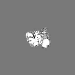
基本情報 マップデータ
 試料
試料 キーワード
キーワード 機能・相同性情報
 Vibrio vulnificus (バクテリア) /
  Homo sapiens (ヒト)
 データ登録者
 ドイツ, 1件
  引用
引用
 構造の表示
構造の表示
 ダウンロードとリンク
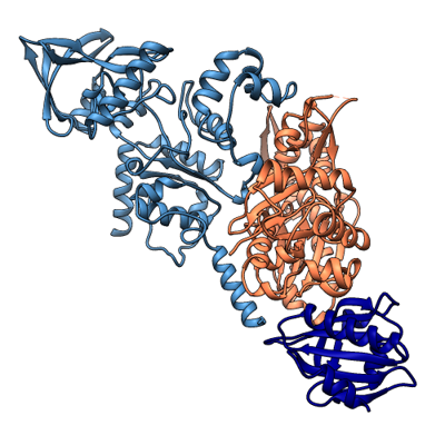 emd_13159.png
 http://ftp.pdbj.org/pub/emdb/structures/EMD-13159


 Z (Sec.)
 Y (Row.)
 X (Col.)


 試料の構成要素
 Trichoplusia ni (イラクサキンウワバ)
 解析
解析 電子顕微鏡法
 FIELD EMISSION GUN