 Movie
Movie Controller

[English] 日本語
 Yorodumi
- PDB-6se3: Crystal Structure of Ancestral Flavin-containing monooxygenase (F... -
+ Open data

- Basic information
| Entry | Database: PDB / ID: 6se3 | ||||||
|---|---|---|---|---|---|---|---|
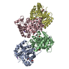
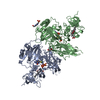
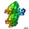
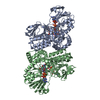
| Title | Crystal Structure of Ancestral Flavin-containing monooxygenase (FMO) 3-6 | ||||||
 Components Components | Ancestral Flavin-containing monooxygenase (FMO) 3-6 | ||||||
 Keywords Keywords | MEMBRANE PROTEIN / Flavin / enzyme / Ancestral Sequence Reconstruction | ||||||
| Function / homology | FLAVIN-ADENINE DINUCLEOTIDE / NADP NICOTINAMIDE-ADENINE-DINUCLEOTIDE PHOSPHATE / OXYGEN MOLECULE Function and homology information Function and homology information | ||||||
| Biological species | synthetic construct (others) | ||||||
| Method |  X-RAY DIFFRACTION / SYNCHROTRON / MOLECULAR REPLACEMENT / Resolution: 2.8 Å X-RAY DIFFRACTION / SYNCHROTRON / MOLECULAR REPLACEMENT / Resolution: 2.8 Å | ||||||
 Authors Authors | Nicoll, C. / Bailleul, G. / Fiorentini, F. / Mascotti, M.L. / Fraaije, M. / Mattevi, A. | ||||||
| Funding support | 1items
| ||||||
 Citation Citation | Journal: Nat.Struct.Mol.Biol. / Year: 2020 Title: Ancestral-sequence reconstruction unveils the structural basis of function in mammalian FMOs. Authors: Nicoll, C.R. / Bailleul, G. / Fiorentini, F. / Mascotti, M.L. / Fraaije, M.W. / Mattevi, A. | ||||||
| History |
|
- Structure visualization
| Structure viewer | Molecule: MolmilJmol/JSmol |
|---|
- Downloads & links
-Download
| PDBx/mmCIF format | 6se3.cif.gz | 643 KB | Display | PDBx/mmCIF format |
|---|---|---|---|---|
| PDB format | pdb6se3.ent.gz | 531.8 KB | Display | PDB format |
| PDBx/mmJSON format | 6se3.json.gz | Tree view | PDBx/mmJSON format | |
| Others |  Other downloads Other downloads |
-Validation report
| Arichive directory | https://data.pdbj.org/pub/pdb/validation_reports/se/6se3ftp://data.pdbj.org/pub/pdb/validation_reports/se/6se3 | HTTPS FTP |
|---|
-Related structure data
| Related structure data | 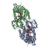 6sekC 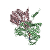 6semC 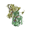 6sf0C 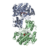 5nmwS S: Starting model for refinement C: citing same article ( |
|---|---|
| Similar structure data |


-Links
 PDBj
PDBj


- Assembly
| Deposited unit | 
| ||||||||
|---|---|---|---|---|---|---|---|---|---|
| 1 | 
| ||||||||
| 2 | 
| ||||||||
| 3 | 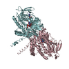
| ||||||||
| Unit cell |
|
-Components
| #1: Protein | Mass: 60127.992 Da / Num. of mol.: 6 Source method: isolated from a genetically manipulated source Details: Residues at the N and C termini were removed from the structural coordinates due to a lack of clear electron density Source: (gene. exp.) synthetic construct (others) / Production host:  #2: Chemical | ChemComp-FAD /   Mass: 785.550 Da / Num. of mol.: 6 / Source method: obtained synthetically / Formula: C27H33N9O15P2 / Comment: FAD*YM Mass: 785.550 Da / Num. of mol.: 6 / Source method: obtained synthetically / Formula: C27H33N9O15P2 / Comment: FAD*YM#3: Chemical | ChemComp-NAP /   Mass: 743.405 Da / Num. of mol.: 6 / Source method: obtained synthetically / Formula: C21H28N7O17P3 Mass: 743.405 Da / Num. of mol.: 6 / Source method: obtained synthetically / Formula: C21H28N7O17P3#4: Chemical | ChemComp-OXY /   Mass: 31.999 Da / Num. of mol.: 6 / Source method: obtained synthetically / Formula: O2 Mass: 31.999 Da / Num. of mol.: 6 / Source method: obtained synthetically / Formula: O2#5: Water | ChemComp-HOH / |  Mass: 18.015 Da / Num. of mol.: 357 / Source method: isolated from a natural source / Formula: H2O Mass: 18.015 Da / Num. of mol.: 357 / Source method: isolated from a natural source / Formula: H2OHas ligand of interest | N | |
|---|
-Experimental details
-Experiment
| Experiment | Method: X-RAY DIFFRACTION / Number of used crystals: 1 |
|---|
- Sample preparation
| Crystal | Density Matthews: 3.64 Å3/Da / Density % sol: 66.23 % |
|---|---|
| Crystal grow | Temperature: 293 K / Method: vapor diffusion, sitting drop / pH: 5.5 / Details: PEG 4000, Sodium Acetate |
-Data collection
| Diffraction | Mean temperature: 100 K / Serial crystal experiment: N |
|---|---|
| Diffraction source | Source: SYNCHROTRON / Site: SLS  / Beamline: X06DA / Wavelength: 0.98 Å / Beamline: X06DA / Wavelength: 0.98 Å |
| Detector | Type: DECTRIS PILATUS 2M-F / Detector: PIXEL / Date: Dec 15, 2018 |
| Radiation | Protocol: SINGLE WAVELENGTH / Monochromatic (M) / Laue (L): M / Scattering type: x-ray |
| Radiation wavelength | Wavelength: 0.98 Å / Relative weight: 1 |
| Reflection | Resolution: 2.8→49.3 Å / Num. obs: 129389 / % possible obs: 100 % / Redundancy: 20.3 % / CC1/2: 0.998 / Rmerge(I) obs: 0.235 / Net I/σ(I): 14.3 |
| Reflection shell | Resolution: 2.8→2.85 Å / Rmerge(I) obs: 2.876 / Mean I/σ(I) obs: 1.3 / Num. unique obs: 6372 / CC1/2: 0.409 |
- Processing
| Software |
| ||||||||||||||||||||
|---|---|---|---|---|---|---|---|---|---|---|---|---|---|---|---|---|---|---|---|---|---|
| Refinement | Method to determine structure: MOLECULAR REPLACEMENT Starting model: 5nmw Resolution: 2.8→49.3 Å / Cross valid method: THROUGHOUT
| ||||||||||||||||||||
| Displacement parameters | Biso mean: 72 Å2 | ||||||||||||||||||||
| Refinement step | Cycle: LAST / Resolution: 2.8→49.3 Å
|
