Movie
Movie Controller


+ Open data

- Basic information
| Entry | Database: EMDB / ID: EMD-13159 | |||||||||
|---|---|---|---|---|---|---|---|---|---|---|
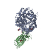
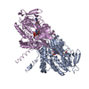
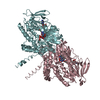
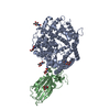
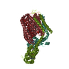
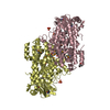
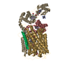
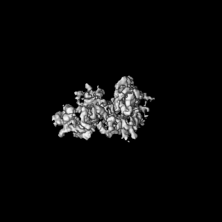
| Title | Structure of the V. vulnificus ExoY-G-actin-profilin complex | |||||||||
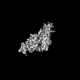
 Map data Map data | Main map, was used to build VvExoY and Actin | |||||||||
 Sample Sample |
| |||||||||
 Keywords Keywords | Bacterial toxin / G-actin / profilin / toxin | |||||||||
| Function / homology |  Function and homology information Function and homology informationcalcium- and calmodulin-responsive adenylate cyclase activity / synapse maturation / adenyl-nucleotide exchange factor activity / negative regulation of actin filament bundle assembly / modification of postsynaptic actin cytoskeleton / positive regulation of norepinephrine uptake / positive regulation of actin filament bundle assembly / regulation of actin filament polymerization / Signaling by ROBO receptors / cellular response to cytochalasin B ...calcium- and calmodulin-responsive adenylate cyclase activity / synapse maturation / adenyl-nucleotide exchange factor activity / negative regulation of actin filament bundle assembly / modification of postsynaptic actin cytoskeleton / positive regulation of norepinephrine uptake / positive regulation of actin filament bundle assembly / regulation of actin filament polymerization / Signaling by ROBO receptors / cellular response to cytochalasin B / Formation of the embryonic stem cell BAF (esBAF) complex / bBAF complex / npBAF complex / brahma complex / nBAF complex / Formation of the canonical BAF (cBAF) complex / regulation of transepithelial transport / Formation of neuronal progenitor and neuronal BAF (npBAF and nBAF) / morphogenesis of a polarized epithelium / Formation of the polybromo-BAF (pBAF) complex / structural constituent of postsynaptic actin cytoskeleton / Formation of annular gap junctions / Formation of the dystrophin-glycoprotein complex (DGC) / Gap junction degradation / Formation of the non-canonical BAF (ncBAF) complex / GBAF complex / protein localization to adherens junction / regulation of G0 to G1 transition / Cell-extracellular matrix interactions / negative regulation of stress fiber assembly / dense body / Folding of actin by CCT/TriC / Tat protein binding / proline-rich region binding / postsynaptic actin cytoskeleton / RSC-type complex / Regulation of CDH1 Function / PCP/CE pathway / regulation of double-strand break repair / Prefoldin mediated transfer of substrate to CCT/TriC / regulation of nucleotide-excision repair / Adherens junctions interactions / adherens junction assembly / RHOF GTPase cycle / apical protein localization / Sensory processing of sound by outer hair cells of the cochlea / SWI/SNF complex / regulation of mitotic metaphase/anaphase transition / tight junction / Sensory processing of sound by inner hair cells of the cochlea / Interaction between L1 and Ankyrins / positive regulation of T cell differentiation / host cell cytosol / positive regulation of ruffle assembly / apical junction complex / positive regulation of double-strand break repair / detection of maltose stimulus / maintenance of blood-brain barrier / regulation of norepinephrine uptake / transporter regulator activity / positive regulation of stem cell population maintenance / NuA4 histone acetyltransferase complex / maltose transport complex / positive regulation of actin filament polymerization / Recycling pathway of L1 / Regulation of MITF-M-dependent genes involved in pigmentation / cortical cytoskeleton / establishment or maintenance of cell polarity / carbohydrate transport / nitric-oxide synthase binding / positive regulation of epithelial cell migration / brush border / regulation of G1/S transition of mitotic cell cycle / EPH-ephrin mediated repulsion of cells / actin monomer binding / negative regulation of cell differentiation / carbohydrate transmembrane transporter activity / regulation of synaptic vesicle endocytosis / maltose binding / positive regulation of myoblast differentiation / RHO GTPases Activate WASPs and WAVEs / maltose transport / maltodextrin transmembrane transport / kinesin binding / regulation of protein localization to plasma membrane / RHO GTPases activate IQGAPs / positive regulation of double-strand break repair via homologous recombination / ATP-binding cassette (ABC) transporter complex, substrate-binding subunit-containing / phosphotyrosine residue binding / phosphatidylinositol-4,5-bisphosphate binding / EPHB-mediated forward signaling / cytoskeleton organization / axonogenesis / substantia nigra development / ATP-binding cassette (ABC) transporter complex / calyx of Held / nitric-oxide synthase regulator activity / FCGR3A-mediated phagocytosis / cell chemotaxis / Translocation of SLC2A4 (GLUT4) to the plasma membrane Similarity search - Function | |||||||||
| Biological species |  Vibrio vulnificus (bacteria) / Vibrio vulnificus (bacteria) /  Homo sapiens (human) Homo sapiens (human) | |||||||||
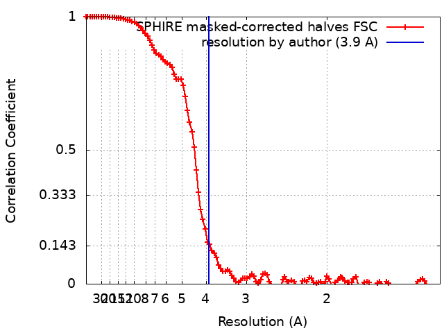
| Method | single particle reconstruction / cryo EM / Resolution: 3.9 Å | |||||||||
 Authors Authors | Belyy A / Merino F | |||||||||
| Funding support |  Germany, 1 items Germany, 1 items
| |||||||||
 Citation Citation | Journal: Nat Commun / Year: 2021 Title: Mechanism of actin-dependent activation of nucleotidyl cyclase toxins from bacterial human pathogens. Authors: Alexander Belyy / Felipe Merino / Undine Mechold / Stefan Raunser /  Abstract: Bacterial human pathogens secrete initially inactive nucleotidyl cyclases that become potent enzymes by binding to actin inside eukaryotic host cells. The underlying molecular mechanism of this ...Bacterial human pathogens secrete initially inactive nucleotidyl cyclases that become potent enzymes by binding to actin inside eukaryotic host cells. The underlying molecular mechanism of this activation is, however, unclear. Here, we report structures of ExoY from Pseudomonas aeruginosa and Vibrio vulnificus bound to their corresponding activators F-actin and profilin-G-actin. The structures reveal that in contrast to the apo-state, two flexible regions become ordered and interact strongly with actin. The specific stabilization of these regions results in an allosteric stabilization of the nucleotide binding pocket and thereby to an activation of the enzyme. Differences in the sequence and conformation of the actin-binding regions are responsible for the selective binding to either F- or G-actin. Other nucleotidyl cyclase toxins that bind to calmodulin rather than actin undergo a similar disordered-to-ordered transition during activation, suggesting that the allosteric activation-by-stabilization mechanism of ExoY is conserved in these enzymes, albeit the different activator. | |||||||||
| History |
|
- Structure visualization
| Movie |
Movie viewer |
|---|---|
| Structure viewer | EM map: SurfViewMolmilJmol/JSmol |
| Supplemental images |

- Downloads & links
-EMDB archive
| Map data | emd_13159.map.gz | 7.5 MB | EMDB map data format | |
|---|---|---|---|---|
| Header (meta data) | emd-13159-v30.xmlemd-13159.xml | 21.1 KB 21.1 KB | Display Display | EMDB header |
| FSC (resolution estimation) | emd_13159_fsc.xml | 12.4 KB | Display | FSC data file |
| Images | 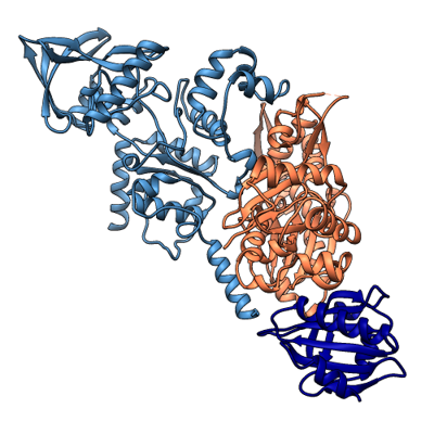 emd_13159.png emd_13159.png | 117.9 KB | ||
| Filedesc metadata | emd-13159.cif.gz | 7.2 KB | ||
| Others | emd_13159_additional_1.map.gz | 7.3 MB | ||
| Archive directory |  http://ftp.pdbj.org/pub/emdb/structures/EMD-13159ftp://ftp.pdbj.org/pub/emdb/structures/EMD-13159 http://ftp.pdbj.org/pub/emdb/structures/EMD-13159ftp://ftp.pdbj.org/pub/emdb/structures/EMD-13159 | HTTPS FTP |
-Related structure data
| Related structure data |  7p1hMC  7p1gC M: atomic model generated by this map C: citing same article ( |
|---|---|
| Similar structure data |


-Links
| EMDB pages | EMDB (EBI/PDBe) / EMDataResource |
|---|---|
| Related items in Molecule of the Month |


-Map
| File | Download / File: emd_13159.map.gz / Format: CCP4 / Size: 125 MB / Type: IMAGE STORED AS FLOATING POINT NUMBER (4 BYTES) | ||||||||||||||||||||||||||||||||||||||||||||||||||||||||||||
|---|---|---|---|---|---|---|---|---|---|---|---|---|---|---|---|---|---|---|---|---|---|---|---|---|---|---|---|---|---|---|---|---|---|---|---|---|---|---|---|---|---|---|---|---|---|---|---|---|---|---|---|---|---|---|---|---|---|---|---|---|---|
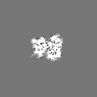
| Annotation | Main map, was used to build VvExoY and Actin | ||||||||||||||||||||||||||||||||||||||||||||||||||||||||||||
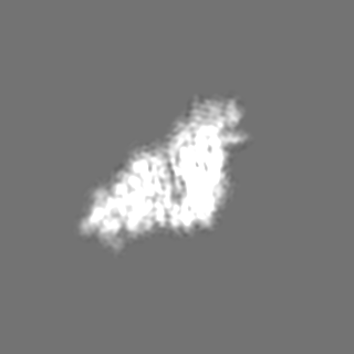
| Projections & slices | Image control
Images are generated by Spider. | ||||||||||||||||||||||||||||||||||||||||||||||||||||||||||||
| Voxel size | X=Y=Z: 0.68 Å | ||||||||||||||||||||||||||||||||||||||||||||||||||||||||||||
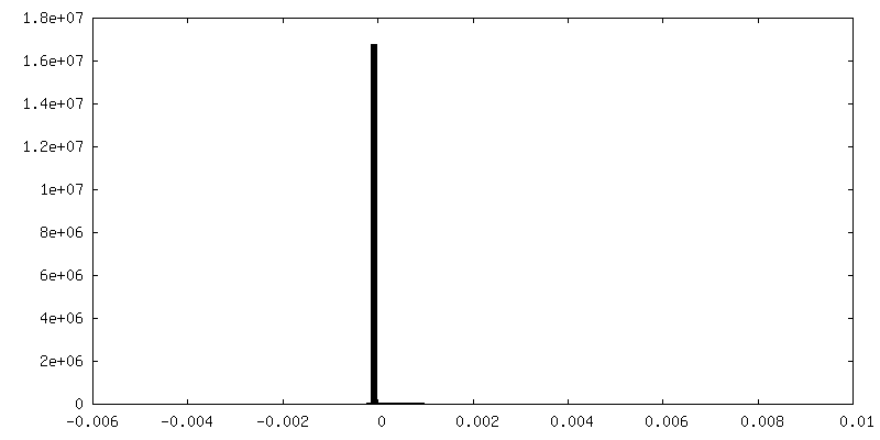
| Density |
| ||||||||||||||||||||||||||||||||||||||||||||||||||||||||||||
| Symmetry | Space group: 1 | ||||||||||||||||||||||||||||||||||||||||||||||||||||||||||||
| Details | EMDB XML:
CCP4 map header:
| ||||||||||||||||||||||||||||||||||||||||||||||||||||||||||||
 Z (Sec.)
 Y (Row.)
 X (Col.)


-Supplemental data
-Additional map: Secondary map, was used to build actin-profilin interface
| File | emd_13159_additional_1.map | ||||||||||||
|---|---|---|---|---|---|---|---|---|---|---|---|---|---|
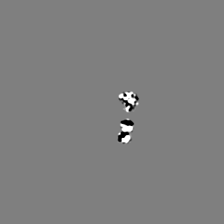
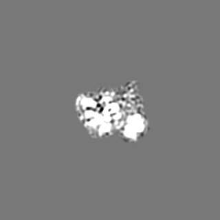
| Annotation | Secondary map, was used to build actin-profilin interface | ||||||||||||
| Projections & Slices |
| ||||||||||||
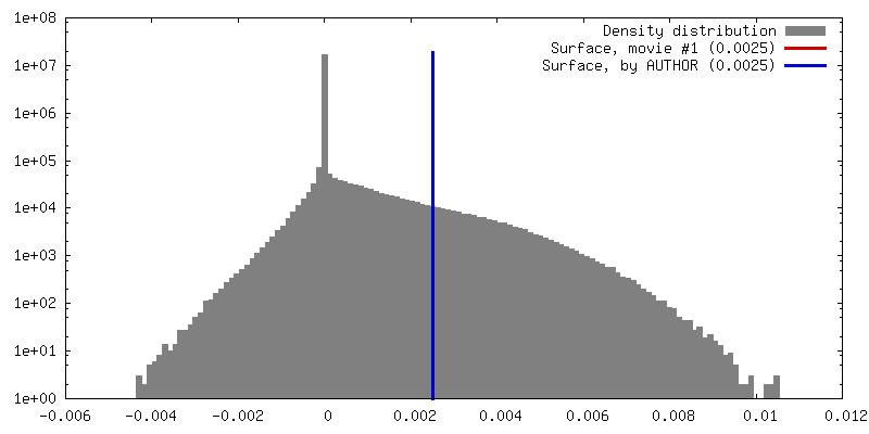
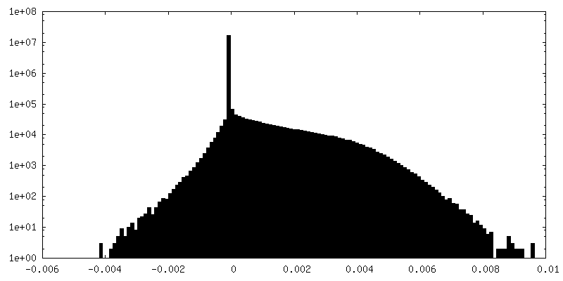
| Density Histograms |
- Sample components
-Entire : Structure of the V. vulnificus ExoY-G-actin-profilin complex
| Entire | Name: Structure of the V. vulnificus ExoY-G-actin-profilin complex |
|---|---|
| Components |
|
-Supramolecule #1: Structure of the V. vulnificus ExoY-G-actin-profilin complex
| Supramolecule | Name: Structure of the V. vulnificus ExoY-G-actin-profilin complex type: complex / ID: 1 / Parent: 0 / Macromolecule list: #1-#3 |
|---|
-Macromolecule #1: Maltose/maltodextrin-binding periplasmic protein,RTX-toxin
| Macromolecule | Name: Maltose/maltodextrin-binding periplasmic protein,RTX-toxin type: protein_or_peptide / ID: 1 / Number of copies: 1 / Enantiomer: LEVO |
|---|---|
| Source (natural) | Organism: Vibrio vulnificus (bacteria) |
| Molecular weight | Theoretical: 91.467539 KDa |
| Recombinant expression | Organism: |
| Sequence | String: MGSSHHHHHH SSGLVPRGSH MKIEEGKLVI WINGDKGYNG LAEVGKKFEK DTGIKVTVEH PDKLEEKFPQ VAATGDGPDI IFWAHDRFG GYAQSGLLAE ITPDKAFQDK LYPFTWDAVR YNGKLIAYPI AVEALSLIYN KDLLPNPPKT WEEIPALDKE L KAKGKSAL ...String: MGSSHHHHHH SSGLVPRGSH MKIEEGKLVI WINGDKGYNG LAEVGKKFEK DTGIKVTVEH PDKLEEKFPQ VAATGDGPDI IFWAHDRFG GYAQSGLLAE ITPDKAFQDK LYPFTWDAVR YNGKLIAYPI AVEALSLIYN KDLLPNPPKT WEEIPALDKE L KAKGKSAL MFNLQEPYFT WPLIAADGGY AFKYENGKYD IKDVGVDNAG AKAGLTFLVD LIKNKHMNAD TDYSIAEAAF NK GETAMTI NGPWAWSNID TSKVNYGVTV LPTFKGQPSK PFVGVLSAGI NAASPNKELA KEFLENYLLT DEGLEAVNKD KPL GAVALK SYEEELVKDP RIAATMENAQ KGEIMPNIPQ MSAFWYAVRT AVINAASGRQ TVDEALKDAQ TNSGSSGSSV EASD TELGT NTDAPHKNYQ SRDLVLEPIV QPETIELGMP DSDQKILAEV AERENVIIGV RPVDEKSKSL IDSKLYSSKG LFVKA KSSD WGPMSGFIPV DQAFAKASAR RDLDKFNGYA EQSIESGNAV SADLYLNQVR IDELVSKYQS LTALEFDAES GMYKTT ATN GDQTVTFFLN KVTVDSKDLW QVHYIKDGKL APFKVIGDPV SKQPMTADYD LLTVMYSYSD LGPQDKLKQP LTWEQWK ES VTYEELTPKY KELYNSEVLY NKKDGASLGV VSDRLKALKD VINTSLGRTD GLEMVHHGAD DANPYAVMAD NFPATFFV P KSFFMEDGLG EGKGSIQTYF NVNEQGAVVI RDPQEFSNFQ QVAINVSYRA SLNDKWNVGL DDPLFTPKSK LSHDFLNAK EEVIKKLSGE VETNVRTTQL LTDNEGL UniProtKB: Maltose/maltodextrin-binding periplasmic protein, RTX-toxin |
-Macromolecule #2: Actin, cytoplasmic 1
| Macromolecule | Name: Actin, cytoplasmic 1 / type: protein_or_peptide / ID: 2 / Number of copies: 1 / Enantiomer: LEVO |
|---|---|
| Source (natural) | Organism: Homo sapiens (human) |
| Molecular weight | Theoretical: 41.402242 KDa |
| Recombinant expression | Organism:  Trichoplusia ni (cabbage looper) Trichoplusia ni (cabbage looper) |
| Sequence | String: DIAALVVDNG SGMCKAGFAG DDAPRAVFPS IVGRPRHQGV MVGMGQKDSY VGDEAQSKRG ILTLKYPIE(HIC) GIVTNW DDM EKIWHHTFYN ELRVAPEEHP VLLTEAPLNP KANREKMTQI MFETFNTPAM YVAIQAVLSL YASGRTTGIV MDSGDGV TH TVPIYEGYAL ...String: DIAALVVDNG SGMCKAGFAG DDAPRAVFPS IVGRPRHQGV MVGMGQKDSY VGDEAQSKRG ILTLKYPIE(HIC) GIVTNW DDM EKIWHHTFYN ELRVAPEEHP VLLTEAPLNP KANREKMTQI MFETFNTPAM YVAIQAVLSL YASGRTTGIV MDSGDGV TH TVPIYEGYAL PHAILRLDLA GRDLTDYLMK ILTERGYSFT TTAEREIVRD IKEKLCYVAL DFEQEMATAA SSSSLEKS Y ELPDGQVITI GNERFRCPEA LFQPSFLGME SAGIHETTFN SIMKCDVDIR KDLYANTVLS GGTTMYPGIA DRMQKEITA LAPSTMKIKI IAPPERKYSV WIGGSILASL STFQQMWISK QEYDESGPSI VHRKCF UniProtKB: Actin, cytoplasmic 1 |
-Macromolecule #3: Profilin-1
| Macromolecule | Name: Profilin-1 / type: protein_or_peptide / ID: 3 / Number of copies: 1 / Enantiomer: LEVO |
|---|---|
| Source (natural) | Organism: Homo sapiens (human) |
| Molecular weight | Theoretical: 15.071222 KDa |
| Recombinant expression | Organism: |
| Sequence | String: MAGWNAYIDN LMADGTCQDA AIVGYKDSPS VWAAVPGKTF VNITPAEVGV LVGKDRSSFY VNGLTLGGQK CSVIRDSLLQ DGEFSMDLR TKSTGGAPTF NVTVTKTDKT LVLLMGKEGV HGGLINKKCY EMASHLRRSQ Y UniProtKB: Profilin-1 |
-Macromolecule #4: CALCIUM ION
| Macromolecule | Name: CALCIUM ION / type: ligand / ID: 4 / Number of copies: 1 / Formula: CA |
|---|---|
| Molecular weight | Theoretical: 40.078 Da |
-Macromolecule #5: ADENOSINE-5'-TRIPHOSPHATE
| Macromolecule | Name: ADENOSINE-5'-TRIPHOSPHATE / type: ligand / ID: 5 / Number of copies: 1 / Formula: ATP |
|---|---|
| Molecular weight | Theoretical: 507.181 Da |
| Chemical component information |  ChemComp-ATP: |
-Experimental details
-Structure determination
| Method | cryo EM |
|---|---|
 Processing Processing | single particle reconstruction |
| Aggregation state | particle |
-Sample preparation
| Buffer | pH: 8 |
|---|---|
| Grid | Model: Quantifoil R2/1 / Material: COPPER / Mesh: 300 |
| Vitrification | Cryogen name: ETHANE / Chamber humidity: 100 % / Instrument: FEI VITROBOT MARK III |
- Electron microscopy
| Microscope | FEI TITAN KRIOS |
|---|---|
| Image recording | Film or detector model: GATAN K3 (6k x 4k) / Detector mode: SUPER-RESOLUTION / Number grids imaged: 1 / Number real images: 6879 / Average exposure time: 2.0 sec. / Average electron dose: 60.0 e/Å2 |
| Electron beam | Acceleration voltage: 300 kV / Electron source:  FIELD EMISSION GUN FIELD EMISSION GUN |
| Electron optics | Illumination mode: SPOT SCAN / Imaging mode: BRIGHT FIELD / Nominal defocus max: 2.5 µm / Nominal defocus min: 1.2 µm |
| Experimental equipment |  Model: Titan Krios / Image courtesy: FEI Company |