 Movie
Movie Controller


+ Open data

- Basic information
| Entry | Database: EMDB / ID: EMD-10731 | |||||||||
|---|---|---|---|---|---|---|---|---|---|---|
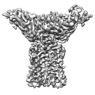
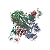
| Title | Structure of full-length CD20 in complex with Rituximab Fab | |||||||||
 Map data Map data | ||||||||||
 Sample Sample |
| |||||||||
 Keywords Keywords | cancer immunotherapy / therapeutic antibody / MEMBRANE PROTEIN | |||||||||
| Function / homology |  Function and homology information Function and homology informationstore-operated calcium entry / calcium ion import into cytosol / positive regulation of calcium ion import across plasma membrane / epidermal growth factor receptor binding / B cell proliferation / humoral immune response / B cell activation / immunoglobulin binding / plasma membrane raft / B cell differentiation ...store-operated calcium entry / calcium ion import into cytosol / positive regulation of calcium ion import across plasma membrane / epidermal growth factor receptor binding / B cell proliferation / humoral immune response / B cell activation / immunoglobulin binding / plasma membrane raft / B cell differentiation / B cell receptor signaling pathway / response to bacterium / protein tetramerization / MHC class II protein complex binding / cell surface receptor signaling pathway / external side of plasma membrane / cell surface / : / extracellular exosome / nucleoplasm / identical protein binding / plasma membrane Similarity search - Function | |||||||||
| Biological species |  Homo sapiens (human) / Homo sapiens (human) /  | |||||||||
| Method | single particle reconstruction / cryo EM / Resolution: 3.69 Å | |||||||||
 Authors Authors | Kumar A / Fronzes R / Reyes N | |||||||||
| Funding support |  France, 1 items France, 1 items
| |||||||||
 Citation Citation | Journal: Science / Year: 2020 Title: Binding mechanisms of therapeutic antibodies to human CD20. Authors: Anand Kumar / Cyril Planchais / Rémi Fronzes / Hugo Mouquet / Nicolas Reyes / Abstract: Monoclonal antibodies (mAbs) targeting human antigen CD20 (cluster of differentiation 20) constitute important immunotherapies for the treatment of B cell malignancies and autoimmune diseases. Type I ...Monoclonal antibodies (mAbs) targeting human antigen CD20 (cluster of differentiation 20) constitute important immunotherapies for the treatment of B cell malignancies and autoimmune diseases. Type I and II therapeutic mAbs differ in B cell binding properties and cytotoxic effects, reflecting differential interaction mechanisms with CD20. Here we present 3.7- to 4.7-angstrom cryo-electron microscopy structures of full-length CD20 in complexes with prototypical type I rituximab and ofatumumab and type II obinutuzumab. The structures and binding thermodynamics demonstrate that upon binding to CD20, type II mAbs form terminal complexes that preclude recruitment of additional mAbs and complement components, whereas type I complexes act as molecular seeds to increase mAb local concentration for efficient complement activation. Among type I mAbs, ofatumumab complexes display optimal geometry for complement recruitment. The uncovered mechanisms should aid rational design of next-generation immunotherapies targeting CD20. | |||||||||
| History |
|
- Structure visualization
| Movie |
Movie viewer |
|---|---|
| Structure viewer | EM map: SurfViewMolmilJmol/JSmol |
| Supplemental images |
- Downloads & links
-EMDB archive
| Map data | emd_10731.map.gz | 52.7 MB | EMDB map data format | |
|---|---|---|---|---|
| Header (meta data) | emd-10731-v30.xmlemd-10731.xml | 25.7 KB 25.7 KB | Display Display | EMDB header |
| Images |  emd_10731.png emd_10731.png | 68.4 KB | ||
| Filedesc metadata | emd-10731.cif.gz | 7.4 KB | ||
| Archive directory |  http://ftp.pdbj.org/pub/emdb/structures/EMD-10731ftp://ftp.pdbj.org/pub/emdb/structures/EMD-10731 http://ftp.pdbj.org/pub/emdb/structures/EMD-10731ftp://ftp.pdbj.org/pub/emdb/structures/EMD-10731 | HTTPS FTP |
-Related structure data
| Related structure data |  6y90MC  6y92C  6y97C  6y9aC C: citing same article ( M: atomic model generated by this map |
|---|---|
| Similar structure data |


-Links
| EMDB pages | EMDB (EBI/PDBe) / EMDataResource |
|---|
-Map
| File | Download / File: emd_10731.map.gz / Format: CCP4 / Size: 103 MB / Type: IMAGE STORED AS FLOATING POINT NUMBER (4 BYTES) | ||||||||||||||||||||||||||||||||||||||||||||||||||||||||||||
|---|---|---|---|---|---|---|---|---|---|---|---|---|---|---|---|---|---|---|---|---|---|---|---|---|---|---|---|---|---|---|---|---|---|---|---|---|---|---|---|---|---|---|---|---|---|---|---|---|---|---|---|---|---|---|---|---|---|---|---|---|---|
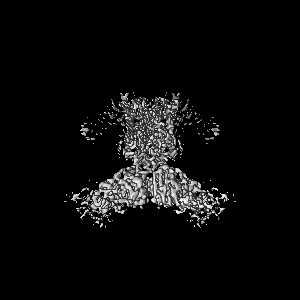
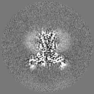
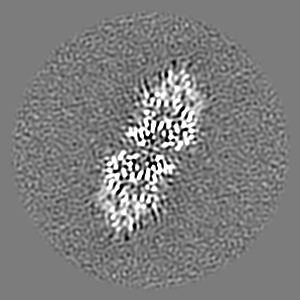
| Projections & slices | Image control
Images are generated by Spider. | ||||||||||||||||||||||||||||||||||||||||||||||||||||||||||||
| Voxel size | X=Y=Z: 0.814 Å | ||||||||||||||||||||||||||||||||||||||||||||||||||||||||||||
| Density |
| ||||||||||||||||||||||||||||||||||||||||||||||||||||||||||||
| Symmetry | Space group: 1 | ||||||||||||||||||||||||||||||||||||||||||||||||||||||||||||
| Details | EMDB XML:
CCP4 map header:
| ||||||||||||||||||||||||||||||||||||||||||||||||||||||||||||
 Z (Sec.)
 Y (Row.)
 X (Col.)


-Supplemental data
- Sample components
-Entire : Full-length human antigen CD20 in complex with Rituximab Fab
| Entire | Name: Full-length human antigen CD20 in complex with Rituximab Fab |
|---|---|
| Components |
|
-Supramolecule #1: Full-length human antigen CD20 in complex with Rituximab Fab
| Supramolecule | Name: Full-length human antigen CD20 in complex with Rituximab Fab type: complex / ID: 1 / Parent: 0 / Macromolecule list: #1-#3 |
|---|
-Supramolecule #2: Full-length human CD20
| Supramolecule | Name: Full-length human CD20 / type: complex / ID: 2 / Parent: 1 / Macromolecule list: #1 |
|---|---|
| Source (natural) | Organism: Homo sapiens (human) |
-Supramolecule #3: Rituximab Fab
| Supramolecule | Name: Rituximab Fab / type: complex / ID: 3 / Parent: 1 / Macromolecule list: #2-#3 |
|---|---|
| Source (natural) | Organism: |
-Macromolecule #1: B-lymphocyte antigen CD20
| Macromolecule | Name: B-lymphocyte antigen CD20 / type: protein_or_peptide / ID: 1 / Details: Full-length wild type Human CD20 / Number of copies: 2 / Enantiomer: LEVO |
|---|---|
| Source (natural) | Organism: Homo sapiens (human) / Cell: B-Lymphocyte |
| Molecular weight | Theoretical: 19.164797 KDa |
| Recombinant expression | Organism: Homo sapiens (human) |
| Sequence | String: FMRESKTLGA VQIMNGLFHI ALGGLLMIPA GIYAPICVTV WYPLWGGIMY IISGSLLAAT EKNSRKCLVK GKMIMNSLSL FAAISGMIL SIMDILNIKI SHFLKMESLN FIRAHTPYIN IYNCEPANPS EKNSPSTQYC YSIQSLFLGI LSVMLIFAFF Q ELVIAGIV ENEW UniProtKB: B-lymphocyte antigen CD20 |
-Macromolecule #2: Rituximab Fab Heavy Chain
| Macromolecule | Name: Rituximab Fab Heavy Chain / type: protein_or_peptide / ID: 2 / Number of copies: 2 / Enantiomer: LEVO |
|---|---|
| Source (natural) | Organism: |
| Molecular weight | Theoretical: 23.733541 KDa |
| Recombinant expression | Organism: Homo sapiens (human) |
| Sequence | String: QVQLQQPGAE LVKPGASVKM SCKASGYTFT SYNMHWVKQT PGRGLEWIGA IYPGNGDTSY NQKFKGKATL TADKSSSTAY MQLSSLTSE DSAVYYCARS TYYGGDWYFN VWGAGTTVTV SAASTKGPSV FPLAPSSKST SGGTAALGCL VKDYFPEPVT V SWNSGALT ...String: QVQLQQPGAE LVKPGASVKM SCKASGYTFT SYNMHWVKQT PGRGLEWIGA IYPGNGDTSY NQKFKGKATL TADKSSSTAY MQLSSLTSE DSAVYYCARS TYYGGDWYFN VWGAGTTVTV SAASTKGPSV FPLAPSSKST SGGTAALGCL VKDYFPEPVT V SWNSGALT SGVHTFPAVL QSSGLYSLSS VVTVPSSSLG TQTYICNVNH KPSNTKVDKK VEPKSC |
-Macromolecule #3: Rituximab Fab Light Chain
| Macromolecule | Name: Rituximab Fab Light Chain / type: protein_or_peptide / ID: 3 / Details: Rituximab Light Chain / Number of copies: 2 / Enantiomer: LEVO |
|---|---|
| Source (natural) | Organism: |
| Molecular weight | Theoretical: 23.078623 KDa |
| Recombinant expression | Organism: Homo sapiens (human) |
| Sequence | String: QIVLSQSPAI LSASPGEKVT MTCRASSSVS YIHWFQQKPG SSPKPWIYAT SNLASGVPVR FSGSGSGTSY SLTISRVEAE DAATYYCQQ WTSNPPTFGG GTKLEIKRTV AAPSVFIFPP SDEQLKSGTA SVVCLLNNFY PREAKVQWKV DNALQSGNSQ E SVTEQDSK ...String: QIVLSQSPAI LSASPGEKVT MTCRASSSVS YIHWFQQKPG SSPKPWIYAT SNLASGVPVR FSGSGSGTSY SLTISRVEAE DAATYYCQQ WTSNPPTFGG GTKLEIKRTV AAPSVFIFPP SDEQLKSGTA SVVCLLNNFY PREAKVQWKV DNALQSGNSQ E SVTEQDSK DSTYSLSSTL TLSKADYEKH KVYACEVTHQ GLSSPVTKSF NRGEC |
-Macromolecule #4: CHOLESTEROL HEMISUCCINATE
| Macromolecule | Name: CHOLESTEROL HEMISUCCINATE / type: ligand / ID: 4 / Number of copies: 8 / Formula: Y01 |
|---|---|
| Molecular weight | Theoretical: 486.726 Da |
| Chemical component information |  ChemComp-Y01: |
-Macromolecule #5: 1,2-DIACYL-SN-GLYCERO-3-PHOSPHOCHOLINE
| Macromolecule | Name: 1,2-DIACYL-SN-GLYCERO-3-PHOSPHOCHOLINE / type: ligand / ID: 5 / Number of copies: 2 / Formula: PC1 |
|---|---|
| Molecular weight | Theoretical: 790.145 Da |
| Chemical component information |  ChemComp-PC1: |
-Macromolecule #6: PENTADECANE
| Macromolecule | Name: PENTADECANE / type: ligand / ID: 6 / Number of copies: 10 / Formula: MYS |
|---|---|
| Molecular weight | Theoretical: 212.415 Da |
| Chemical component information |  ChemComp-MYS: |
-Experimental details
-Structure determination
| Method | cryo EM |
|---|---|
 Processing Processing | single particle reconstruction |
| Aggregation state | particle |
-Sample preparation
| Concentration | 1.5 mg/mL | |||||||||||||||
|---|---|---|---|---|---|---|---|---|---|---|---|---|---|---|---|---|
| Buffer | pH: 7.4 Component:
| |||||||||||||||
| Grid | Model: Quantifoil R1.2/1.3 / Material: GOLD / Mesh: 300 / Pretreatment - Type: GLOW DISCHARGE / Pretreatment - Time: 20 sec. / Pretreatment - Atmosphere: OTHER | |||||||||||||||
| Vitrification | Cryogen name: ETHANE / Chamber humidity: 100 % / Chamber temperature: 277.15 K / Instrument: FEI VITROBOT MARK IV |
- Electron microscopy
| Microscope | FEI TITAN KRIOS |
|---|---|
| Image recording | Film or detector model: GATAN K2 SUMMIT (4k x 4k) / Detector mode: COUNTING / Digitization - Frames/image: 1-40 / Number grids imaged: 1 / Number real images: 9263 / Average exposure time: 8.0 sec. / Average electron dose: 41.3 e/Å2 |
| Electron beam | Acceleration voltage: 300 kV / Electron source:  FIELD EMISSION GUN FIELD EMISSION GUN |
| Electron optics | C2 aperture diameter: 50.0 µm / Calibrated defocus max: 2.0 µm / Calibrated defocus min: 0.8 µm / Illumination mode: FLOOD BEAM / Imaging mode: BRIGHT FIELD / Cs: 2.7 mm |
| Sample stage | Cooling holder cryogen: NITROGEN |
| Experimental equipment |  Model: Titan Krios / Image courtesy: FEI Company |
+Image processing

-Atomic model buiding 1
| Initial model |
| ||||||
|---|---|---|---|---|---|---|---|
| Details | The CD20-Rituximab fab model was built by placing CD20 peptide (residues 167-186) and Rituximab fab from PDB 2OSL into the EM map. The initial model was fitted manually and extended to a full CD20 model encompassing residues 45-216. | ||||||
| Refinement | Space: REAL / Protocol: AB INITIO MODEL | ||||||
| Output model | PDB-6y90: |
