stringent response / translational termination / transcriptional attenuation / endoribonuclease inhibitor activity / positive regulation of ribosome biogenesis / RNA-binding transcription regulator activity / negative regulation of cytoplasmic translation / DnaA-L2 complex / translation repressor activity / negative regulation of DNA-templated DNA replication initiation ...stringent response / translational termination / transcriptional attenuation / endoribonuclease inhibitor activity / positive regulation of ribosome biogenesis / RNA-binding transcription regulator activity / negative regulation of cytoplasmic translation / DnaA-L2 complex / translation repressor activity / negative regulation of DNA-templated DNA replication initiation / mRNA regulatory element binding translation repressor activity / response to reactive oxygen species / cytosolic ribosome assembly / ribosome assembly / assembly of large subunit precursor of preribosome / regulation of cell growth / DNA-templated transcription termination / response to radiation / mRNA 5'-UTR binding / large ribosomal subunit / transferase activity / ribosome binding / 5S rRNA binding / ribosomal large subunit assembly / large ribosomal subunit rRNA binding / cytosolic large ribosomal subunit / cytoplasmic translation / tRNA binding / negative regulation of translation / rRNA binding / structural constituent of ribosome / ribosome / translation / response to antibiotic / negative regulation of DNA-templated transcription / mRNA binding / DNA binding / RNA binding / zinc ion binding / cytoplasm / cytosol Similarity search - Function
Ribosomal protein L10, eubacterial, conserved site / Ribosomal protein L10 signature. / Ribosomal protein L10 / : / Ribosomal protein L11, bacterial-type / Ribosomal protein L25, short-form / Ribosomal protein L11, conserved site / Ribosomal protein L11 signature. / Ribosomal protein L10-like domain superfamily / Ribosomal protein L10P ...Ribosomal protein L10, eubacterial, conserved site / Ribosomal protein L10 signature. / Ribosomal protein L10 / : / Ribosomal protein L11, bacterial-type / Ribosomal protein L25, short-form / Ribosomal protein L11, conserved site / Ribosomal protein L11 signature. / Ribosomal protein L10-like domain superfamily / Ribosomal protein L10P / Ribosomal protein L10 / Ribosomal protein L16 signature 1. / Ribosomal protein L6, conserved site / Ribosomal protein L6 signature 1. / Ribosomal protein L11, N-terminal / Ribosomal protein L11, N-terminal domain / Ribosomal protein L21, conserved site / : / Ribosomal protein L21 signature. / Ribosomal protein L11/L12 / Ribosomal protein L11, C-terminal / Ribosomal protein L11, C-terminal domain superfamily / Ribosomal protein L11/L12, N-terminal domain superfamily / Ribosomal protein L11/L12 / Ribosomal protein L11, RNA binding domain / Ribosomal protein L16 signature 2. / Ribosomal protein L16, conserved site / Ribosomal protein L17 signature. / Ribosomal L25p family / Ribosomal protein L25 / Ribosomal protein L36 signature. / Ribosomal protein L25/Gln-tRNA synthetase, N-terminal / Ribosomal protein L25/Gln-tRNA synthetase, anti-codon-binding domain superfamily / : / Ribosomal protein L28/L24 superfamily / Ribosomal protein L33, conserved site / Ribosomal protein L33 signature. / Ribosomal protein L32p, bacterial type / Ribosomal protein L35, conserved site / Ribosomal protein L35 signature. / Ribosomal protein L28 / Ribosomal protein L35, non-mitochondrial / Ribosomal protein L18, bacterial-type / : / Ribosomal protein L6, bacterial-type / Ribosomal protein L5, bacterial-type / Ribosomal protein L19, conserved site / Ribosomal protein L19 signature. / : / Ribosomal protein L36 / Ribosomal protein L36 superfamily / Ribosomal protein L36 / Ribosomal protein L20 signature. / Ribosomal protein L34, conserved site / Ribosomal protein L34 signature. / Ribosomal protein L14P, bacterial-type / Ribosomal protein L27, conserved site / Ribosomal protein L27 signature. / Ribosomal protein L35 / Ribosomal protein L35 superfamily / Ribosomal protein L22, bacterial/chloroplast-type / Ribosomal protein L35 / Ribosomal protein L2, bacterial/organellar-type / Ribosomal protein L33 / Ribosomal protein L18 / Ribosomal L18 of archaea, bacteria, mitoch. and chloroplast / Ribosomal protein L33 / Ribosomal L28 family / Ribosomal protein L33 superfamily / Ribosomal protein L28/L24 / Ribosomal protein L30, bacterial-type / L28p-like / Ribosomal protein L16 / Ribosomal protein L20 / Ribosomal protein L20 / Ribosomal protein L20, C-terminal / Ribosomal protein L19 / Ribosomal protein L19 / Ribosomal protein L19 superfamily / : / Large ribosomal subunit protein uL24, C-terminal domain / Ribosomal protein L17 / Ribosomal protein L17 superfamily / Ribosomal protein L17 / Ribosomal protein L27 / Ribosomal L27 protein / Ribosomal protein L34 / Ribosomal protein L34 / Ribosomal protein L24 / Ribosomal L32p protein family / Ribosomal protein L21 / Ribosomal protein L32p / Ribosomal protein L21-like / L21-like superfamily / Ribosomal prokaryotic L21 protein / Ribosomal protein L3, bacterial/organelle-type / Ribosomal protein L15, bacterial-type / 50S ribosomal protein uL4 / Ribosomal protein L13, bacterial-type / Ribosomal protein L23/L25, conserved site Similarity search - Domain/homology
Large ribosomal subunit protein uL15 / Large ribosomal subunit protein uL10 / Large ribosomal subunit protein uL11 / Large ribosomal subunit protein bL19 / Large ribosomal subunit protein bL20 / Large ribosomal subunit protein bL27 / Large ribosomal subunit protein bL28 / Large ribosomal subunit protein uL29 / Large ribosomal subunit protein bL32 / Large ribosomal subunit protein bL33 ...Large ribosomal subunit protein uL15 / Large ribosomal subunit protein uL10 / Large ribosomal subunit protein uL11 / Large ribosomal subunit protein bL19 / Large ribosomal subunit protein bL20 / Large ribosomal subunit protein bL27 / Large ribosomal subunit protein bL28 / Large ribosomal subunit protein uL29 / Large ribosomal subunit protein bL32 / Large ribosomal subunit protein bL33 / Large ribosomal subunit protein bL34 / Large ribosomal subunit protein bL35 / Large ribosomal subunit protein bL36A / Large ribosomal subunit protein uL13 / Large ribosomal subunit protein uL14 / Large ribosomal subunit protein uL16 / Large ribosomal subunit protein uL23 / Large ribosomal subunit protein bL17 / Large ribosomal subunit protein bL21 / Large ribosomal subunit protein uL30 / Large ribosomal subunit protein uL6 / Large ribosomal subunit protein uL18 / Large ribosomal subunit protein uL2 / Large ribosomal subunit protein uL3 / Large ribosomal subunit protein uL24 / Large ribosomal subunit protein uL4 / Large ribosomal subunit protein uL22 / Large ribosomal subunit protein uL5 / Large ribosomal subunit protein bL25 Similarity search - Component
National Institutes of Health/National Institute of General Medical Sciences (NIH/NIGMS)
R01-GM132302
United States
National Institutes of Health/National Institute Of Allergy and Infectious Diseases (NIH/NIAID)
R21-AI137584
United States
Russian Foundation for Basic Research
17-00-00366
Russian Federation
Russian Science Foundation
18-44-04005
Russian Federation
Citation
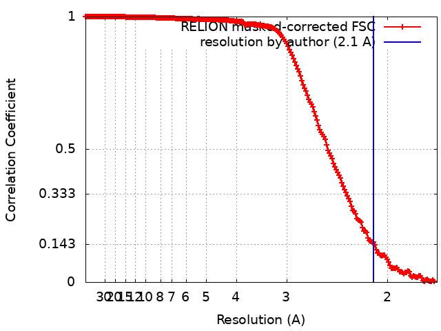
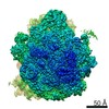
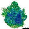
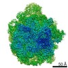
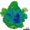
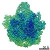
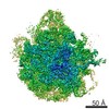
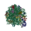
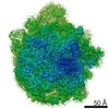
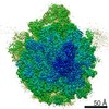
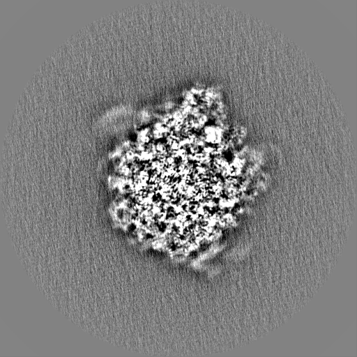
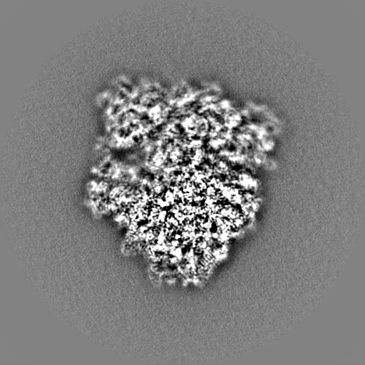
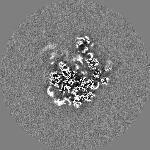
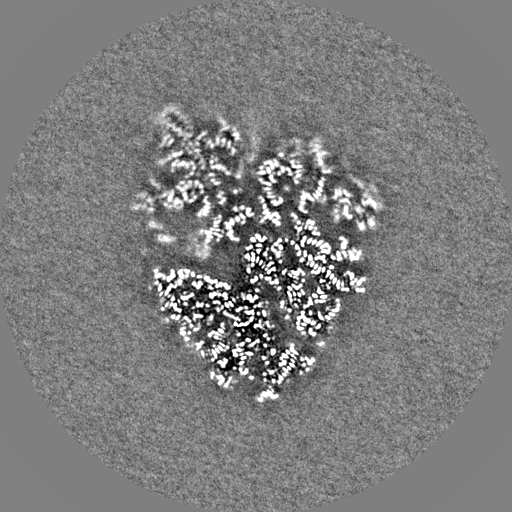
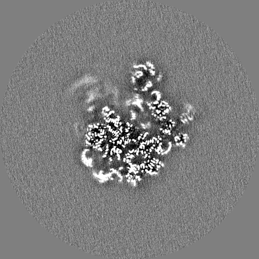
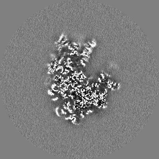
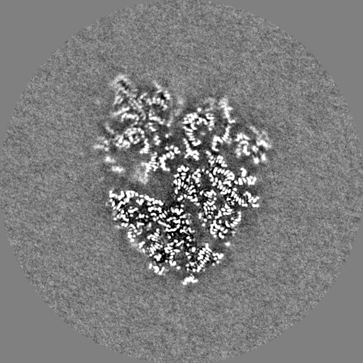
Journal: RNA / Year: 2020 Title: Insights into the improved macrolide inhibitory activity from the high-resolution cryo-EM structure of dirithromycin bound to the 70S ribosome. Authors: Evgeny B Pichkur / Alena Paleskava / Andrey G Tereshchenkov / Pavel Kasatsky / Ekaterina S Komarova / Dmitrii I Shiriaev / Alexey A Bogdanov / Olga A Dontsova / Ilya A Osterman / Petr V ...Authors: Evgeny B Pichkur / Alena Paleskava / Andrey G Tereshchenkov / Pavel Kasatsky / Ekaterina S Komarova / Dmitrii I Shiriaev / Alexey A Bogdanov / Olga A Dontsova / Ilya A Osterman / Petr V Sergiev / Yury S Polikanov / Alexander G Myasnikov / Andrey L Konevega / Abstract: Macrolides are one of the most successful and widely used classes of antibacterials, which kill or stop the growth of pathogenic bacteria by binding near the active site of the ribosome and ...Macrolides are one of the most successful and widely used classes of antibacterials, which kill or stop the growth of pathogenic bacteria by binding near the active site of the ribosome and interfering with protein synthesis. Dirithromycin is a derivative of the prototype macrolide erythromycin with additional hydrophobic side chain. In our recent study, we have discovered that the side chain of dirithromycin forms lone pair-π stacking interaction with the aromatic imidazole ring of the His69 residue in ribosomal protein uL4 of the 70S ribosome. In the current work, we found that neither the presence of the side chain, nor the additional contact with the ribosome, improve the binding affinity of dirithromycin to the ribosome. Nevertheless, we found that dirithromycin is a more potent inhibitor of in vitro protein synthesis in comparison with its parent compound, erythromycin. Using high-resolution cryo-electron microscopy, we determined the structure of the dirithromycin bound to the translating 70S ribosome, which suggests that the better inhibitory properties of the drug could be rationalized by the side chain of dirithromycin pointing into the lumen of the nascent peptide exit tunnel, where it can interfere with the normal passage of the growing polypeptide chain.
History
Deposition
Feb 3, 2020
-
Header (metadata) release
Jul 22, 2020
-
Map release
Jul 22, 2020
-
Update
Mar 12, 2025
-
Current status
Mar 12, 2025
Processing site: PDBe / Status: Released
-
Structure visualization
Movie
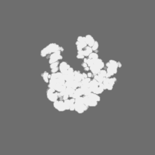
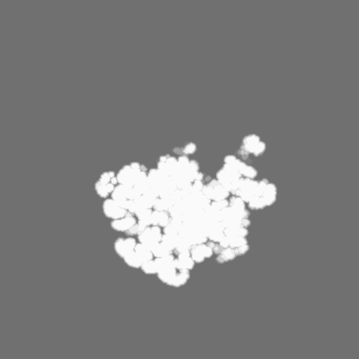
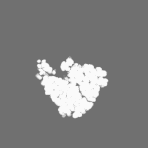
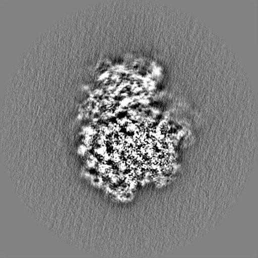
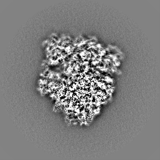
Surface view with section colored by density value
Model: Quantifoil R2/2 / Material: COPPER/RHODIUM / Mesh: 300 / Support film - Material: CARBON / Support film - topology: CONTINUOUS / Support film - Film thickness: 10 / Pretreatment - Type: GLOW DISCHARGE / Pretreatment - Time: 45 sec. / Pretreatment - Atmosphere: AIR
Vitrification
Cryogen name: ETHANE
-
Electron microscopy
Microscope
FEI TITAN KRIOS
Image recording
Film or detector model: FEI FALCON II (4k x 4k) / Detector mode: INTEGRATING / Digitization - Frames/image: 2-27 / Average exposure time: 1.4 sec. / Average electron dose: 80.0 e/Å2
Electron beam
Acceleration voltage: 300 kV / Electron source: FIELD EMISSION GUN
In the structure databanks used in Yorodumi, some data are registered as the other names, "COVID-19 virus" and "2019-nCoV". Here are the details of the virus and the list of structure data.
Jan 31, 2019. EMDB accession codes are about to change! (news from PDBe EMDB page)
EMDB accession codes are about to change! (news from PDBe EMDB page)
The allocation of 4 digits for EMDB accession codes will soon come to an end. Whilst these codes will remain in use, new EMDB accession codes will include an additional digit and will expand incrementally as the available range of codes is exhausted. The current 4-digit format prefixed with “EMD-” (i.e. EMD-XXXX) will advance to a 5-digit format (i.e. EMD-XXXXX), and so on. It is currently estimated that the 4-digit codes will be depleted around Spring 2019, at which point the 5-digit format will come into force.
The EM Navigator/Yorodumi systems omit the EMD- prefix.
Related info.:Q: What is EMD? / ID/Accession-code notation in Yorodumi/EM Navigator
Yorodumi is a browser for structure data from EMDB, PDB, SASBDB, etc.
This page is also the successor to EM Navigator detail page, and also detail information page/front-end page for Omokage search.
The word "yorodu" (or yorozu) is an old Japanese word meaning "ten thousand". "mi" (miru) is to see.
Related info.:EMDB / PDB / SASBDB / Comparison of 3 databanks / Yorodumi Search / Aug 31, 2016. New EM Navigator & Yorodumi / Yorodumi Papers / Jmol/JSmol / Function and homology information / Changes in new EM Navigator and Yorodumi
 Movie
Movie Controller

 Yorodumi
 Open data
 Basic information
 Map data
 Sample
 Keywords
 Function and homology information

 Authors
 Russian Federation,
  United States, 5 items
  Citation
 Structure visualization
 Downloads & links
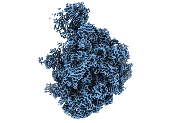 emd_10655.png
 http://ftp.pdbj.org/pub/emdb/structures/EMD-10655


 Z (Sec.)
 Y (Row.)
 X (Col.)


 Sample components

 Processing
 Electron microscopy
 FIELD EMISSION GUN