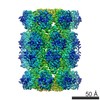
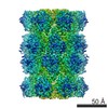
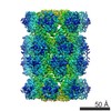
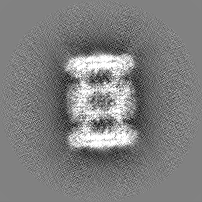
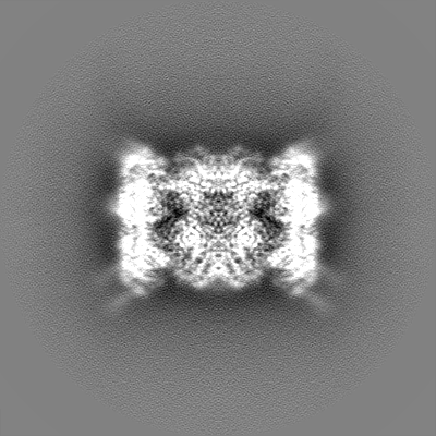
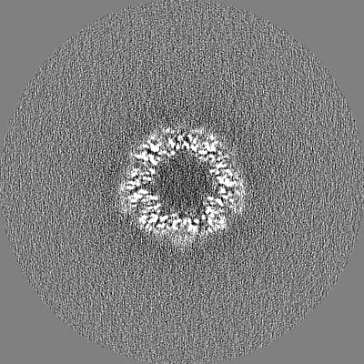
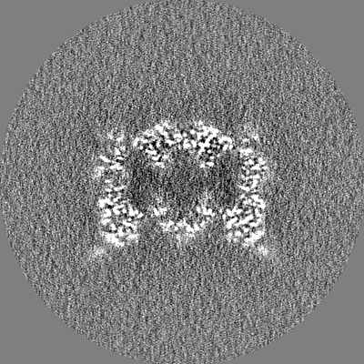
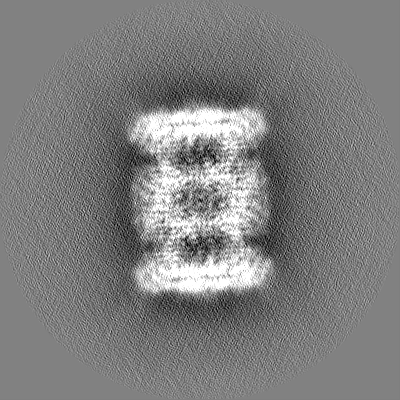
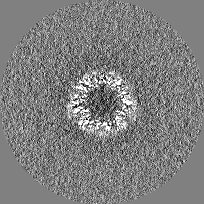
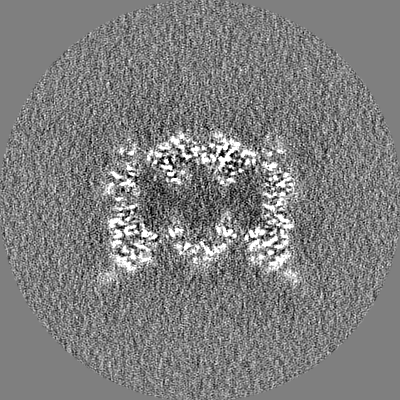
endogenous rat liver 20S proteasome, local resolution filtered
試料
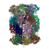
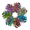
複合体: Rat liver 20S proteasome
タンパク質・ペプチド: x 14種
キーワード
PROTEASOME / HYDROLASE
機能・相同性
機能・相同性情報
Cross-presentation of soluble exogenous antigens (endosomes) / SCF-beta-TrCP mediated degradation of Emi1 / Regulation of ornithine decarboxylase (ODC) / Assembly of the pre-replicative complex / GSK3B and BTRC:CUL1-mediated-degradation of NFE2L2 / Oxygen-dependent proline hydroxylation of Hypoxia-inducible Factor Alpha / Autodegradation of Cdh1 by Cdh1:APC/C / APC/C:Cdc20 mediated degradation of Securin / APC/C:Cdh1 mediated degradation of Cdc20 and other APC/C:Cdh1 targeted proteins in late mitosis/early G1 / Cdc20:Phospho-APC/C mediated degradation of Cyclin A ...Cross-presentation of soluble exogenous antigens (endosomes) / SCF-beta-TrCP mediated degradation of Emi1 / Regulation of ornithine decarboxylase (ODC) / Assembly of the pre-replicative complex / GSK3B and BTRC:CUL1-mediated-degradation of NFE2L2 / Oxygen-dependent proline hydroxylation of Hypoxia-inducible Factor Alpha / Autodegradation of Cdh1 by Cdh1:APC/C / APC/C:Cdc20 mediated degradation of Securin / APC/C:Cdh1 mediated degradation of Cdc20 and other APC/C:Cdh1 targeted proteins in late mitosis/early G1 / Cdc20:Phospho-APC/C mediated degradation of Cyclin A / SCF(Skp2)-mediated degradation of p27/p21 / Autodegradation of the E3 ubiquitin ligase COP1 / Asymmetric localization of PCP proteins / Degradation of DVL / Hedgehog ligand biogenesis / Dectin-1 mediated noncanonical NF-kB signaling / Degradation of GLI1 by the proteasome / Hedgehog 'on' state / TNFR2 non-canonical NF-kB pathway / NIK-->noncanonical NF-kB signaling / UCH proteinases / CDK-mediated phosphorylation and removal of Cdc6 / G2/M Checkpoints / Ubiquitin Mediated Degradation of Phosphorylated Cdc25A / Ubiquitin-dependent degradation of Cyclin D / The role of GTSE1 in G2/M progression after G2 checkpoint / FBXL7 down-regulates AURKA during mitotic entry and in early mitosis / RUNX1 regulates transcription of genes involved in differentiation of HSCs / Regulation of RUNX3 expression and activity / GLI3 is processed to GLI3R by the proteasome / Activation of NF-kappaB in B cells / Degradation of beta-catenin by the destruction complex / Degradation of AXIN / Regulation of RAS by GAPs / Orc1 removal from chromatin / Neddylation / AUF1 (hnRNP D0) binds and destabilizes mRNA / MAPK6/MAPK4 signaling / Regulation of PTEN stability and activity / KEAP1-NFE2L2 pathway / Separation of Sister Chromatids / Antigen processing: Ubiquitination & Proteasome degradation / ABC-family proteins mediated transport / Ub-specific processing proteases / proteasome core complex / Neutrophil degranulation / immune system process / myofibril / NF-kappaB binding / proteasome endopeptidase complex / proteasome core complex, beta-subunit complex / proteasome core complex, alpha-subunit complex / threonine-type endopeptidase activity / negative regulation of inflammatory response to antigenic stimulus / : / sarcomere / proteasome complex / ciliary basal body / proteolysis involved in protein catabolic process / lipopolysaccharide binding / P-body / response to virus / response to organic cyclic compound / nuclear matrix / peptidase activity / positive regulation of NF-kappaB transcription factor activity / postsynapse / proteasome-mediated ubiquitin-dependent protein catabolic process / endopeptidase activity / response to oxidative stress / ribosome / centrosome / ubiquitin protein ligase binding / synapse / proteolysis / RNA binding / nucleoplasm / identical protein binding / nucleus / cytoplasm / cytosol 類似検索 - 分子機能
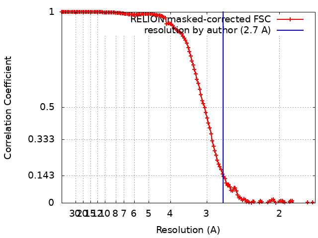
ジャーナル: ACS Cent Sci / 年: 2020 タイトル: Comparative Structural Analysis of 20S Proteasome Ortholog Protein Complexes by Native Mass Spectrometry. 著者: Shay Vimer / Gili Ben-Nissan / David Morgenstern / Fanindra Kumar-Deshmukh / Caley Polkinghorn / Royston S Quintyn / Yury V Vasil'ev / Joseph S Beckman / Nadav Elad / Vicki H Wysocki / Michal Sharon / 要旨: Ortholog protein complexes are responsible for equivalent functions in different organisms. However, during evolution, each organism adapts to meet its physiological needs and the environmental ...Ortholog protein complexes are responsible for equivalent functions in different organisms. However, during evolution, each organism adapts to meet its physiological needs and the environmental challenges imposed by its niche. This selection pressure leads to structural diversity in protein complexes, which are often difficult to specify, especially in the absence of high-resolution structures. Here, we describe a multilevel experimental approach based on native mass spectrometry (MS) tools for elucidating the structural preservation and variations among highly related protein complexes. The 20S proteasome, an essential protein degradation machinery, served as our model system, wherein we examined five complexes isolated from different organisms. We show that throughout evolution, from the archaeal prokaryotic complex to the eukaryotic 20S proteasomes in yeast () and mammals (rat - , rabbit - and human - HEK293 cells), the proteasome increased both in size and stability. Native MS structural signatures of the rat and rabbit 20S proteasomes, which heretofore lacked high-resolution, three-dimensional structures, highly resembled that of the human complex. Using cryoelectron microscopy single-particle analysis, we were able to obtain a high-resolution structure of the rat 20S proteasome, allowing us to validate the MS-based results. Our study also revealed that the yeast complex, and not those in mammals, was the largest in size and displayed the greatest degree of kinetic stability. Moreover, we also identified a new proteoform of the PSMA7 subunit that resides within the rat and rabbit complexes, which to our knowledge have not been previously described. Altogether, our strategy enables elucidation of the unique structural properties of protein complexes that are highly similar to one another, a framework that is valid not only to ortholog protein complexes, but also for other highly related protein assemblies.
名称: Rat liver 20S proteasome / タイプ: complex / ID: 1 / 親要素: 0 / 含まれる分子: all 詳細: Endogenous 20S proteasome purified from the rat livers by anion-exchange chromatography
Preliminary grid screening was performed on Talos Arctica
撮影
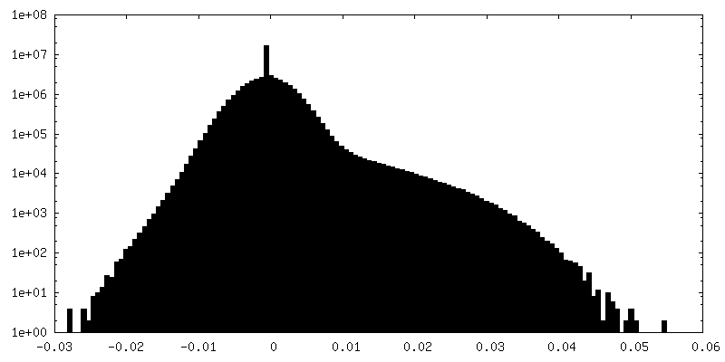
フィルム・検出器のモデル: GATAN K3 BIOQUANTUM (6k x 4k) 撮影したグリッド数: 1 / 実像数: 2234 / 平均露光時間: 1.5 sec. / 平均電子線量: 37.0 e/Å2 詳細: Images were collected in movie-mode at 45 frames per second
 ムービー
ムービー コントローラー


 データを開く
 基本情報
基本情報 マップデータ
 試料
試料 キーワード
キーワード 機能・相同性情報
 データ登録者
 イスラエル, 2件
  引用
引用
 構造の表示
構造の表示
 ダウンロードとリンク
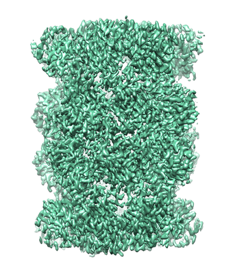 emd_10586.png
 http://ftp.pdbj.org/pub/emdb/structures/EMD-10586
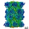


 Z
Z Y
Y X
X


 試料の構成要素
 解析
解析 電子顕微鏡法
 FIELD EMISSION GUN