- EMDB-10046: Needle complex of the Shigella type 3 secretion system -
+
Open data
ID or keywords:
Loading...
-
Basic information
Entry
Database: EMDB / ID: EMD-10046
Title
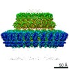
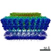
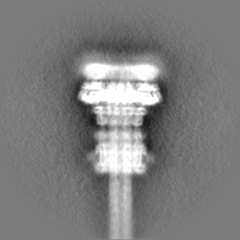
Needle complex of the Shigella type 3 secretion system
Map data
Masked C1 reconstruction of needle complex of the Shigella type 3 secretion system, post-refined sharpened (b=-162), low-pass filtered at 5.11 A.
Sample
Complex: Needle complex of the Shigella type 3 secretion system
Function / homology
Function and homology information
type III protein secretion system complex / protein secretion by the type III secretion system / protein secretion / protein targeting / cell surface / extracellular region / identical protein binding / plasma membrane Similarity search - Function
Type III secretion protein SpaR/YscT / Type III secretion protein HrpO / Yop virulence translocation protein R / Type III secretion, needle-protein-like / Type III secretion, needle-protein-like superfamily / Type III secretion needle MxiH, YscF, SsaG, EprI, PscF, EscF / Type III secretion system, needle protein / Type III secretion system inner membrane R protein / Bacterial export protein family 3 / Bacterial export proteins, family 1 ...Type III secretion protein SpaR/YscT / Type III secretion protein HrpO / Yop virulence translocation protein R / Type III secretion, needle-protein-like / Type III secretion, needle-protein-like superfamily / Type III secretion needle MxiH, YscF, SsaG, EprI, PscF, EscF / Type III secretion system, needle protein / Type III secretion system inner membrane R protein / Bacterial export protein family 3 / Bacterial export proteins, family 1 / Bacterial export proteins, family 3 / Flagella transport protein fliP family signature 1. / Type III secretion system inner membrane P protein / FliP family / Flagella transport protein fliP family signature 2. Similarity search - Domain/homology
Surface presentation of antigens protein SpaP / Surface presentation of antigens protein SpaQ / Surface presentation of antigens protein SpaR / Type 3 secretion system needle filament protein Similarity search - Component
Biological species
Shigella flexneri (bacteria)
Method
single particle reconstruction / cryo EM / Resolution: 5.11 Å
Journal: PLoS Pathog / Year: 2020 Title: Cryo-EM structure of the Shigella type III needle complex. Authors: Michele Lunelli / Antje Kamprad / Jörg Bürger / Thorsten Mielke / Christian M T Spahn / Michael Kolbe / Abstract: The Type III Secretion Systems (T3SS) needle complex is a conserved syringe-shaped protein translocation nanomachine with a mass of about 3.5 MDa essential for the survival and virulence of many Gram- ...The Type III Secretion Systems (T3SS) needle complex is a conserved syringe-shaped protein translocation nanomachine with a mass of about 3.5 MDa essential for the survival and virulence of many Gram-negative bacterial pathogens. This system is composed of a membrane-embedded basal body and an extracellular needle that deliver effector proteins into host cells. High-resolution structures of the T3SS from different organisms and infection stages are needed to understand the underlying molecular mechanisms of effector translocation. Here, we present the cryo-electron microscopy structure of the isolated Shigella T3SS needle complex. The inner membrane (IM) region of the basal body adopts 24-fold rotational symmetry and forms a channel system that connects the bacterial periplasm with the export apparatus cage. The secretin oligomer adopts a heterogeneous architecture with 16- and 15-fold cyclic symmetry in the periplasmic N-terminal connector and C-terminal outer membrane ring, respectively. Two out of three IM subunits bind the secretin connector via a β-sheet augmentation. The cryo-EM map also reveals the helical architecture of the export apparatus core, the inner rod, the needle and their intervening interfaces.
History
Deposition
Jun 6, 2019
-
Header (metadata) release
Feb 12, 2020
-
Map release
Feb 12, 2020
-
Update
Nov 25, 2020
-
Current status
Nov 25, 2020
Processing site: PDBe / Status: Released
-
Structure visualization
Movie
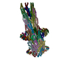
Surface view with section colored by density value
Model: Quantifoil R2/1 / Material: COPPER / Support film - Material: CARBON / Support film - topology: CONTINUOUS / Support film - Film thickness: 5.0 nm / Pretreatment - Type: GLOW DISCHARGE / Pretreatment - Atmosphere: AIR
Vitrification
Cryogen name: ETHANE / Chamber humidity: 100 % / Chamber temperature: 295 K / Instrument: FEI VITROBOT MARK IV Details: Sample applied on grid 5 ul, incubation time 5 min on ice, then moved into Vitrobot and 5 ul sample applied again. Blot time: 2 sec Blot force: -2 Drain time: 0 sec.
Details
Isolated needle complex in detergent solution
-
Electron microscopy
Microscope
FEI TITAN KRIOS
Image recording
Film or detector model: FEI FALCON II (4k x 4k) / Digitization - Dimensions - Width: 4096 pixel / Digitization - Dimensions - Height: 4096 pixel / Number grids imaged: 1 / Number real images: 5238 / Average exposure time: 1.5 sec. / Average electron dose: 25.0 e/Å2
Electron beam
Acceleration voltage: 300 kV / Electron source: FIELD EMISSION GUN
Electron optics
Illumination mode: OTHER / Imaging mode: BRIGHT FIELD / Cs: 2.7 mm / Nominal defocus max: 0.004 µm / Nominal defocus min: 0.0015 µm / Nominal magnification: 101179
In the structure databanks used in Yorodumi, some data are registered as the other names, "COVID-19 virus" and "2019-nCoV". Here are the details of the virus and the list of structure data.
Jan 31, 2019. EMDB accession codes are about to change! (news from PDBe EMDB page)
EMDB accession codes are about to change! (news from PDBe EMDB page)
The allocation of 4 digits for EMDB accession codes will soon come to an end. Whilst these codes will remain in use, new EMDB accession codes will include an additional digit and will expand incrementally as the available range of codes is exhausted. The current 4-digit format prefixed with “EMD-” (i.e. EMD-XXXX) will advance to a 5-digit format (i.e. EMD-XXXXX), and so on. It is currently estimated that the 4-digit codes will be depleted around Spring 2019, at which point the 5-digit format will come into force.
The EM Navigator/Yorodumi systems omit the EMD- prefix.
Related info.:Q: What is EMD? / ID/Accession-code notation in Yorodumi/EM Navigator
Yorodumi is a browser for structure data from EMDB, PDB, SASBDB, etc.
This page is also the successor to EM Navigator detail page, and also detail information page/front-end page for Omokage search.
The word "yorodu" (or yorozu) is an old Japanese word meaning "ten thousand". "mi" (miru) is to see.
Related info.:EMDB / PDB / SASBDB / Comparison of 3 databanks / Yorodumi Search / Aug 31, 2016. New EM Navigator & Yorodumi / Yorodumi Papers / Jmol/JSmol / Function and homology information / Changes in new EM Navigator and Yorodumi
 Movie
Movie Controller


 Open data
 Basic information
 Map data
 Sample
 Function and homology information
 Shigella flexneri (bacteria)
 Authors
 Citation
 Structure visualization
 Downloads & links
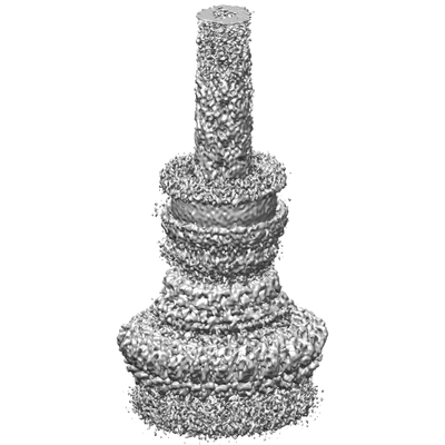 emd_10046.png
 http://ftp.pdbj.org/pub/emdb/structures/EMD-10046


 Z (Sec.)
 Y (Row.)
 X (Col.)


 Sample components
 Processing
 Electron microscopy
 FIELD EMISSION GUN

