+データを開く
-基本情報
登録情報 | データベース: EMDB / ID: EMD-0274 | |||||||||
---|---|---|---|---|---|---|---|---|---|---|
タイトル | Cryo-EM structure of cardiac amyloid fibrils from an immunoglobulin light chain (AL) amyloidosis patient. | |||||||||
マップデータ | 3D helical reconstrutcion of an amyloid fibrils from an immunoglobulin light chain (AL) amyloidosis patient. | |||||||||
試料 |
| |||||||||
生物種 | Homo sapiens (ヒト) | |||||||||
手法 | らせん対称体再構成法 / クライオ電子顕微鏡法 / 解像度: 4.0 Å | |||||||||
データ登録者 | Swuec P | |||||||||
引用 | 
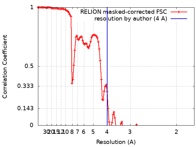
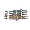
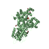
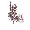
ジャーナル: Nat Commun / 年: 2019 タイトル: Cryo-EM structure of cardiac amyloid fibrils from an immunoglobulin light chain AL amyloidosis patient. 著者: Paolo Swuec / Francesca Lavatelli / Masayoshi Tasaki / Cristina Paissoni / Paola Rognoni / Martina Maritan / Francesca Brambilla / Paolo Milani / Pierluigi Mauri / Carlo Camilloni / Giovanni ...著者: Paolo Swuec / Francesca Lavatelli / Masayoshi Tasaki / Cristina Paissoni / Paola Rognoni / Martina Maritan / Francesca Brambilla / Paolo Milani / Pierluigi Mauri / Carlo Camilloni / Giovanni Palladini / Giampaolo Merlini / Stefano Ricagno / Martino Bolognesi / 要旨: Systemic light chain amyloidosis (AL) is a life-threatening disease caused by aggregation and deposition of monoclonal immunoglobulin light chains (LC) in target organs. Severity of heart ...Systemic light chain amyloidosis (AL) is a life-threatening disease caused by aggregation and deposition of monoclonal immunoglobulin light chains (LC) in target organs. Severity of heart involvement is the most important factor determining prognosis. Here, we report the 4.0 Å resolution cryo-electron microscopy map and molecular model of amyloid fibrils extracted from the heart of an AL amyloidosis patient with severe amyloid cardiomyopathy. The helical fibrils are composed of a single protofilament, showing typical 4.9 Å stacking and cross-β architecture. Two distinct polypeptide stretches (total of 77 residues) from the LC variable domain (V) fit the fibril density. Despite V high sequence variability, residues stabilizing the fibril core are conserved through different cardiotoxic V, highlighting structural motifs that may be common to misfolding-prone LCs. Our data shed light on the architecture of LC amyloids, correlate amino acid sequences with fibril assembly, providing the grounds for development of innovative medicines. | |||||||||
履歴 |
|
-構造の表示
ムービー |
ムービービューア |
---|---|
構造ビューア | EMマップ: SurfViewMolmilJmol/JSmol |
添付画像 |
-ダウンロードとリンク
-EMDBアーカイブ
マップデータ | emd_0274.map.gz | 4.6 MB | EMDBマップデータ形式 | |
---|---|---|---|---|
ヘッダ (付随情報) | emd-0274-v30.xml emd-0274.xml | 11.9 KB 11.9 KB | 表示 表示 | EMDBヘッダ |
FSC (解像度算出) | emd_0274_fsc.xml | 11.4 KB | 表示 | FSCデータファイル |
画像 | emd_0274.png | 121.4 KB | ||
アーカイブディレクトリ | http://ftp.pdbj.org/pub/emdb/structures/EMD-0274 ftp://ftp.pdbj.org/pub/emdb/structures/EMD-0274 | HTTPS FTP |
-検証レポート
文書・要旨 | emd_0274_validation.pdf.gz | 227.1 KB | 表示 | EMDB検証レポート |
---|---|---|---|---|
文書・詳細版 | emd_0274_full_validation.pdf.gz | 226.2 KB | 表示 | |
XML形式データ | emd_0274_validation.xml.gz | 12.3 KB | 表示 | |
アーカイブディレクトリ | https://ftp.pdbj.org/pub/emdb/validation_reports/EMD-0274 ftp://ftp.pdbj.org/pub/emdb/validation_reports/EMD-0274 | HTTPS FTP |
-関連構造データ
-リンク
EMDBのページ | EMDB (EBI/PDBe) / EMDataResource |
---|---|
「今月の分子」の関連する項目 |
-マップ
ファイル | ダウンロード / ファイル: emd_0274.map.gz / 形式: CCP4 / 大きさ: 125 MB / タイプ: IMAGE STORED AS FLOATING POINT NUMBER (4 BYTES) | ||||||||||||||||||||||||||||||||||||||||||||||||||||||||||||||||||||
---|---|---|---|---|---|---|---|---|---|---|---|---|---|---|---|---|---|---|---|---|---|---|---|---|---|---|---|---|---|---|---|---|---|---|---|---|---|---|---|---|---|---|---|---|---|---|---|---|---|---|---|---|---|---|---|---|---|---|---|---|---|---|---|---|---|---|---|---|---|
注釈 | 3D helical reconstrutcion of an amyloid fibrils from an immunoglobulin light chain (AL) amyloidosis patient. | ||||||||||||||||||||||||||||||||||||||||||||||||||||||||||||||||||||
ボクセルのサイズ | X=Y=Z: 0.887 Å | ||||||||||||||||||||||||||||||||||||||||||||||||||||||||||||||||||||
密度 |
| ||||||||||||||||||||||||||||||||||||||||||||||||||||||||||||||||||||
対称性 | 空間群: 1 | ||||||||||||||||||||||||||||||||||||||||||||||||||||||||||||||||||||
詳細 | EMDB XML:
CCP4マップ ヘッダ情報:
|
-添付データ
-試料の構成要素
-全体 : Cardiac amyloid fibril from an immunoglobulin light chain (AL) am...
全体 | 名称: Cardiac amyloid fibril from an immunoglobulin light chain (AL) amyloidosis patient |
---|---|
要素 |
|
-超分子 #1: Cardiac amyloid fibril from an immunoglobulin light chain (AL) am...
超分子 | 名称: Cardiac amyloid fibril from an immunoglobulin light chain (AL) amyloidosis patient タイプ: complex / ID: 1 / 親要素: 0 / 含まれる分子: all |
---|---|
由来(天然) | 生物種: Homo sapiens (ヒト) / 組織: Heart |
-分子 #1: Monoclonal immunoglobulin light chains (LC)
分子 | 名称: Monoclonal immunoglobulin light chains (LC) / タイプ: protein_or_peptide / ID: 1 / コピー数: 5 / 光学異性体: LEVO |
---|---|
由来(天然) | 生物種: Homo sapiens (ヒト) / 組織: Heart |
分子量 | 理論値: 14.620864 KDa |
配列 | 文字列: NFMLTQPHSV SESPGKTLTI SCTGSSASIA SHYVQWYQQR PGGAPTTLIY ENDQRPSEVP DRFSGSIDSS SNSASLTISG LKTEDEADY YCQSYDGNNH WVFGGGTKLT VLSQPKAAPS VTLFPPSSEE LQANKAT |
-実験情報
-構造解析
手法 | クライオ電子顕微鏡法 |
---|---|
解析 | らせん対称体再構成法 |
試料の集合状態 | filament |
-試料調製
緩衝液 | pH: 7 |
---|---|
グリッド | モデル: Quantifoil R1.2/1.3 / 材質: COPPER / メッシュ: 300 / 前処理 - タイプ: GLOW DISCHARGE / 前処理 - 雰囲気: AIR / 前処理 - 気圧: 0.01 kPa |
凍結 | 凍結剤: ETHANE / チャンバー内湿度: 100 % / チャンバー内温度: 278 K / 装置: FEI VITROBOT MARK IV |
-電子顕微鏡法
顕微鏡 | FEI TALOS ARCTICA |
---|---|
撮影 | フィルム・検出器のモデル: FEI FALCON III (4k x 4k) 検出モード: INTEGRATING / 実像数: 1680 / 平均露光時間: 1.0 sec. / 平均電子線量: 50.0 e/Å2 |
電子線 | 加速電圧: 200 kV / 電子線源: FIELD EMISSION GUN |
電子光学系 | C2レンズ絞り径: 50.0 µm / 照射モード: SPOT SCAN / 撮影モード: BRIGHT FIELD / Cs: 2.7 mm / 倍率(公称値): 120000 |
試料ステージ | 試料ホルダーモデル: OTHER / ホルダー冷却材: NITROGEN |
実験機器 | モデル: Talos Arctica / 画像提供: FEI Company |
+画像解析
-原子モデル構築 1
精密化 | 空間: REAL |
---|---|
得られたモデル | PDB-6hud: |