 Movie
Movie Controller

[English] 日本語
 Yorodumi
- EMDB-8334: Structural basis for dynamic regulation of the human 26S proteasome -
+ Open data

- Basic information
| Entry | Database: EMDB / ID: EMD-8334 | |||||||||
|---|---|---|---|---|---|---|---|---|---|---|
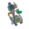
| Title | Structural basis for dynamic regulation of the human 26S proteasome | |||||||||
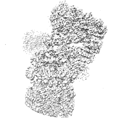
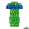
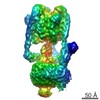
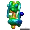
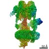
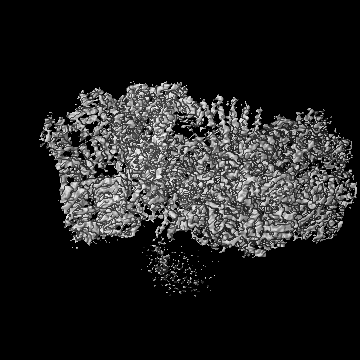
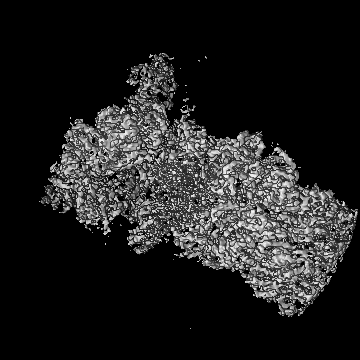
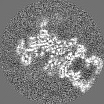
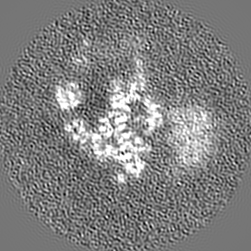
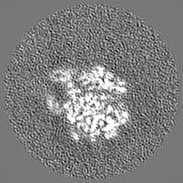
 Map data Map data | The human half 26S proteasome reconstruction in the SA state, focusing on the individual regulatory particle, from 139236 particles of half 26S | |||||||||
 Sample Sample |
| |||||||||
| Function / homology |  Function and homology information Function and homology informationthyrotropin-releasing hormone receptor binding / nuclear proteasome complex / host-mediated perturbation of viral transcription / positive regulation of inclusion body assembly / Impaired BRCA2 translocation to the nucleus / Impaired BRCA2 binding to SEM1 (DSS1) / proteasome accessory complex / purine ribonucleoside triphosphate binding / integrator complex / proteasome regulatory particle ...thyrotropin-releasing hormone receptor binding / nuclear proteasome complex / host-mediated perturbation of viral transcription / positive regulation of inclusion body assembly / Impaired BRCA2 translocation to the nucleus / Impaired BRCA2 binding to SEM1 (DSS1) / proteasome accessory complex / purine ribonucleoside triphosphate binding / integrator complex / proteasome regulatory particle / CD8-positive, alpha-beta T cell differentiation / thymic T cell selection / CD8-positive, alpha-beta T cell homeostasis / cytosolic proteasome complex / positive regulation of proteasomal protein catabolic process / proteasome-activating activity / Antigen processing: Ub, ATP-independent proteasomal degradation / proteasome regulatory particle, lid subcomplex / proteasome regulatory particle, base subcomplex / negative regulation of regulatory T cell differentiation / T-helper 1 cell differentiation / protein K63-linked deubiquitination / metal-dependent deubiquitinase activity / cellular response to type I interferon / negative regulation of programmed cell death / Regulation of ornithine decarboxylase (ODC) / proteasome core complex / Proteasome assembly / T-helper 17 cell differentiation / retrograde vesicle-mediated transport, Golgi to endoplasmic reticulum / Cross-presentation of soluble exogenous antigens (endosomes) / transcription factor binding / K63-linked deubiquitinase activity / Somitogenesis / flagellated sperm motility / Homologous DNA Pairing and Strand Exchange / Defective homologous recombination repair (HRR) due to BRCA1 loss of function / Defective HDR through Homologous Recombination Repair (HRR) due to PALB2 loss of BRCA1 binding function / Defective HDR through Homologous Recombination Repair (HRR) due to PALB2 loss of BRCA2/RAD51/RAD51C binding function / Resolution of D-loop Structures through Synthesis-Dependent Strand Annealing (SDSA) / Resolution of D-loop Structures through Holliday Junction Intermediates / proteasome binding / Impaired BRCA2 binding to RAD51 / myofibril / positive regulation of RNA polymerase II transcription preinitiation complex assembly / proteasomal ubiquitin-independent protein catabolic process / general transcription initiation factor binding / Presynaptic phase of homologous DNA pairing and strand exchange / proteasome storage granule / AMPK-induced ERAD and lysosome mediated degradation of PD-L1(CD274) / protein deubiquitination / polyubiquitin modification-dependent protein binding / proteasome endopeptidase complex / NF-kappaB binding / proteasome core complex, beta-subunit complex / endopeptidase activator activity / threonine-type endopeptidase activity / GSK3B-mediated proteasomal degradation of PD-L1(CD274) / proteasome core complex, alpha-subunit complex / SPOP-mediated proteasomal degradation of PD-L1(CD274) / proteasome assembly / mRNA export from nucleus / SARS-CoV-1 targets host intracellular signalling and regulatory pathways / immune system process / regulation of G1/S transition of mitotic cell cycle / regulation of macroautophagy / enzyme regulator activity / Ribosome Quality Control (RQC) complex extracts and degrades nascent peptide / positive regulation of interleukin-2 production / ciliary tip / ERAD pathway / response to type II interferon / inclusion body / TBP-class protein binding / proteasome complex / : / stem cell differentiation / regulation of proteasomal protein catabolic process / sperm end piece / sarcomere / Regulation of activated PAK-2p34 by proteasome mediated degradation / ubiquitin binding / proteasomal protein catabolic process / Autodegradation of Cdh1 by Cdh1:APC/C / negative regulation of inflammatory response to antigenic stimulus / APC/C:Cdc20 mediated degradation of Securin / N-glycan trimming in the ER and Calnexin/Calreticulin cycle / Asymmetric localization of PCP proteins / Ubiquitin-dependent degradation of Cyclin D / lipopolysaccharide binding / SCF-beta-TrCP mediated degradation of Emi1 / NIK-->noncanonical NF-kB signaling / AUF1 (hnRNP D0) binds and destabilizes mRNA / TNFR2 non-canonical NF-kB pathway / Assembly of the pre-replicative complex / P-body / Vpu mediated degradation of CD4 / Cdc20:Phospho-APC/C mediated degradation of Cyclin A / Dectin-1 mediated noncanonical NF-kB signaling / Degradation of DVL Similarity search - Function | |||||||||
| Biological species |  Homo sapiens (human) Homo sapiens (human) | |||||||||
| Method | single particle reconstruction / cryo EM / Resolution: 4.4 Å | |||||||||
 Authors Authors | Chen S / Wu J / Lu Y / Ma YB / Lee BH / Yu Z / Ouyang Q / Finley D / Kirschner MW / Mao Y | |||||||||
 Citation Citation | Journal: Proc Natl Acad Sci U S A / Year: 2016 Title: Structural basis for dynamic regulation of the human 26S proteasome. Authors: Shuobing Chen / Jiayi Wu / Ying Lu / Yong-Bei Ma / Byung-Hoon Lee / Zhou Yu / Qi Ouyang / Daniel J Finley / Marc W Kirschner / Youdong Mao /   Abstract: The proteasome is the major engine of protein degradation in all eukaryotic cells. At the heart of this machine is a heterohexameric ring of AAA (ATPases associated with diverse cellular activities) ...The proteasome is the major engine of protein degradation in all eukaryotic cells. At the heart of this machine is a heterohexameric ring of AAA (ATPases associated with diverse cellular activities) proteins that unfolds ubiquitylated target proteins that are concurrently translocated into a proteolytic chamber and degraded into peptides. Using cryoelectron microscopy, we determined a near-atomic-resolution structure of the 2.5-MDa human proteasome in its ground state, as well as subnanometer-resolution structures of the holoenzyme in three alternative conformational states. The substrate-unfolding AAA-ATPase channel is narrowed by 10 inward-facing pore loops arranged into two helices that run in parallel with each other, one hydrophobic in character and the other highly charged. The gate of the core particle was unexpectedly found closed in the ground state and open in only one of the alternative states. Coordinated, stepwise conformational changes of the regulatory particle couple ATP hydrolysis to substrate translocation and regulate gating of the core particle, leading to processive degradation. | |||||||||
| History |
|
- Structure visualization
| Movie |
Movie viewer |
|---|---|
| Structure viewer | EM map: SurfViewMolmilJmol/JSmol |
| Supplemental images |
- Downloads & links
-EMDB archive
| Map data | emd_8334.map.gz | 160.9 MB | EMDB map data format | |
|---|---|---|---|---|
| Header (meta data) | emd-8334-v30.xmlemd-8334.xml | 14.8 KB 14.8 KB | Display Display | EMDB header |
| Images |  emd_8334.png emd_8334.png | 62.8 KB | ||
| Archive directory |  http://ftp.pdbj.org/pub/emdb/structures/EMD-8334ftp://ftp.pdbj.org/pub/emdb/structures/EMD-8334 http://ftp.pdbj.org/pub/emdb/structures/EMD-8334ftp://ftp.pdbj.org/pub/emdb/structures/EMD-8334 | HTTPS FTP |
-Related structure data
| Related structure data |  5t0gMC  8332C  8333C 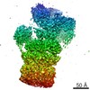 8335C 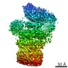 8336C 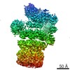 8337C 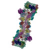 5t0cC 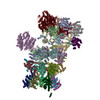 5t0hC  5t0iC  5t0jC C: citing same article ( M: atomic model generated by this map |
|---|---|
| Similar structure data | |
| EM raw data | EMPIAR-10072 (Title: Structural basis for dynamic regulation of the human 26S proteasome Data size: 2.0 TB Data #1: Drift-corrected micrographs of human 26S proteasome holoenzyme [micrographs - single frame] Data #2: Classified particle datasets for the human proteasome in four conformational states [picked particles - multiframe - processed]) |
-Links
| EMDB pages | EMDB (EBI/PDBe) / EMDataResource |
|---|---|
| Related items in Molecule of the Month |


-Map
| File | Download / File: emd_8334.map.gz / Format: CCP4 / Size: 178 MB / Type: IMAGE STORED AS FLOATING POINT NUMBER (4 BYTES) | ||||||||||||||||||||||||||||||||||||||||||||||||||||||||||||||||||||
|---|---|---|---|---|---|---|---|---|---|---|---|---|---|---|---|---|---|---|---|---|---|---|---|---|---|---|---|---|---|---|---|---|---|---|---|---|---|---|---|---|---|---|---|---|---|---|---|---|---|---|---|---|---|---|---|---|---|---|---|---|---|---|---|---|---|---|---|---|---|
| Annotation | The human half 26S proteasome reconstruction in the SA state, focusing on the individual regulatory particle, from 139236 particles of half 26S | ||||||||||||||||||||||||||||||||||||||||||||||||||||||||||||||||||||
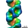
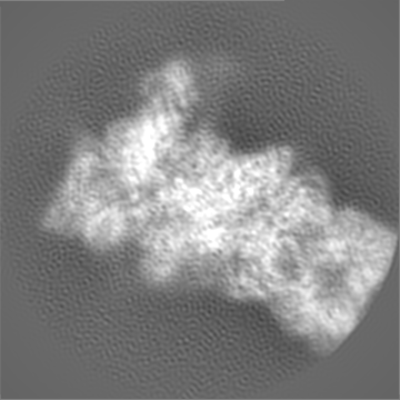
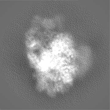
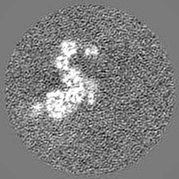
| Projections & slices | Image control
Images are generated by Spider. | ||||||||||||||||||||||||||||||||||||||||||||||||||||||||||||||||||||
| Voxel size | X=Y=Z: 0.86 Å | ||||||||||||||||||||||||||||||||||||||||||||||||||||||||||||||||||||
| Density |
| ||||||||||||||||||||||||||||||||||||||||||||||||||||||||||||||||||||
| Symmetry | Space group: 1 | ||||||||||||||||||||||||||||||||||||||||||||||||||||||||||||||||||||
| Details | EMDB XML:
CCP4 map header:
| ||||||||||||||||||||||||||||||||||||||||||||||||||||||||||||||||||||
 Z (Sec.)
 Y (Row.)
 X (Col.)


-Supplemental data
- Sample components
-Entire : 26S proteasome holoenzyme
| Entire | Name: 26S proteasome holoenzyme |
|---|---|
| Components |
|
-Supramolecule #1: 26S proteasome holoenzyme
| Supramolecule | Name: 26S proteasome holoenzyme / type: complex / ID: 1 / Parent: 0 / Macromolecule list: #1 |
|---|---|
| Source (natural) | Organism: Homo sapiens (human) |
| Molecular weight | Experimental: 2.5 MDa |
-Experimental details
-Structure determination
| Method | cryo EM |
|---|---|
 Processing Processing | single particle reconstruction |
| Aggregation state | particle |
-Sample preparation
| Concentration | 1.5 mg/mL | ||||||||||||||||||
|---|---|---|---|---|---|---|---|---|---|---|---|---|---|---|---|---|---|---|---|
| Buffer | pH: 7.5 Component:
| ||||||||||||||||||
| Grid | Model: C-Flat R1/1 / Material: COPPER / Mesh: 400 / Support film - Material: CARBON / Support film - topology: HOLEY ARRAY / Support film - Film thickness: 50.0 nm / Pretreatment - Type: GLOW DISCHARGE / Pretreatment - Atmosphere: AIR | ||||||||||||||||||
| Vitrification | Cryogen name: ETHANE / Chamber humidity: 100 % / Chamber temperature: 277 K / Instrument: FEI VITROBOT MARK IV / Details: blotted for 2 seconds, blotting force 3. |
- Electron microscopy
| Microscope | FEI TECNAI ARCTICA |
|---|---|
| Image recording | Film or detector model: GATAN K2 SUMMIT (4k x 4k) / Detector mode: SUPER-RESOLUTION / Digitization - Dimensions - Width: 7420 pixel / Digitization - Dimensions - Height: 7676 pixel / Digitization - Sampling interval: 5.0 µm / Digitization - Frames/image: 3-20 / Number grids imaged: 12 / Number real images: 10367 / Average exposure time: 9.0 sec. / Average electron dose: 30.0 e/Å2 |
| Electron beam | Acceleration voltage: 200 kV / Electron source:  FIELD EMISSION GUN FIELD EMISSION GUN |
| Electron optics | C2 aperture diameter: 50.0 µm / Calibrated magnification: 28736 / Illumination mode: FLOOD BEAM / Imaging mode: BRIGHT FIELD / Cs: 2.7 mm / Nominal defocus max: -3.0 µm / Nominal defocus min: -1.0 µm / Nominal magnification: 21000 |
| Sample stage | Specimen holder model: FEI TITAN KRIOS AUTOGRID HOLDER / Cooling holder cryogen: NITROGEN |
| Experimental equipment |  Model: Talos Arctica / Image courtesy: FEI Company |
+Image processing
-Atomic model buiding 1
| Refinement | Space: RECIPROCAL / Protocol: AB INITIO MODEL / Overall B value: 70 |
|---|---|
| Output model | PDB-5t0g: |
