 Movie
Movie Controller

[English] 日本語
 Yorodumi
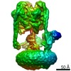
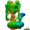
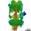
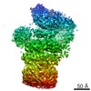
- EMDB-1590: Structure of the Manduca sexta V-ATPase by cryo-electron microscopy -
+ Open data

- Basic information
| Entry | Database: EMDB / ID: EMD-1590 | |||||||||
|---|---|---|---|---|---|---|---|---|---|---|
| Title | Structure of the Manduca sexta V-ATPase by cryo-electron microscopy | |||||||||
 Map data Map data | Manduca sexta vacuolar ATPase | |||||||||
 Sample Sample |
| |||||||||
 Keywords Keywords | H-ATPase / V-ATPase / cryo-electron microscopy / vacuolar membrane | |||||||||
| Biological species |  Manduca sexta (tobacco hornworm) Manduca sexta (tobacco hornworm) | |||||||||
| Method | single particle reconstruction / cryo EM / Resolution: 17.0 Å | |||||||||
 Authors Authors | Muench SP / Huss M / Phillips C / Song CF / Wieczorek H / Trinick J / Harrison MA | |||||||||
 Citation Citation | Journal: J Mol Biol / Year: 2009 Title: Cryo-electron microscopy of the vacuolar ATPase motor reveals its mechanical and regulatory complexity. Authors: Stephen P Muench / Markus Huss / Chun Feng Song / Clair Phillips / Helmut Wieczorek / John Trinick / Michael A Harrison /  Abstract: The vacuolar H+-ATPase (V-ATPase) is an ATP-driven rotary molecular motor that is a transmembrane proton pump in all eukaryotic cells. Although its activity is fundamental to many physiological ...The vacuolar H+-ATPase (V-ATPase) is an ATP-driven rotary molecular motor that is a transmembrane proton pump in all eukaryotic cells. Although its activity is fundamental to many physiological processes, our understanding of the structure and mechanism of the V-ATPase is poor. Using cryo-electron microscopy of the tobacco hornworm (Manduca sexta) enzyme, we have calculated the first 3D reconstruction of the intact pump in its native state. The resolution of 16.5 A is significantly higher than that of previous cryo-electron microscopy models of either V-ATPase or the related F1F0-ATPase. A network of four stalk structures connecting the V1 catalytic domain and the V0 membrane domain is now fully resolved, demonstrating substantially greater complexity than that found in the F-ATPase. Three peripheral stator stalks connect these domains to a horizontal collar that partly encircles the region between V1 and V0. The fourth stalk is a central axle that connects to V0 but makes minimal contact with V1. Several subunit crystal structures can be fit accurately into the reconstruction. The model thus provides new insights into the organisation of key components involved in mechanical coupling between the domains and regulation of activity. | |||||||||
| History |
|
- Structure visualization
| Movie |
 Movie viewer Movie viewer |
|---|---|
| Structure viewer | EM map: SurfViewMolmilJmol/JSmol |
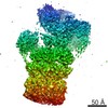
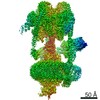
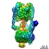
| Supplemental images |

- Downloads & links
-EMDB archive
| Map data | emd_1590.map.gz | 3.5 MB | EMDB map data format | |
|---|---|---|---|---|
| Header (meta data) | emd-1590-v30.xmlemd-1590.xml | 9.4 KB 9.4 KB | Display Display | EMDB header |
| Images | 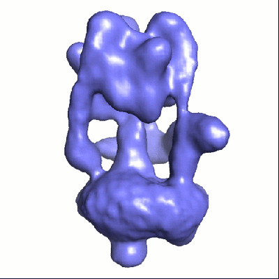 1590.gif 1590.gif | 50.5 KB | ||
| Archive directory |  http://ftp.pdbj.org/pub/emdb/structures/EMD-1590ftp://ftp.pdbj.org/pub/emdb/structures/EMD-1590 http://ftp.pdbj.org/pub/emdb/structures/EMD-1590ftp://ftp.pdbj.org/pub/emdb/structures/EMD-1590 | HTTPS FTP |
-Related structure data
| Similar structure data |
|---|

-Links
| EMDB pages | EMDB (EBI/PDBe) / EMDataResource |
|---|
-Map
| File | Download / File: emd_1590.map.gz / Format: CCP4 / Size: 3.7 MB / Type: IMAGE STORED AS FLOATING POINT NUMBER (4 BYTES) | ||||||||||||||||||||||||||||||||||||||||||||||||||||||||||||||||||||
|---|---|---|---|---|---|---|---|---|---|---|---|---|---|---|---|---|---|---|---|---|---|---|---|---|---|---|---|---|---|---|---|---|---|---|---|---|---|---|---|---|---|---|---|---|---|---|---|---|---|---|---|---|---|---|---|---|---|---|---|---|---|---|---|---|---|---|---|---|---|
| Annotation | Manduca sexta vacuolar ATPase | ||||||||||||||||||||||||||||||||||||||||||||||||||||||||||||||||||||
| Projections & slices | Image control
Images are generated by Spider. | ||||||||||||||||||||||||||||||||||||||||||||||||||||||||||||||||||||
| Voxel size | X=Y=Z: 4.36 Å | ||||||||||||||||||||||||||||||||||||||||||||||||||||||||||||||||||||
| Density |
| ||||||||||||||||||||||||||||||||||||||||||||||||||||||||||||||||||||
| Symmetry | Space group: 1 | ||||||||||||||||||||||||||||||||||||||||||||||||||||||||||||||||||||
| Details | EMDB XML:
CCP4 map header:
| ||||||||||||||||||||||||||||||||||||||||||||||||||||||||||||||||||||
 Z (Sec.)
 Y (Row.)
 X (Col.)


-Supplemental data
- Sample components
-Entire : Manduca sexta vacuolar ATPase complex
| Entire | Name: Manduca sexta vacuolar ATPase complex |
|---|---|
| Components |
|
-Supramolecule #1000: Manduca sexta vacuolar ATPase complex
| Supramolecule | Name: Manduca sexta vacuolar ATPase complex / type: sample / ID: 1000 / Details: The sample was monodisperse / Oligomeric state: monomer / Number unique components: 1 |
|---|---|
| Molecular weight | Experimental: 900 KDa / Theoretical: 900 KDa |
-Supramolecule #1: membrane proton pump
| Supramolecule | Name: membrane proton pump / type: organelle_or_cellular_component / ID: 1 / Name.synonym: V-ATPase / Oligomeric state: monomer / Recombinant expression: No |
|---|---|
| Source (natural) | Organism: Manduca sexta (tobacco hornworm) / synonym: tobacco hornworm / Tissue: midgut |
-Experimental details
-Structure determination
| Method | cryo EM |
|---|---|
 Processing Processing | single particle reconstruction |
| Aggregation state | particle |
-Sample preparation
| Concentration | 2.0 mg/mL |
|---|---|
| Buffer | pH: 8.1 Details: 150 mM NaCl, 9.6 mM B mercaptoethanol, 20 mM TrisHCl. Solubilised in C12E10 detergent |
| Grid | Details: 300 mesh Cu Lacey grid |
| Vitrification | Cryogen name: ETHANE / Chamber temperature: 22 K / Instrument: HOMEMADE PLUNGER Details: Vitrification instrument: Computer controlled blotting device Method: A small vial of ethane was placed inside a larger liquid nitrogen reservoir. 3ul of protein sample was then applied to a lacey grid which had been glow discharged for 30 seconds prior to use. ...Method: A small vial of ethane was placed inside a larger liquid nitrogen reservoir. 3ul of protein sample was then applied to a lacey grid which had been glow discharged for 30 seconds prior to use. The grid was then blotted (1.6 seconds) and quickly frozen in liquid ethane using a computer operated device as described in White et al., 2003, J. Struct, Biol 144 246-252 |
- Electron microscopy
| Microscope | FEI TECNAI F20 |
|---|---|
| Temperature | Average: 22 K |
| Alignment procedure | Legacy - Astigmatism: astigmatism corrected at 100,000 magnification |
| Date | Nov 10, 2007 |
| Image recording | Category: CCD / Film or detector model: GENERIC GATAN (4k x 4k) / Digitization - Sampling interval: 15.0 µm / Number real images: 320 / Average electron dose: 15 e/Å2 |
| Tilt angle min | 0 |
| Tilt angle max | 0 |
| Electron beam | Acceleration voltage: 200 kV / Electron source:  FIELD EMISSION GUN FIELD EMISSION GUN |
| Electron optics | Calibrated magnification: 69000 / Illumination mode: FLOOD BEAM / Imaging mode: BRIGHT FIELD / Cs: 2.0 mm / Nominal defocus max: 4.5 µm / Nominal defocus min: 1.5 µm / Nominal magnification: 50000 |
| Sample stage | Specimen holder: Gatan side entry cryo holder / Specimen holder model: GATAN LIQUID NITROGEN |
| Experimental equipment |  Model: Tecnai F20 / Image courtesy: FEI Company |
-Image processing
| Details | Particles were picked using BOXER |
|---|---|
| CTF correction | Details: phase flipping each particle |
| Final reconstruction | Applied symmetry - Point group: C1 (asymmetric) / Algorithm: OTHER / Resolution.type: BY AUTHOR / Resolution: 17.0 Å / Resolution method: FSC 0.5 CUT-OFF / Software - Name: Imagic Eman / Number images used: 11742 |
