 Movie
Movie Controller


+ Open data

- Basic information
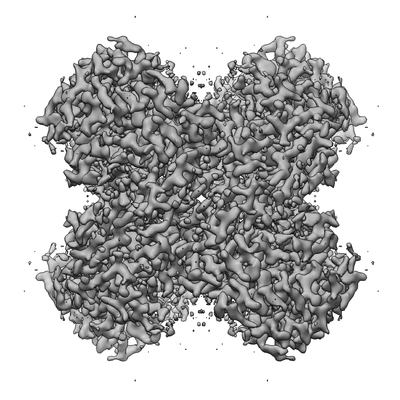
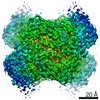
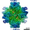
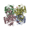
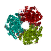
| Entry | Database: EMDB / ID: EMD-30809 | |||||||||
|---|---|---|---|---|---|---|---|---|---|---|
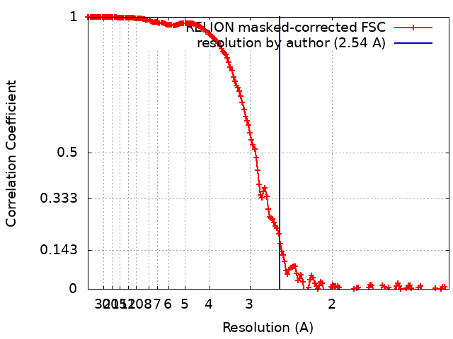
| Title | Cryo-EM structure of DfgA-B at 2.54 angstrom resolution | |||||||||
 Map data Map data | ||||||||||
 Sample Sample |
| |||||||||
| Function / homology |  Function and homology information Function and homology informationLyases; Carbon-carbon lyases; Other carbon-carbon lyases / lyase activity / metal ion binding Similarity search - Function | |||||||||
| Biological species |  Eubacterium cellulosolvens (bacteria) / [Eubacterium] cellulosolvens (bacteria) Eubacterium cellulosolvens (bacteria) / [Eubacterium] cellulosolvens (bacteria) | |||||||||
| Method | single particle reconstruction / cryo EM / Resolution: 2.54 Å | |||||||||
 Authors Authors | Mori T / Moriya T / Adachi N / Kawasaki M / Senda T / Abe I | |||||||||
| Funding support |  Japan, 1 items Japan, 1 items
| |||||||||
 Citation Citation | Journal: Nat Commun / Year: 2021 Title: C-Glycoside metabolism in the gut and in nature: Identification, characterization, structural analyses and distribution of C-C bond-cleaving enzymes. Authors: Takahiro Mori / Takuto Kumano / Haibing He / Satomi Watanabe / Miki Senda / Toshio Moriya / Naruhiko Adachi / Sanae Hori / Yuzu Terashita / Masato Kawasaki / Yoshiteru Hashimoto / Takayoshi ...Authors: Takahiro Mori / Takuto Kumano / Haibing He / Satomi Watanabe / Miki Senda / Toshio Moriya / Naruhiko Adachi / Sanae Hori / Yuzu Terashita / Masato Kawasaki / Yoshiteru Hashimoto / Takayoshi Awakawa / Toshiya Senda / Ikuro Abe / Michihiko Kobayashi / Abstract: C-Glycosides, in which a sugar moiety is linked via a carbon-carbon (C-C) bond to a non-sugar moiety (aglycone), are found in our food and medicine. The C-C bond is cleaved by intestinal microbes and ...C-Glycosides, in which a sugar moiety is linked via a carbon-carbon (C-C) bond to a non-sugar moiety (aglycone), are found in our food and medicine. The C-C bond is cleaved by intestinal microbes and the resulting aglycones exert various bioactivities. Although the enzymes responsible for the reactions have been identified, their catalytic mechanisms and the generality of the reactions in nature remain to be explored. Here, we present the identification and structural basis for the activation of xenobiotic C-glycosides by heterocomplex C-deglycosylation enzymes from intestinal and soil bacteria. They are found to be metal-dependent enzymes exhibiting broad substrate specificity toward C-glycosides. X-ray crystallographic and cryo-electron microscopic analyses, as well as structure-based mutagenesis, reveal the structural details of these enzymes and the detailed catalytic mechanisms of their remarkable C-C bond cleavage reactions. Furthermore, bioinformatic and biochemical analyses suggest that the C-deglycosylation enzymes are widely distributed in the gut, soil, and marine bacteria. | |||||||||
| History |
|
- Structure visualization
| Movie |
Movie viewer |
|---|---|
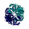
| Structure viewer | EM map: SurfViewMolmilJmol/JSmol |
| Supplemental images |
- Downloads & links
-EMDB archive
| Map data | emd_30809.map.gz | 392.1 MB | EMDB map data format | |
|---|---|---|---|---|
| Header (meta data) | emd-30809-v30.xmlemd-30809.xml | 19.4 KB 19.4 KB | Display Display | EMDB header |
| FSC (resolution estimation) | emd_30809_fsc.xml | 17 KB | Display | FSC data file |
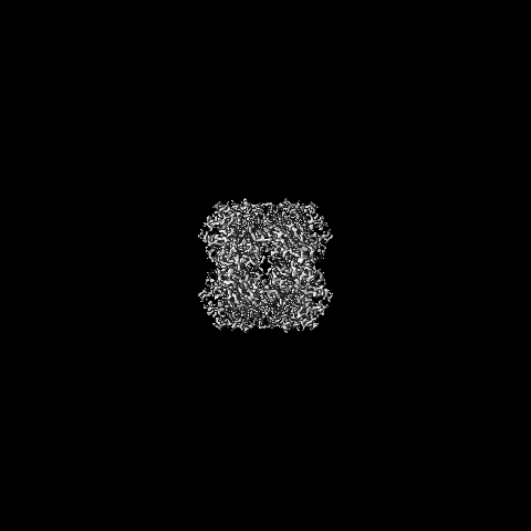
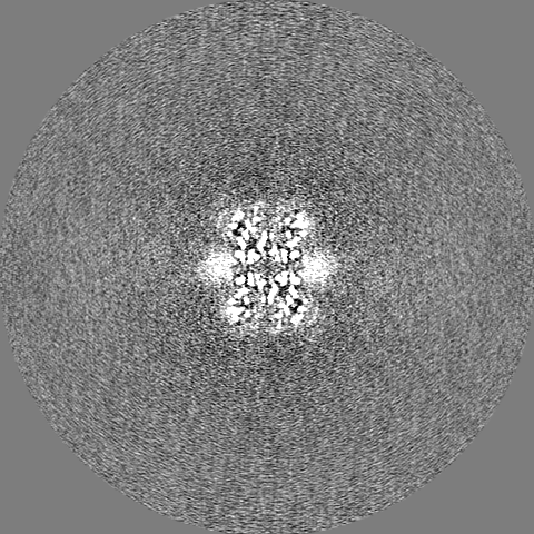
| Images |  emd_30809.png emd_30809.png | 81.7 KB | ||
| Masks | emd_30809_msk_1.map | 421.9 MB | Mask map | |
| Others | emd_30809_half_map_1.map.gzemd_30809_half_map_2.map.gz | 337.4 MB 338.2 MB | ||
| Archive directory |  http://ftp.pdbj.org/pub/emdb/structures/EMD-30809ftp://ftp.pdbj.org/pub/emdb/structures/EMD-30809 http://ftp.pdbj.org/pub/emdb/structures/EMD-30809ftp://ftp.pdbj.org/pub/emdb/structures/EMD-30809 | HTTPS FTP |
-Related structure data
| Related structure data |  7dreMC  7bvrC 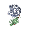 7bvsC 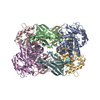 7drdC 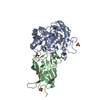 7exbC  7exzC C: citing same article ( M: atomic model generated by this map |
|---|---|
| Similar structure data |


-Links
| EMDB pages | EMDB (EBI/PDBe) / EMDataResource |
|---|---|
| Related items in Molecule of the Month |

-Map
| File | Download / File: emd_30809.map.gz / Format: CCP4 / Size: 421.9 MB / Type: IMAGE STORED AS FLOATING POINT NUMBER (4 BYTES) | ||||||||||||||||||||||||||||||||||||||||||||||||||||||||||||
|---|---|---|---|---|---|---|---|---|---|---|---|---|---|---|---|---|---|---|---|---|---|---|---|---|---|---|---|---|---|---|---|---|---|---|---|---|---|---|---|---|---|---|---|---|---|---|---|---|---|---|---|---|---|---|---|---|---|---|---|---|---|
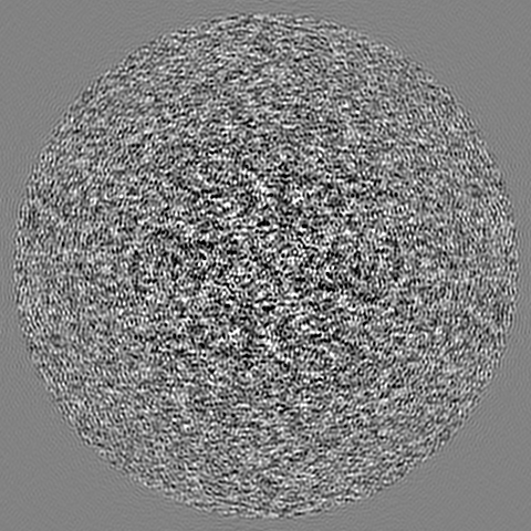
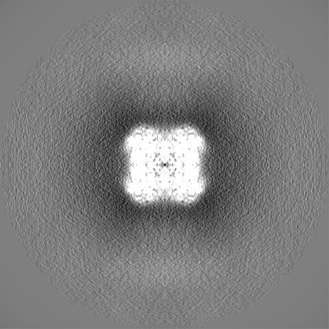
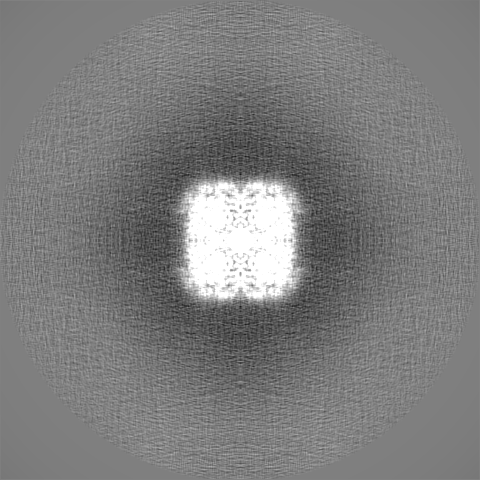
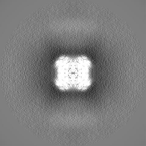
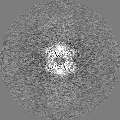
| Projections & slices | Image control
Images are generated by Spider. | ||||||||||||||||||||||||||||||||||||||||||||||||||||||||||||
| Voxel size | X=Y=Z: 0.676 Å | ||||||||||||||||||||||||||||||||||||||||||||||||||||||||||||
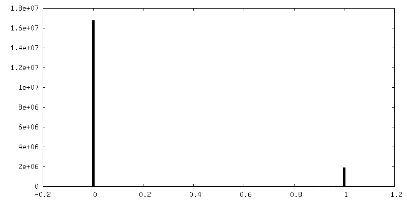
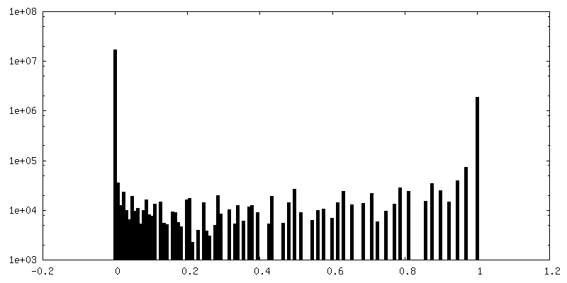
| Density |
| ||||||||||||||||||||||||||||||||||||||||||||||||||||||||||||
| Symmetry | Space group: 1 | ||||||||||||||||||||||||||||||||||||||||||||||||||||||||||||
| Details | EMDB XML:
CCP4 map header:
| ||||||||||||||||||||||||||||||||||||||||||||||||||||||||||||
 Z (Sec.)
 Y (Row.)
 X (Col.)


-Supplemental data
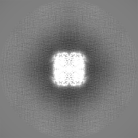
-Mask #1
| File | emd_30809_msk_1.map | ||||||||||||
|---|---|---|---|---|---|---|---|---|---|---|---|---|---|
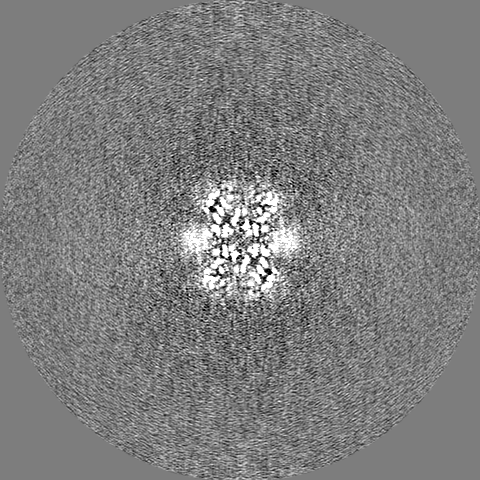
| Projections & Slices |
| ||||||||||||
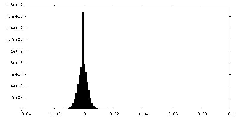
| Density Histograms |


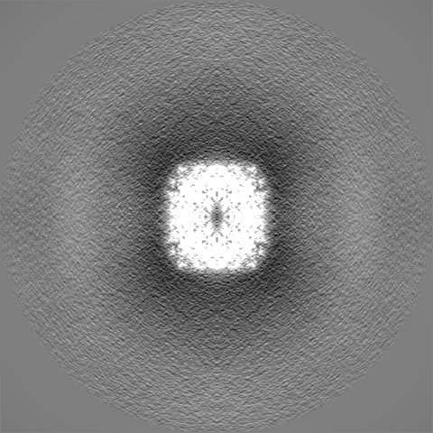
-Half map: #2
| File | emd_30809_half_map_1.map | ||||||||||||
|---|---|---|---|---|---|---|---|---|---|---|---|---|---|
| Projections & Slices |
| ||||||||||||
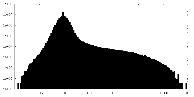
| Density Histograms |


-Half map: #1
| File | emd_30809_half_map_2.map | ||||||||||||
|---|---|---|---|---|---|---|---|---|---|---|---|---|---|
| Projections & Slices |
| ||||||||||||
| Density Histograms |

- Sample components
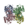
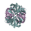
-Entire : DfgA-B
| Entire | Name: DfgA-B |
|---|---|
| Components |
|
-Supramolecule #1: DfgA-B
| Supramolecule | Name: DfgA-B / type: complex / ID: 1 / Parent: 0 / Macromolecule list: all / Details: heterodimer complex of DfgA and DfgB |
|---|---|
| Source (natural) | Organism: Eubacterium cellulosolvens (bacteria) |
| Recombinant expression | Organism: |
| Molecular weight | Theoretical: 250 KDa |
-Macromolecule #1: DfgA
| Macromolecule | Name: DfgA / type: protein_or_peptide / ID: 1 / Enantiomer: LEVO |
|---|---|
| Source (natural) | Organism: [Eubacterium] cellulosolvens (bacteria) |
| Recombinant expression | Organism: |
| Sequence | String: MYRYEKKGPK RGVALYSYSA EFGLTKTLED CFEDLHDMGA HGIEILANTH IENYPYPTDE WVEKWWRLCD KYEIVPVEYG NWIDSHVLGD RDLTTEESVE MLKRDIRLAH RLGFTVMRTK MPVINDLLEP VENWKEIIKG ALPLAEELGI KMCPEIHTPS NLKGKLVNDF ...String: MYRYEKKGPK RGVALYSYSA EFGLTKTLED CFEDLHDMGA HGIEILANTH IENYPYPTDE WVEKWWRLCD KYEIVPVEYG NWIDSHVLGD RDLTTEESVE MLKRDIRLAH RLGFTVMRTK MPVINDLLEP VENWKEIIKG ALPLAEELGI KMCPEIHTPS NLKGKLVNDF VEFIKETGTK NFGLNIDFSV FRTSFAEGEW VDPNYTPNKP EDIIPLLPYV YCCHAKFIHM SDDFKETTIP YEEVVKTMED NGYEGYLLSE YEGADKYDEG YEVGQTLRKH HILLKNLLGD |
-Macromolecule #2: DfgB
| Macromolecule | Name: DfgB / type: protein_or_peptide / ID: 2 / Enantiomer: LEVO |
|---|---|
| Source (natural) | Organism: [Eubacterium] cellulosolvens (bacteria) |
| Recombinant expression | Organism: |
| Sequence | String: MEKQVIQSVG FRNIKNGNGE ITGFQFKVKL PYYRGVFLSQ IRPGTLFVDG QKIEKDQITW TINGEEYTNQ EMRGDFKTHW ATTKPAVLKV KMPGGLAQGY HDLKYGFCFT SSYMPPIIQD GLDPDKESMV YMPEFGHHVN ERRLLIVKEE AAALEHHHHH H |
-Experimental details
-Structure determination
| Method | cryo EM |
|---|---|
 Processing Processing | single particle reconstruction |
| Aggregation state | particle |
-Sample preparation
| Concentration | 2 mg/mL | |||||||||
|---|---|---|---|---|---|---|---|---|---|---|
| Buffer | pH: 7.5 Component:
| |||||||||
| Grid | Model: Quantifoil R1.2/1.3 / Material: COPPER / Mesh: 300 / Support film - Material: CARBON / Support film - topology: HOLEY / Pretreatment - Type: GLOW DISCHARGE / Pretreatment - Atmosphere: AIR / Details: The grid was washed by acetone prior to use. | |||||||||
| Vitrification | Cryogen name: ETHANE / Chamber humidity: 100 % / Chamber temperature: 291 K / Instrument: FEI VITROBOT MARK IV / Details: Blotting time was 20 seconds (blot force 0). | |||||||||
| Details | This sample was mono-disperse. |
- Electron microscopy
| Microscope | TFS TALOS |
|---|---|
| Image recording | Film or detector model: FEI FALCON III (4k x 4k) / Detector mode: COUNTING / Number grids imaged: 1 / Number real images: 1664 / Average exposure time: 62.06 sec. / Average electron dose: 50.0 e/Å2 |
| Electron beam | Acceleration voltage: 200 kV / Electron source:  FIELD EMISSION GUN FIELD EMISSION GUN |
| Electron optics | C2 aperture diameter: 50.0 µm / Illumination mode: FLOOD BEAM / Imaging mode: BRIGHT FIELD / Cs: 2.7 mm / Nominal defocus max: 1.5 µm / Nominal defocus min: 0.6 µm / Nominal magnification: 150000 |
| Sample stage | Cooling holder cryogen: NITROGEN |
+Image processing
-Atomic model buiding 1
| Refinement | Space: REAL / Protocol: OTHER |
|---|---|
| Output model | PDB-7dre: |
