 ムービー
ムービー コントローラー


+ データを開く

- 基本情報
基本情報
| 登録情報 | データベース: EMDB / ID: EMD-2982 | |||||||||
|---|---|---|---|---|---|---|---|---|---|---|
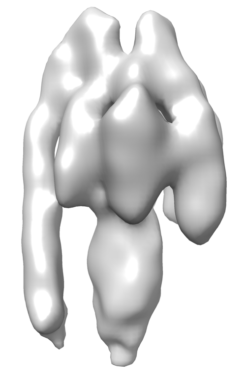
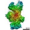
| タイトル | Sub-tomogram average of a mammalian F-type ATP synthase monomer | |||||||||
 マップデータ マップデータ | Sub-tomogram average of bovine F-type ATP synthase in a 2D crystal | |||||||||
 試料 試料 |
| |||||||||
 キーワード キーワード | F-type ATP synthase / Mitochondria Bovine heart | |||||||||
| 生物種 |  | |||||||||
| 手法 | サブトモグラム平均法 / クライオ電子顕微鏡法 / ネガティブ染色法 / 解像度: 24.0 Å | |||||||||
 データ登録者 データ登録者 | Jiko C / Davies KM / Shinzawa-Itoh K / Tani K / Maeda S / Mills DJ / Tsukihara T / Fujiyoshi Y / Kuehlbrandt W / Gerle C | |||||||||
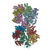
 引用 引用 | ジャーナル: Elife / 年: 2015 タイトル: Bovine F1Fo ATP synthase monomers bend the lipid bilayer in 2D membrane crystals. 著者: Chimari Jiko / Karen M Davies / Kyoko Shinzawa-Itoh / Kazutoshi Tani / Shintaro Maeda / Deryck J Mills / Tomitake Tsukihara / Yoshinori Fujiyoshi / Werner Kühlbrandt / Christoph Gerle /   要旨: We have used a combination of electron cryo-tomography, subtomogram averaging, and electron crystallographic image processing to analyse the structure of intact bovine F(1)F(o) ATP synthase in 2D ...We have used a combination of electron cryo-tomography, subtomogram averaging, and electron crystallographic image processing to analyse the structure of intact bovine F(1)F(o) ATP synthase in 2D membrane crystals. ATPase assays and mass spectrometry analysis of the 2D crystals confirmed that the enzyme complex was complete and active. The structure of the matrix-exposed region was determined at 24 Å resolution by subtomogram averaging and repositioned into the tomographic volume to reveal the crystal packing. F(1)F(o) ATP synthase complexes are inclined by 16° relative to the crystal plane, resulting in a zigzag topology of the membrane and indicating that monomeric bovine heart F(1)F(o) ATP synthase by itself is sufficient to deform lipid bilayers. This local membrane curvature is likely to be instrumental in the formation of ATP synthase dimers and dimer rows, and thus for the shaping of mitochondrial cristae. | |||||||||
| 履歴 |
|
- 構造の表示
構造の表示
| ムービー |
 ムービービューア ムービービューア |
|---|---|
| 構造ビューア | EMマップ: SurfViewMolmilJmol/JSmol |
| 添付画像 |
- ダウンロードとリンク
-EMDBアーカイブ
| マップデータ | emd_2982.map.gz | 339 KB | EMDBマップデータ形式 | |
|---|---|---|---|---|
| ヘッダ (付随情報) | emd-2982-v30.xmlemd-2982.xml | 11.3 KB 11.3 KB | 表示 表示 | EMDBヘッダ |
| 画像 |  EMD-2982.png EMD-2982.png | 90.5 KB | ||
| アーカイブディレクトリ |  http://ftp.pdbj.org/pub/emdb/structures/EMD-2982ftp://ftp.pdbj.org/pub/emdb/structures/EMD-2982 http://ftp.pdbj.org/pub/emdb/structures/EMD-2982ftp://ftp.pdbj.org/pub/emdb/structures/EMD-2982 | HTTPS FTP |
-検証レポート
| 文書・要旨 | emd_2982_validation.pdf.gz | 192.9 KB | 表示 | EMDB検証レポート |
|---|---|---|---|---|
| 文書・詳細版 | emd_2982_full_validation.pdf.gz | 192 KB | 表示 | |
| XML形式データ | emd_2982_validation.xml.gz | 4.5 KB | 表示 | |
| アーカイブディレクトリ | https://ftp.pdbj.org/pub/emdb/validation_reports/EMD-2982ftp://ftp.pdbj.org/pub/emdb/validation_reports/EMD-2982 | HTTPS FTP |
-関連構造データ
| 類似構造データ | |
|---|---|
| 電子顕微鏡画像生データ | EMPIAR-10027 (タイトル: Sub-tomogram average of a mammalian F-type ATP synthase monomer Data size: 8.9 / Data #1: 2dxtal_A [class averages] / Data #2: 2dxtal_B [class averages]) |


-リンク
| EMDBのページ | EMDB (EBI/PDBe) / EMDataResource |
|---|
-マップ
| ファイル | ダウンロード / ファイル: emd_2982.map.gz / 形式: CCP4 / 大きさ: 429.7 KB / タイプ: IMAGE STORED AS FLOATING POINT NUMBER (4 BYTES) | ||||||||||||||||||||||||||||||||||||||||||||||||||||||||||||
|---|---|---|---|---|---|---|---|---|---|---|---|---|---|---|---|---|---|---|---|---|---|---|---|---|---|---|---|---|---|---|---|---|---|---|---|---|---|---|---|---|---|---|---|---|---|---|---|---|---|---|---|---|---|---|---|---|---|---|---|---|---|
| 注釈 | Sub-tomogram average of bovine F-type ATP synthase in a 2D crystal | ||||||||||||||||||||||||||||||||||||||||||||||||||||||||||||
| ボクセルのサイズ | X=Y=Z: 3.3 Å | ||||||||||||||||||||||||||||||||||||||||||||||||||||||||||||
| 密度 |
| ||||||||||||||||||||||||||||||||||||||||||||||||||||||||||||
| 対称性 | 空間群: 1 | ||||||||||||||||||||||||||||||||||||||||||||||||||||||||||||
| 詳細 | EMDB XML:
CCP4マップ ヘッダ情報:
| ||||||||||||||||||||||||||||||||||||||||||||||||||||||||||||
-添付データ
- 試料の構成要素
-全体 : 2D crystal of bovine F-type ATP synthase monomers
| 全体 | 名称: 2D crystal of bovine F-type ATP synthase monomers |
|---|---|
| 要素 |
|
-超分子 #1000: 2D crystal of bovine F-type ATP synthase monomers
| 超分子 | 名称: 2D crystal of bovine F-type ATP synthase monomers / タイプ: sample / ID: 1000 詳細: 2D Membrane crystals lipids for reconstitution: 1,2-dimyristoyl-sn-glycero-3-phosphocholine 集合状態: 1 / Number unique components: 1 |
|---|---|
| 分子量 | 理論値: 600 KDa |
-分子 #1: F-type ATP synthase
| 分子 | 名称: F-type ATP synthase / タイプ: protein_or_peptide / ID: 1 / コピー数: 1 / 集合状態: monomer / 組換発現: No |
|---|---|
| 由来(天然) | 生物種: |
| 分子量 | 理論値: 600 KDa |
-実験情報
-構造解析
| 手法 | ネガティブ染色法, クライオ電子顕微鏡法 |
|---|---|
 解析 解析 | サブトモグラム平均法 |
| 試料の集合状態 | 2D array |
-試料調製
| 濃度 | 10 mg/mL |
|---|---|
| 緩衝液 | pH: 8.2 詳細: 40mM Tris-HCL pH8.2 100mM NaCl 0.02%(w/v)NaN3, 0.5mM ADP, 5mM MgCl2, 0.1mM DTT, 0.1mM EDTA |
| 染色 | タイプ: NEGATIVE 詳細: Plunge frozen in liquid ethane using home-made freezing device |
| グリッド | 詳細: Glow discharged R2/2, 300 copper mesh quantifoil grids |
| 凍結 | 凍結剤: ETHANE / チャンバー内温度: 100 K / 装置: HOMEMADE PLUNGER 手法: 2D crystals were mixed 1:1 (v/v) with 6nm collodial gold particles. Three microliters of protein/gold sample was applied to R2/2 300 copper mesh quantifoil grids. Access liquid was removed by ...手法: 2D crystals were mixed 1:1 (v/v) with 6nm collodial gold particles. Three microliters of protein/gold sample was applied to R2/2 300 copper mesh quantifoil grids. Access liquid was removed by blotting for 3s with Whatman #4 paper before plunge-freezing in liquid ethane |
| 詳細 | 10mg/ml ATP synthase were mixed with 1,2-dimyristoyl-sn-glycero-3-phosphocholine (Avanti Polar Lipids) using a lipid to protein ratio of 0.2. Detergent was removed by dialysis using 20 microliter Hampton dialysis buttons and 15kDa membrane cuttoff. |
- 電子顕微鏡法
| 顕微鏡 | FEI TITAN KRIOS |
|---|---|
| 温度 | 最低: 80 K / 最高: 100 K / 平均: 90 K |
| アライメント法 | Legacy - 非点収差: Objection lend astigmation was corrected on K2 at magnification used for imaging. |
| 特殊光学系 | エネルギーフィルター - 名称: quantum エネルギーフィルター - エネルギー下限: 0.0 eV エネルギーフィルター - エネルギー上限: 20.0 eV |
| 詳細 | tomographic tilt series |
| 日付 | 2013年2月1日 |
| 撮影 | カテゴリ: CCD / フィルム・検出器のモデル: GATAN K2 (4k x 4k) / 実像数: 80 / 平均電子線量: 60 e/Å2 |
| 電子線 | 加速電圧: 300 kV / 電子線源:  FIELD EMISSION GUN FIELD EMISSION GUN |
| 電子光学系 | 倍率(補正後): 14942 / 照射モード: FLOOD BEAM / 撮影モード: BRIGHT FIELD / Cs: 2.7 mm / 最大 デフォーカス(公称値): 3.0 µm / 最小 デフォーカス(公称値): 2.0 µm / 倍率(公称値): 42000 |
| 試料ステージ | 試料ホルダー: Nitrogen cooled 試料ホルダーモデル: FEI TITAN KRIOS AUTOGRID HOLDER Tilt series - Axis1 - Min angle: -60 ° / Tilt series - Axis1 - Max angle: 60 ° |
| 実験機器 |  モデル: Titan Krios / 画像提供: FEI Company |
-画像解析
| 詳細 | Subtomograms were selected manually and averaged using PEET (Particle Estimation for Electron Tomography, Boulder) |
|---|---|
| 最終 再構成 | 想定した対称性 - 点群: C1 (非対称) / 解像度のタイプ: BY AUTHOR / 解像度: 24.0 Å / 解像度の算出法: OTHER / ソフトウェア - 名称: IMOD / 使用したサブトモグラム数: 2100 |
| CTF補正 | 詳細: IMOD |
| 結晶パラメータ | 単位格子 - A: 179.1 Å / 単位格子 - B: 171.4 Å / 単位格子 - γ: 94.9 ° / 面群: P 1 |
