Movie
Movie Controller

[English] 日本語
 Yorodumi
- EMDB-2788: 3D structure of horse spleen apoferritin determined by electron c... -
+ Open data

- Basic information
| Entry | Database: EMDB / ID: EMD-2788 | |||||||||
|---|---|---|---|---|---|---|---|---|---|---|
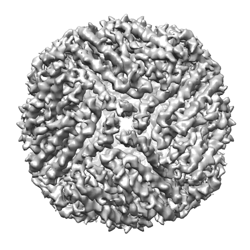
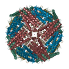
| Title | 3D structure of horse spleen apoferritin determined by electron cryomicroscopy | |||||||||
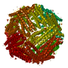
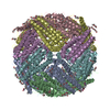
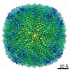
 Map data Map data | Reconstructed density map of horse spleen apoferritin | |||||||||
 Sample Sample |
| |||||||||
 Keywords Keywords | iron storage | |||||||||
| Function / homology |  Function and homology information Function and homology informationferritin complex / autolysosome / ferric iron binding / autophagosome / iron ion transport / ferrous iron binding / cytoplasmic vesicle / intracellular iron ion homeostasis / iron ion binding / cytoplasm Similarity search - Function | |||||||||
| Biological species |  | |||||||||
| Method | single particle reconstruction / cryo EM / Resolution: 4.7 Å | |||||||||
 Authors Authors | Russo CJ / Passmore LA | |||||||||
 Citation Citation | Journal: Science / Year: 2014 Title: Electron microscopy: Ultrastable gold substrates for electron cryomicroscopy. Authors: Christopher J Russo / Lori A Passmore /  Abstract: Despite recent advances, the structures of many proteins cannot be determined by electron cryomicroscopy because the individual proteins move during irradiation. This blurs the images so that they ...Despite recent advances, the structures of many proteins cannot be determined by electron cryomicroscopy because the individual proteins move during irradiation. This blurs the images so that they cannot be aligned with each other to calculate a three-dimensional density. Much of this movement stems from instabilities in the carbon substrates used to support frozen samples in the microscope. Here we demonstrate a gold specimen support that nearly eliminates substrate motion during irradiation. This increases the subnanometer image contrast such that α helices of individual proteins are resolved. With this improvement, we determine the structure of apoferritin, a smooth octahedral shell of α-helical subunits that is particularly difficult to solve by electron microscopy. This advance in substrate design will enable the solution of currently intractable protein structures. | |||||||||
| History |
|
- Structure visualization
| Movie |
Movie viewer |
|---|---|
| Structure viewer | EM map: SurfViewMolmilJmol/JSmol |
| Supplemental images |
- Downloads & links
-EMDB archive
| Map data | emd_2788.map.gz | 2.2 MB | EMDB map data format | |
|---|---|---|---|---|
| Header (meta data) | emd-2788-v30.xmlemd-2788.xml | 10 KB 10 KB | Display Display | EMDB header |
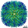
| Images |  emd_2788.png emd_2788.png | 192.6 KB | ||
| Archive directory |  http://ftp.pdbj.org/pub/emdb/structures/EMD-2788ftp://ftp.pdbj.org/pub/emdb/structures/EMD-2788 http://ftp.pdbj.org/pub/emdb/structures/EMD-2788ftp://ftp.pdbj.org/pub/emdb/structures/EMD-2788 | HTTPS FTP |
-Related structure data
| Related structure data | 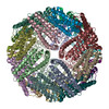 4v1wMC M: atomic model generated by this map C: citing same article ( |
|---|---|
| Similar structure data | |
| EM raw data | EMPIAR-10026 (Title: Raw data for the 3D structure of horse spleen apoferritin determined by electron cryomicroscopy Data size: 180.0 Data #1: Raw unprocessed micrograph movies [micrographs - multiframe] Data #2: Final selected particles [picked particles - multiframe - processed]) |


-Links
| EMDB pages | EMDB (EBI/PDBe) / EMDataResource |
|---|---|
| Related items in Molecule of the Month |


-Map
| File | Download / File: emd_2788.map.gz / Format: CCP4 / Size: 8.6 MB / Type: IMAGE STORED AS FLOATING POINT NUMBER (4 BYTES) | ||||||||||||||||||||||||||||||||||||||||||||||||||||||||||||||||||||
|---|---|---|---|---|---|---|---|---|---|---|---|---|---|---|---|---|---|---|---|---|---|---|---|---|---|---|---|---|---|---|---|---|---|---|---|---|---|---|---|---|---|---|---|---|---|---|---|---|---|---|---|---|---|---|---|---|---|---|---|---|---|---|---|---|---|---|---|---|---|
| Annotation | Reconstructed density map of horse spleen apoferritin | ||||||||||||||||||||||||||||||||||||||||||||||||||||||||||||||||||||
| Projections & slices | Image control
Images are generated by Spider. | ||||||||||||||||||||||||||||||||||||||||||||||||||||||||||||||||||||
| Voxel size | X=Y=Z: 1.346 Å | ||||||||||||||||||||||||||||||||||||||||||||||||||||||||||||||||||||
| Density |
| ||||||||||||||||||||||||||||||||||||||||||||||||||||||||||||||||||||
| Symmetry | Space group: 1 | ||||||||||||||||||||||||||||||||||||||||||||||||||||||||||||||||||||
| Details | EMDB XML:
CCP4 map header:
| ||||||||||||||||||||||||||||||||||||||||||||||||||||||||||||||||||||
 Z (Sec.)
 Y (Row.)
 X (Col.)


-Supplemental data
- Sample components
-Entire : Horse spleen apoferritin
| Entire | Name: Horse spleen apoferritin |
|---|---|
| Components |
|
-Supramolecule #1000: Horse spleen apoferritin
| Supramolecule | Name: Horse spleen apoferritin / type: sample / ID: 1000 Oligomeric state: 24 subunits combine to form one octahedral complex Number unique components: 1 |
|---|---|
| Molecular weight | Theoretical: 440 KDa |
-Macromolecule #1: FERRITIN LIGHT CHAIN
| Macromolecule | Name: FERRITIN LIGHT CHAIN / type: protein_or_peptide / ID: 1 / Number of copies: 24 / Oligomeric state: 24mer / Recombinant expression: No |
|---|---|
| Source (natural) | Organism: |
| Molecular weight | Theoretical: 18.5 KDa |
| Sequence | UniProtKB: Ferritin light chain |
-Experimental details
-Structure determination
| Method | cryo EM |
|---|---|
 Processing Processing | single particle reconstruction |
| Aggregation state | particle |
-Sample preparation
| Concentration | 3.5 mg/mL |
|---|---|
| Buffer | pH: 7.4 / Details: PBS |
| Grid | Details: MRC-LMB all gold grid |
| Vitrification | Cryogen name: ETHANE / Chamber humidity: 100 % / Instrument: FEI VITROBOT MARK III |
- Electron microscopy
| Microscope | FEI POLARA 300 |
|---|---|
| Date | Mar 8, 2013 |
| Image recording | Category: CCD / Film or detector model: FEI FALCON II (4k x 4k) |
| Electron beam | Acceleration voltage: 300 kV / Electron source:  FIELD EMISSION GUN FIELD EMISSION GUN |
| Electron optics | Illumination mode: FLOOD BEAM / Imaging mode: BRIGHT FIELD |
| Sample stage | Specimen holder model: OTHER |
| Experimental equipment |  Model: Tecnai Polara / Image courtesy: FEI Company |
-Image processing
| Final reconstruction | Applied symmetry - Point group: O (octahedral) / Resolution.type: BY AUTHOR / Resolution: 4.7 Å / Resolution method: OTHER / Software - Name: RELION / Number images used: 483 |
|---|
-Atomic model buiding 1
| Initial model | PDB ID: Chain - #0 - Chain ID: A / Chain - #1 - Chain ID: B / Chain - #2 - Chain ID: C / Chain - #3 - Chain ID: D / Chain - #4 - Chain ID: E / Chain - #5 - Chain ID: F / Chain - #6 - Chain ID: G / Chain - #7 - Chain ID: H / Chain - #8 - Chain ID: I / Chain - #9 - Chain ID: J / Chain - #10 - Chain ID: K / Chain - #11 - Chain ID: L / Chain - #12 - Chain ID: M / Chain - #13 - Chain ID: N / Chain - #14 - Chain ID: O / Chain - #15 - Chain ID: P / Chain - #16 - Chain ID: Q / Chain - #17 - Chain ID: R / Chain - #18 - Chain ID: S / Chain - #19 - Chain ID: T / Chain - #20 - Chain ID: U / Chain - #21 - Chain ID: V / Chain - #22 - Chain ID: W / Chain - #23 - Chain ID: X |
|---|---|
| Software | Name: RefMac |
| Refinement | Space: REAL / Protocol: FLEXIBLE FIT |
| Output model | PDB-4v1w: |