 Movie
Movie Controller

[English] 日本語
 Yorodumi
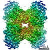
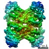
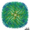
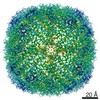
- EMDB-24797: Mouse heavy chain apoferritin determined in presence of 20% glyce... -
+ Open data

- Basic information
| Entry | Database: EMDB / ID: EMD-24797 | |||||||||
|---|---|---|---|---|---|---|---|---|---|---|
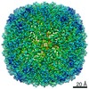
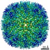
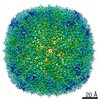
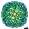
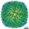
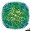
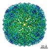
| Title | Mouse heavy chain apoferritin determined in presence of 20% glycerol (300keV) | |||||||||
 Map data Map data | Mouse heavy chain apoferritin in buffer with 20% glycerol. | |||||||||
 Sample Sample |
| |||||||||
| Function / homology |  Function and homology information Function and homology informationautolysosome / ferric iron binding / iron ion transport / cytoplasmic vesicle / intracellular iron ion homeostasis Similarity search - Function | |||||||||
| Biological species |  | |||||||||
| Method | single particle reconstruction / cryo EM / Resolution: 2.1 Å | |||||||||
 Authors Authors | Hirschi M / Basanta B / Lander GC | |||||||||
| Funding support |  United States, 2 items United States, 2 items
| |||||||||
 Citation Citation | Journal: Acta Crystallogr D Struct Biol / Year: 2022 Title: A case for glycerol as an acceptable additive for single-particle cryoEM samples. Authors: Benjamin Basanta / Marscha M Hirschi / Danielle A Grotjahn / Gabriel C Lander / Abstract: Buffer-composition and sample-preparation guidelines for cryo-electron microscopy are geared towards maximizing imaging contrast and reducing electron-beam-induced motion. These pursuits often ...Buffer-composition and sample-preparation guidelines for cryo-electron microscopy are geared towards maximizing imaging contrast and reducing electron-beam-induced motion. These pursuits often involve the minimization or the complete removal of additives that are commonly used to facilitate proper protein folding and minimize aggregation. Among these admonished additives is glycerol, a widely used osmolyte that aids protein stability. In this work, it is shown that the inclusion of glycerol does not preclude high-resolution structure determination by cryoEM, as demonstrated by an ∼2.3 Å resolution reconstruction of mouse apoferritin (∼500 kDa) and an ∼3.3 Å resolution reconstruction of rabbit muscle aldolase (∼160 kDa) in the presence of 20%(v/v) glycerol. While it was found that generating thin ice that is amenable to high-resolution imaging requires long blot times, the addition of glycerol did not result in increased beam-induced motion or an inability to pick particles. Overall, these findings indicate that glycerol should not be discounted as a cryoEM sample-buffer additive, particularly for large, fragile complexes that are prone to disassembly or aggregation upon its removal. | |||||||||
| History |
|
- Structure visualization
| Movie |
Movie viewer |
|---|---|
| Structure viewer | EM map: SurfViewMolmilJmol/JSmol |
| Supplemental images |

- Downloads & links
-EMDB archive
| Map data | emd_24797.map.gz | 9.6 MB | EMDB map data format | |
|---|---|---|---|---|
| Header (meta data) | emd-24797-v30.xmlemd-24797.xml | 18.8 KB 18.8 KB | Display Display | EMDB header |
| FSC (resolution estimation) | emd_24797_fsc.xml | 6.8 KB | Display | FSC data file |
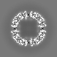
| Images |  emd_24797.png emd_24797.png | 123.9 KB | ||
| Masks | emd_24797_msk_1.map | 27 MB | Mask map | |
| Others | emd_24797_half_map_1.map.gzemd_24797_half_map_2.map.gz | 17.6 MB 17.6 MB | ||
| Archive directory |  http://ftp.pdbj.org/pub/emdb/structures/EMD-24797ftp://ftp.pdbj.org/pub/emdb/structures/EMD-24797 http://ftp.pdbj.org/pub/emdb/structures/EMD-24797ftp://ftp.pdbj.org/pub/emdb/structures/EMD-24797 | HTTPS FTP |
-Related structure data
| Related structure data | C: citing same article ( |
|---|---|
| Similar structure data | |
| EM raw data | EMPIAR-10861 (Title: Apoferritin 20% glycerol, 300kV / Data size: 477.7 Data #1: Unaligned multiframe micrographs from mouse apoferritin in 20% glcyerol, imaged at 300kev [micrographs - multiframe]) |


-Links
| EMDB pages | EMDB (EBI/PDBe) / EMDataResource |
|---|---|
| Related items in Molecule of the Month |


-Map
| File | Download / File: emd_24797.map.gz / Format: CCP4 / Size: 27 MB / Type: IMAGE STORED AS FLOATING POINT NUMBER (4 BYTES) | ||||||||||||||||||||||||||||||||||||||||||||||||||||||||||||||||||||
|---|---|---|---|---|---|---|---|---|---|---|---|---|---|---|---|---|---|---|---|---|---|---|---|---|---|---|---|---|---|---|---|---|---|---|---|---|---|---|---|---|---|---|---|---|---|---|---|---|---|---|---|---|---|---|---|---|---|---|---|---|---|---|---|---|---|---|---|---|---|
| Annotation | Mouse heavy chain apoferritin in buffer with 20% glycerol. | ||||||||||||||||||||||||||||||||||||||||||||||||||||||||||||||||||||
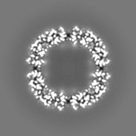
| Projections & slices | Image control
Images are generated by Spider. | ||||||||||||||||||||||||||||||||||||||||||||||||||||||||||||||||||||
| Voxel size | X=Y=Z: 1.03 Å | ||||||||||||||||||||||||||||||||||||||||||||||||||||||||||||||||||||
| Density |
| ||||||||||||||||||||||||||||||||||||||||||||||||||||||||||||||||||||
| Symmetry | Space group: 1 | ||||||||||||||||||||||||||||||||||||||||||||||||||||||||||||||||||||
| Details | EMDB XML:
CCP4 map header:
| ||||||||||||||||||||||||||||||||||||||||||||||||||||||||||||||||||||
 Z (Sec.)
 Y (Row.)
 X (Col.)


-Supplemental data
-Mask #1
| File | emd_24797_msk_1.map | ||||||||||||
|---|---|---|---|---|---|---|---|---|---|---|---|---|---|
| Projections & Slices |
| ||||||||||||
| Density Histograms |


-Half map: Mouse heavy chain apoferritin in buffer with 20%...
| File | emd_24797_half_map_1.map | ||||||||||||
|---|---|---|---|---|---|---|---|---|---|---|---|---|---|
| Annotation | Mouse heavy chain apoferritin in buffer with 20% glycerol. Half map 2. | ||||||||||||
| Projections & Slices |
| ||||||||||||
| Density Histograms |


-Half map: Mouse heavy chain apoferritin in buffer with 20%...
| File | emd_24797_half_map_2.map | ||||||||||||
|---|---|---|---|---|---|---|---|---|---|---|---|---|---|
| Annotation | Mouse heavy chain apoferritin in buffer with 20% glycerol. Half map 1. | ||||||||||||
| Projections & Slices |
| ||||||||||||
| Density Histograms |


- Sample components
-Entire : Apoferritin
| Entire | Name: Apoferritin |
|---|---|
| Components |
|
-Supramolecule #1: Apoferritin
| Supramolecule | Name: Apoferritin / type: complex / ID: 1 / Parent: 0 / Macromolecule list: all |
|---|---|
| Source (natural) | Organism: |
| Recombinant expression | Organism:  |
| Molecular weight | Theoretical: 487.32 KDa |
-Macromolecule #1: Apoferritin
| Macromolecule | Name: Apoferritin / type: protein_or_peptide / ID: 1 / Enantiomer: LEVO |
|---|---|
| Source (natural) | Organism: |
| Recombinant expression | Organism: |
| Sequence | String: SPSQVRQNYH QDAEAAINRQ INLELYASYV YLSMSCYFDR DDVALKNFAK YFLHQSHEER EHAEKLMKLQ NQRGGRIFLQ DIKKPDRDDW ESGLNAMECA LHLEKSVNQS LLELHKLATD KNDPHLCDFI ETYYLSEQVK SIKELGDHVT NLRKMGAPEA GMAEYLFDKH TLGH |
-Experimental details
-Structure determination
| Method | cryo EM |
|---|---|
 Processing Processing | single particle reconstruction |
| Aggregation state | particle |
-Sample preparation
| Concentration | 5 mg/mL | |||||||||||||||
|---|---|---|---|---|---|---|---|---|---|---|---|---|---|---|---|---|
| Buffer | pH: 7.5 Component:
| |||||||||||||||
| Grid | Model: UltrAuFoil R1.2/1.3 / Material: GOLD / Mesh: 300 / Support film - Material: GOLD / Support film - topology: HOLEY ARRAY / Support film - Film thickness: 50.0 nm / Pretreatment - Type: PLASMA CLEANING / Pretreatment - Atmosphere: OTHER / Pretreatment - Pressure: 9.33257 kPa | |||||||||||||||
| Vitrification | Cryogen name: ETHANE / Chamber humidity: 95 % / Chamber temperature: 277 K / Instrument: HOMEMADE PLUNGER |
- Electron microscopy
| Microscope | FEI TITAN KRIOS |
|---|---|
| Temperature | Min: 77.0 K / Max: 77.0 K |
| Image recording | Film or detector model: GATAN K2 SUMMIT (4k x 4k) / Detector mode: COUNTING / Digitization - Dimensions - Width: 3710 pixel / Digitization - Dimensions - Height: 3838 pixel / Digitization - Frames/image: 1-65 / Number grids imaged: 1 / Number real images: 1859 / Average exposure time: 6.5 sec. / Average electron dose: 55.25 e/Å2 Details: Images were collected using stage position navigation to the center of 16 holes and then beam shift was used to target exposures. |
| Electron beam | Acceleration voltage: 300 kV / Electron source:  FIELD EMISSION GUN FIELD EMISSION GUN |
| Electron optics | C2 aperture diameter: 70.0 µm / Calibrated defocus max: 1.5 µm / Calibrated defocus min: 0.5 µm / Calibrated magnification: 48732 / Illumination mode: FLOOD BEAM / Imaging mode: BRIGHT FIELD / Cs: 2.7 mm / Nominal defocus max: 1.5 µm / Nominal defocus min: 0.5 µm / Nominal magnification: 29000 |
| Sample stage | Specimen holder model: FEI TITAN KRIOS AUTOGRID HOLDER / Cooling holder cryogen: NITROGEN |
| Experimental equipment |  Model: Titan Krios / Image courtesy: FEI Company |

