Movie
Movie Controller

[English] 日本語
 Yorodumi
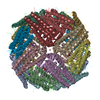
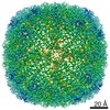
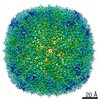
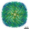
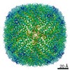
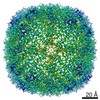
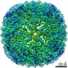
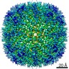
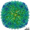
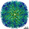
- EMDB-12358: Mouse heavy chain apoferritin collected on cryoARM300 with coma-c... -
+ Open data

- Basic information
| Entry | Database: EMDB / ID: EMD-12358 | |||||||||
|---|---|---|---|---|---|---|---|---|---|---|
| Title | Mouse heavy chain apoferritin collected on cryoARM300 with coma-corrected beam-image shift | |||||||||
 Map data Map data | ||||||||||
 Sample Sample |
| |||||||||
| Function / homology |  Function and homology information Function and homology informationIron uptake and transport / Golgi Associated Vesicle Biogenesis / ferroxidase / autolysosome / negative regulation of ferroptosis / ferroxidase activity / negative regulation of fibroblast proliferation / endocytic vesicle lumen / Neutrophil degranulation / ferric iron binding ...Iron uptake and transport / Golgi Associated Vesicle Biogenesis / ferroxidase / autolysosome / negative regulation of ferroptosis / ferroxidase activity / negative regulation of fibroblast proliferation / endocytic vesicle lumen / Neutrophil degranulation / ferric iron binding / autophagosome / iron ion transport / ferrous iron binding / intracellular iron ion homeostasis / immune response / iron ion binding / negative regulation of cell population proliferation / mitochondrion / extracellular region / membrane / identical protein binding / cytosol / cytoplasm Similarity search - Function | |||||||||
| Biological species |  | |||||||||
| Method | single particle reconstruction / cryo EM / Resolution: 1.7 Å | |||||||||
 Authors Authors | Efremov R / Stroobants A | |||||||||
| Funding support |  Belgium, 1 items Belgium, 1 items
| |||||||||
 Citation Citation | Journal: Acta Crystallogr D Struct Biol / Year: 2021 Title: Coma-corrected rapid single-particle cryo-EM data collection on the CRYO ARM 300. Authors: Rouslan G Efremov / Annelore Stroobants / Abstract: Single-particle cryogenic electron microscopy has recently become a major method for determining the structures of proteins and protein complexes. This has markedly increased the demand for ...Single-particle cryogenic electron microscopy has recently become a major method for determining the structures of proteins and protein complexes. This has markedly increased the demand for throughput of high-resolution electron microscopes, which are required to produce high-resolution images at high rates. An increase in data-collection throughput can be achieved by using large beam-image shifts combined with off-axis coma correction, enabling the acquisition of multiple images from a large area of the EM grid without moving the microscope stage. Here, the optical properties of the JEOL CRYO ARM 300 electron microscope equipped with a K3 camera were characterized under off-axis illumination conditions. It is shown that efficient coma correction can be achieved for beam-image shifts with an amplitude of at least 10 µm, enabling a routine throughput for data collection of between 6000 and 9000 images per day. Use of the benchmark for the rapid data-collection procedure (with beam-image shifts of up to 7 µm) on apoferritin resulted in a reconstruction at a resolution of 1.7 Å. This demonstrates that the rapid automated acquisition of high-resolution micrographs is possible using a CRYO ARM 300. | |||||||||
| History |
|
- Structure visualization
| Movie |
Movie viewer |
|---|---|
| Structure viewer | EM map: SurfViewMolmilJmol/JSmol |
| Supplemental images |

- Downloads & links
-EMDB archive
| Map data | emd_12358.map.gz | 15.4 MB | EMDB map data format | |
|---|---|---|---|---|
| Header (meta data) | emd-12358-v30.xmlemd-12358.xml | 16.8 KB 16.8 KB | Display Display | EMDB header |
| FSC (resolution estimation) | emd_12358_fsc.xml | 9.9 KB | Display | FSC data file |
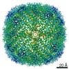
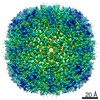
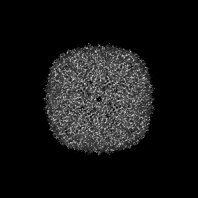
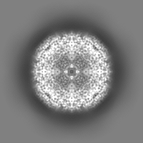
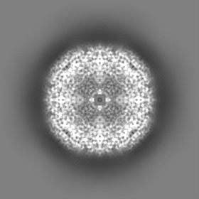
| Images | 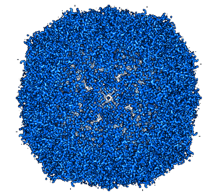 emd_12358.png emd_12358.png | 314.6 KB | ||
| Masks | emd_12358_msk_1.map | 83.7 MB | Mask map | |
| Others | emd_12358_half_map_1.map.gzemd_12358_half_map_2.map.gz | 61.1 MB 61.1 MB | ||
| Archive directory |  http://ftp.pdbj.org/pub/emdb/structures/EMD-12358ftp://ftp.pdbj.org/pub/emdb/structures/EMD-12358 http://ftp.pdbj.org/pub/emdb/structures/EMD-12358ftp://ftp.pdbj.org/pub/emdb/structures/EMD-12358 | HTTPS FTP |
-Related structure data
| Similar structure data | |
|---|---|
| EM raw data | EMPIAR-10639 (Title: Single particle cryo-EM dataset of mouse heavy chain apoferritin collected on cryoARM300 with beam-image shift of 7 um Data size: 695.6 Data #1: Unaligned multi frame micrographs of mouse heavy chain apoferritin collected on cryoARM300 with image shift 7um [micrographs - multiframe]) |


-Links
| EMDB pages | EMDB (EBI/PDBe) / EMDataResource |
|---|---|
| Related items in Molecule of the Month |


-Map
| File | Download / File: emd_12358.map.gz / Format: CCP4 / Size: 83.7 MB / Type: IMAGE STORED AS FLOATING POINT NUMBER (4 BYTES) | ||||||||||||||||||||||||||||||||||||||||||||||||||||||||||||
|---|---|---|---|---|---|---|---|---|---|---|---|---|---|---|---|---|---|---|---|---|---|---|---|---|---|---|---|---|---|---|---|---|---|---|---|---|---|---|---|---|---|---|---|---|---|---|---|---|---|---|---|---|---|---|---|---|---|---|---|---|---|
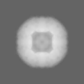
| Projections & slices | Image control
Images are generated by Spider. | ||||||||||||||||||||||||||||||||||||||||||||||||||||||||||||
| Voxel size | X=Y=Z: 0.753 Å | ||||||||||||||||||||||||||||||||||||||||||||||||||||||||||||
| Density |
| ||||||||||||||||||||||||||||||||||||||||||||||||||||||||||||
| Symmetry | Space group: 1 | ||||||||||||||||||||||||||||||||||||||||||||||||||||||||||||
| Details | EMDB XML:
CCP4 map header:
| ||||||||||||||||||||||||||||||||||||||||||||||||||||||||||||
 Z (Sec.)
 Y (Row.)
 X (Col.)


-Supplemental data
-Mask #1
| File | emd_12358_msk_1.map | ||||||||||||
|---|---|---|---|---|---|---|---|---|---|---|---|---|---|
| Projections & Slices |
| ||||||||||||
| Density Histograms |


-Half map: #1
| File | emd_12358_half_map_1.map | ||||||||||||
|---|---|---|---|---|---|---|---|---|---|---|---|---|---|
| Projections & Slices |
| ||||||||||||
| Density Histograms |


-Half map: #2
| File | emd_12358_half_map_2.map | ||||||||||||
|---|---|---|---|---|---|---|---|---|---|---|---|---|---|
| Projections & Slices |
| ||||||||||||
| Density Histograms |


- Sample components
-Entire : mouse heavy chain apoferritin
| Entire | Name: mouse heavy chain apoferritin |
|---|---|
| Components |
|
-Supramolecule #1: mouse heavy chain apoferritin
| Supramolecule | Name: mouse heavy chain apoferritin / type: complex / ID: 1 / Parent: 0 / Macromolecule list: all / Details: Wilde type, octamer |
|---|---|
| Source (natural) | Organism: |
| Recombinant expression | Organism:  |
| Molecular weight | Theoretical: 506 KDa |
-Macromolecule #1: mouse heavy chain apoferritin
| Macromolecule | Name: mouse heavy chain apoferritin / type: protein_or_peptide / ID: 1 / Enantiomer: LEVO / EC number: ferroxidase |
|---|---|
| Source (natural) | Organism: |
| Recombinant expression | Organism: |
| Sequence | String: MTTASPSQVR QNYHQDAEAA INRQINLELY ASYVYLSMSC YFDRDDVALK NFAKYFLHQS HEEREHAEK LMKLQNQRGG RIFLQDIKKP DRDDWESGLN AMECALHLEK SVNQSLLELH K LATDKNDP HLCDFIETYY LSEQVKSIKE LGDHVTNLRK MGAPEAGMAE YLFDKHTLGH GD ES |
-Experimental details
-Structure determination
| Method | cryo EM |
|---|---|
 Processing Processing | single particle reconstruction |
| Aggregation state | particle |
-Sample preparation
| Concentration | 3.6 mg/mL |
|---|---|
| Buffer | pH: 7.5 / Details: 20 mM Hepes pH 7.5, 300 mM NaCl, 1mM TCEP |
| Grid | Model: Quantifoil / Material: COPPER / Mesh: 200 / Support film - Material: CARBON / Support film - topology: HOLEY / Pretreatment - Type: GLOW DISCHARGE |
| Vitrification | Cryogen name: ETHANE / Chamber humidity: 98 % / Chamber temperature: 298 K / Instrument: GATAN CRYOPLUNGE 3 / Details: 5 seconds blotting. |
- Electron microscopy
| Microscope | JEOL CRYO ARM 300 |
|---|---|
| Alignment procedure | Coma free - Residual tilt: 0.9 mrad |
| Specialist optics | Energy filter - Name: In-column Omega Filter / Energy filter - Slit width: 20 eV |
| Image recording | Film or detector model: GATAN K3 (6k x 4k) / Detector mode: COUNTING / Number grids imaged: 1 / Number real images: 2639 / Average exposure time: 3.37 sec. / Average electron dose: 59.0 e/Å2 |
| Electron beam | Acceleration voltage: 300 kV / Electron source:  FIELD EMISSION GUN FIELD EMISSION GUN |
| Electron optics | Illumination mode: FLOOD BEAM / Imaging mode: BRIGHT FIELD / Cs: 2.55 mm |
| Sample stage | Specimen holder model: JEOL CRYOSPECPORTER / Cooling holder cryogen: NITROGEN |
+Image processing

-Atomic model buiding 1
| Initial model | PDB ID: |
|---|---|
| Refinement | Space: REAL / Protocol: OTHER / Target criteria: correlation coefficient |