 Movie
Movie Controller

[English] 日本語
 Yorodumi
- EMDB-1200: Automated cryoEM data acquisition and analysis of 284742 particle... -
+ Open data

- Basic information
| Entry | Database: EMDB / ID: EMD-1200 | |||||||||
|---|---|---|---|---|---|---|---|---|---|---|
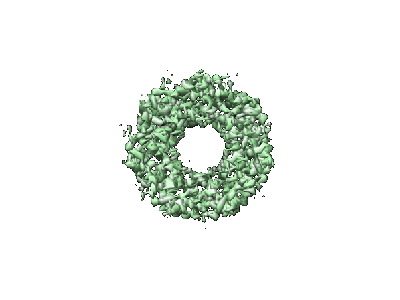
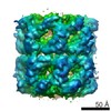
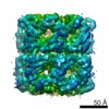
| Title | Automated cryoEM data acquisition and analysis of 284742 particles of GroEL. | |||||||||
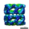
 Map data Map data | This is a three-dimensional volume of GroEL reconstructed to 7.8 Angstroms resolution | |||||||||
 Sample Sample |
| |||||||||
| Biological species |  | |||||||||
| Method | single particle reconstruction / cryo EM / Resolution: 7.8 Å | |||||||||
 Authors Authors | Stagg SM / Pulokas J / Fellmann D / Cheng A / Quispe JD / Mallick SP / Avila RM / Carragher B / Potter CS | |||||||||
 Citation Citation | Journal: J Struct Biol / Year: 2006 Title: Automated cryoEM data acquisition and analysis of 284742 particles of GroEL. Authors: Scott M Stagg / Gabriel C Lander / James Pulokas / Denis Fellmann / Anchi Cheng / Joel D Quispe / Satya P Mallick / Radomir M Avila / Bridget Carragher / Clinton S Potter /  Abstract: One of the goals in developing our automated electron microscopy data acquisition system, Leginon, was to improve both the ease of use and the throughput of the process of acquiring low dose images ...One of the goals in developing our automated electron microscopy data acquisition system, Leginon, was to improve both the ease of use and the throughput of the process of acquiring low dose images of macromolecular specimens embedded in vitreous ice. In this article, we demonstrate the potential of the Leginon system for high-throughput data acquisition by describing an experiment in which we acquired images of more than 280,000 particles of GroEL in a single 25 h session at the microscope. We also demonstrate the potential for an automated pipeline for molecular microscopy by showing that these particles can be subjected to completely automated procedures to reconstruct a three-dimensional (3D) density map to a resolution better than 8 A. In generating the 3D maps, we used a variety of metadata associated with the data acquisition and processing steps to sort and select the particles. These metadata provide a number of insights into factors that affect the quality of the acquired images and the resulting reconstructions. In particular, we show that the resolution of the reconstructed 3D density maps improves with decreasing ice thickness. These data provide a basis for assessing the capabilities of high-throughput macromolecular microscopy. | |||||||||
| History |
|
- Structure visualization
| Movie |
 Movie viewer Movie viewer |
|---|---|
| Structure viewer | EM map: SurfViewMolmilJmol/JSmol |
| Supplemental images |
- Downloads & links
-EMDB archive
| Map data | emd_1200.map.gz | 4.9 MB | EMDB map data format | |
|---|---|---|---|---|
| Header (meta data) | emd-1200-v30.xmlemd-1200.xml | 10.3 KB 10.3 KB | Display Display | EMDB header |
| Images |  1200.gif 1200.gif | 18.6 KB | ||
| Archive directory |  http://ftp.pdbj.org/pub/emdb/structures/EMD-1200ftp://ftp.pdbj.org/pub/emdb/structures/EMD-1200 http://ftp.pdbj.org/pub/emdb/structures/EMD-1200ftp://ftp.pdbj.org/pub/emdb/structures/EMD-1200 | HTTPS FTP |
-Related structure data
| Similar structure data | |
|---|---|
| EM raw data | EMPIAR-10018 (Title: GroEL dataset - NRAMM (06jul12a) / Data size: 7.0 / Data #1: Groel micrographs [micrographs - single frame]) |


-Links
| EMDB pages | EMDB (EBI/PDBe) / EMDataResource |
|---|
-Map
| File | Download / File: emd_1200.map.gz / Format: CCP4 / Size: 5.2 MB / Type: IMAGE STORED AS FLOATING POINT NUMBER (4 BYTES) | ||||||||||||||||||||||||||||||||||||||||||||||||||||||||||||||||||||
|---|---|---|---|---|---|---|---|---|---|---|---|---|---|---|---|---|---|---|---|---|---|---|---|---|---|---|---|---|---|---|---|---|---|---|---|---|---|---|---|---|---|---|---|---|---|---|---|---|---|---|---|---|---|---|---|---|---|---|---|---|---|---|---|---|---|---|---|---|---|
| Annotation | This is a three-dimensional volume of GroEL reconstructed to 7.8 Angstroms resolution | ||||||||||||||||||||||||||||||||||||||||||||||||||||||||||||||||||||
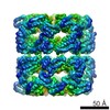
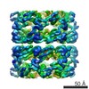
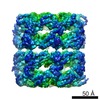
| Projections & slices | Image control
Images are generated by Spider. | ||||||||||||||||||||||||||||||||||||||||||||||||||||||||||||||||||||
| Voxel size | X=Y=Z: 2.263 Å | ||||||||||||||||||||||||||||||||||||||||||||||||||||||||||||||||||||
| Density |
| ||||||||||||||||||||||||||||||||||||||||||||||||||||||||||||||||||||
| Symmetry | Space group: 1 | ||||||||||||||||||||||||||||||||||||||||||||||||||||||||||||||||||||
| Details | EMDB XML:
CCP4 map header:
| ||||||||||||||||||||||||||||||||||||||||||||||||||||||||||||||||||||
 Z (Sec.)
 Y (Row.)
 X (Col.)


-Supplemental data
- Sample components
-Entire : E.coli GroEL
| Entire | Name: E.coli GroEL |
|---|---|
| Components |
|
-Supramolecule #1000: E.coli GroEL
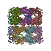
| Supramolecule | Name: E.coli GroEL / type: sample / ID: 1000 / Oligomeric state: homotetradecamer / Number unique components: 1 |
|---|---|
| Molecular weight | Experimental: 800 KDa / Theoretical: 800 KDa / Method: Calculated molecular weight from sequence |
-Macromolecule #1: GroEL
| Macromolecule | Name: GroEL / type: protein_or_peptide / ID: 1 / Number of copies: 14 / Oligomeric state: homotetradecamer / Recombinant expression: Yes |
|---|---|
| Source (natural) | Organism: |
| Molecular weight | Experimental: 800 KDa / Theoretical: 800 KDa |
| Recombinant expression | Organism: |
-Experimental details
-Structure determination
| Method | cryo EM |
|---|---|
 Processing Processing | single particle reconstruction |
| Aggregation state | particle |
-Sample preparation
| Concentration | 3.2 mg/mL |
|---|---|
| Buffer | pH: 7.5 / Details: 100mM Hepes, 10mM Mg(OAc)2, 10mM KOAc, 2mM DTT |
| Grid | Details: Protochips C-flat grid: holey carbon with 2um holes and 2um spacing 400 mesh copper grid |
| Vitrification | Cryogen name: ETHANE / Chamber humidity: 100 % / Chamber temperature: 93 K / Instrument: OTHER Details: Vitrification instrument: FEI Vitrobot. Grid plasma cleaned for 20s with Fischione 1020 plasma cleaner using 75% Argon 25% Oxygen mix. Method: Temperature of chamber was 4 degrees C. 0 seconds drain time. Single blot. 0 mm offset. 4 ul sample applied to grid. Blot for 3.5 seconds before plunging. |
- Electron microscopy
| Microscope | FEI TECNAI F20 |
|---|---|
| Temperature | Average: 94 K |
| Alignment procedure | Legacy - Astigmatism: objective lens astigmatism was corrected at 50,000X magnification |
| Date | May 19, 2005 |
| Image recording | Category: CCD / Film or detector model: GATAN ULTRASCAN 4000 (4k x 4k) / Number real images: 552 / Average electron dose: 11.5 e/Å2 / Bits/pixel: 16 |
| Tilt angle min | 0 |
| Tilt angle max | 0 |
| Electron beam | Acceleration voltage: 120 kV / Electron source:  FIELD EMISSION GUN FIELD EMISSION GUN |
| Electron optics | Illumination mode: FLOOD BEAM / Imaging mode: BRIGHT FIELD / Cs: 2 mm / Nominal defocus max: 2.28 µm / Nominal defocus min: 0.5 µm / Nominal magnification: 50000 |
| Sample stage | Specimen holder: Gatan 626 side entry cryo-stage / Specimen holder model: GATAN LIQUID NITROGEN |
| Experimental equipment |  Model: Tecnai F20 / Image courtesy: FEI Company |
-Image processing
| Details | The images were acquired using the Leginon automated data aquisition system. The particles were automatically selected using the Selexon package. The CTF was automatically estimated using the ACE package |
|---|---|
| CTF correction | Details: Phase correction for each particle. Amplitude correction for the final volume |
| Final reconstruction | Applied symmetry - Point group: D7 (2x7 fold dihedral) / Algorithm: OTHER / Resolution.type: BY AUTHOR / Resolution: 7.8 Å / Resolution method: FSC 0.5 CUT-OFF / Software - Name: EMAN / Number images used: 158390 |
| Final two d classification | Number classes: 333 |
