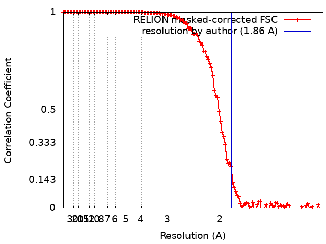
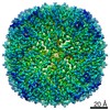
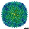
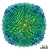
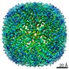
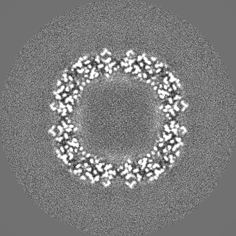
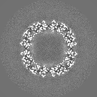
Journal: IUCrJ / Year: 2020 Title: Assessing the JEOL CRYO ARM 300 for high-throughput automated single-particle cryo-EM in a multiuser environment. Authors: Marcus Fislage / Alexander V Shkumatov / Annelore Stroobants / Rouslan G Efremov / Abstract: Single-particle cryo-EM has become an indispensable structural biology method. It requires regular access to high-resolution electron cryogenic microscopes. To fully utilize the capacity of the ...Single-particle cryo-EM has become an indispensable structural biology method. It requires regular access to high-resolution electron cryogenic microscopes. To fully utilize the capacity of the expensive high-resolution instruments, the time used for data acquisition and the rate of data collection have to be maximized. This in turn requires high stability and high uptime of the instrument. One of the first 300 kV JEOL CRYO ARM 300 microscopes has been installed at the cryo-EM facility BECM at VIB-VUB, Brussels, where the microscope is used for continuous data collection on multiple projects. Here, the suitability and performance of the microscope is assessed for high-throughput single-particle data collection. In particular, the properties of the illumination system, the stage stability and ice contamination rates are reported. The microscope was benchmarked using mouse heavy-chain apoferritin which was reconstructed to a resolution of 1.9 Å. Finally, uptime and throughput statistics of the instrument accumulated during the first six months of the facility operation in user access mode are reported.
History
Deposition
Feb 17, 2020
-
Header (metadata) release
Mar 4, 2020
-
Map release
Jun 24, 2020
-
Update
Aug 5, 2020
-
Current status
Aug 5, 2020
Processing site: PDBe / Status: Released
-
Structure visualization
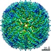
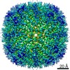
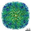
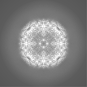
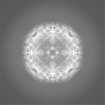
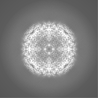
Movie
Surface view with section colored by density value
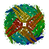
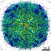
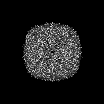
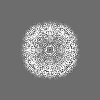
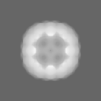
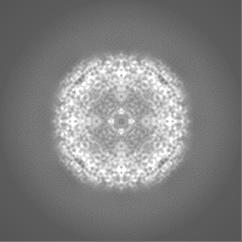
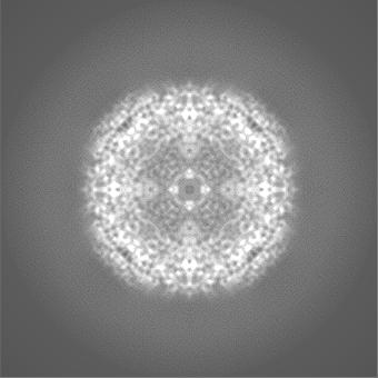
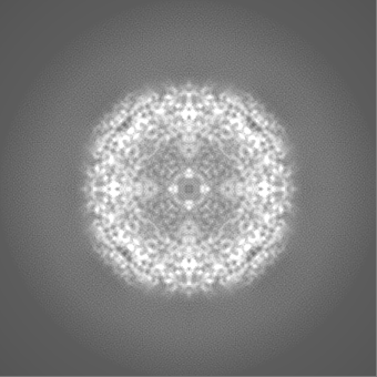
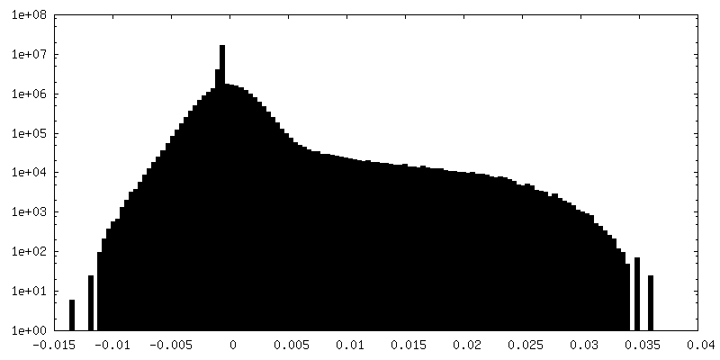
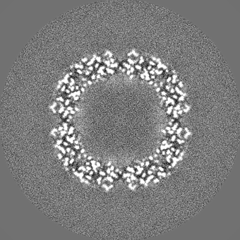
EMPIAR-10408 (Title: Single particle cryo-EM dataset of mouse heavy chain apoferritin collected on cryoARM300 Data size: 486.7 Data #1: raw uncorrected multiframe micrographs of mouse heavy chain apoferritin [micrographs - multiframe])
Energy filter - Name: In-column Omega Filter / Energy filter - Slit width: 20 eV
Image recording
Film or detector model: GATAN K2 SUMMIT (4k x 4k) / Detector mode: COUNTING / Number grids imaged: 1 / Average exposure time: 8.0 sec. / Average electron dose: 48.0 e/Å2
Electron beam
Acceleration voltage: 300 kV / Electron source: FIELD EMISSION GUN
Electron optics
Illumination mode: FLOOD BEAM / Imaging mode: BRIGHT FIELD / Cs: 2.55 mm
In the structure databanks used in Yorodumi, some data are registered as the other names, "COVID-19 virus" and "2019-nCoV". Here are the details of the virus and the list of structure data.
Jan 31, 2019. EMDB accession codes are about to change! (news from PDBe EMDB page)
EMDB accession codes are about to change! (news from PDBe EMDB page)
The allocation of 4 digits for EMDB accession codes will soon come to an end. Whilst these codes will remain in use, new EMDB accession codes will include an additional digit and will expand incrementally as the available range of codes is exhausted. The current 4-digit format prefixed with “EMD-” (i.e. EMD-XXXX) will advance to a 5-digit format (i.e. EMD-XXXXX), and so on. It is currently estimated that the 4-digit codes will be depleted around Spring 2019, at which point the 5-digit format will come into force.
The EM Navigator/Yorodumi systems omit the EMD- prefix.
Related info.:Q: What is EMD? / ID/Accession-code notation in Yorodumi/EM Navigator
Yorodumi is a browser for structure data from EMDB, PDB, SASBDB, etc.
This page is also the successor to EM Navigator detail page, and also detail information page/front-end page for Omokage search.
The word "yorodu" (or yorozu) is an old Japanese word meaning "ten thousand". "mi" (miru) is to see.
Related info.:EMDB / PDB / SASBDB / Comparison of 3 databanks / Yorodumi Search / Aug 31, 2016. New EM Navigator & Yorodumi / Yorodumi Papers / Jmol/JSmol / Function and homology information / Changes in new EM Navigator and Yorodumi
 Movie
Movie Controller


 Open data
 Basic information
 Map data
 Sample
 Function and homology information
 Authors
 Belgium, 2 items
  Citation
 Structure visualization
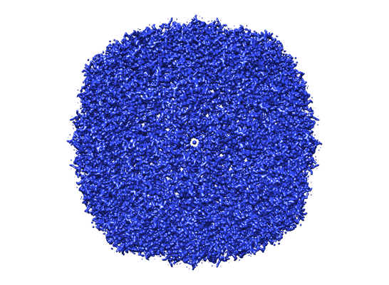
 Downloads & links
 emd_10675.png
 http://ftp.pdbj.org/pub/emdb/structures/EMD-10675
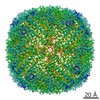


 Z (Sec.)
 Y (Row.)
 X (Col.)


 Sample components
 Processing
 Electron microscopy
 FIELD EMISSION GUN