 Movie
Movie Controller

[English] 日本語
 Yorodumi
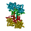
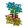
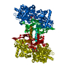
- PDB-1l5v: Crystal Structure of the Maltodextrin Phosphorylase complexed wit... -
+ Open data

- Basic information
| Entry | Database: PDB / ID: 1l5v | ||||||
|---|---|---|---|---|---|---|---|
| Title | Crystal Structure of the Maltodextrin Phosphorylase complexed with Glucose-1-phosphate | ||||||
 Components Components | MALTODEXTRIN PHOSPHORYLASE | ||||||
 Keywords Keywords | TRANSFERASE / phosphorylase / enzymatic catalysis / substrate complex | ||||||
| Function / homology |  Function and homology information Function and homology informationmaltodextrin phosphorylase activity / alpha-glucan catabolic process / glycogen phosphorylase / glycogen phosphorylase activity / glycogen catabolic process / pyridoxal phosphate binding / protein homodimerization activity / cytoplasm / cytosol Similarity search - Function | ||||||
| Biological species |  | ||||||
| Method |  X-RAY DIFFRACTION / SYNCHROTRON / FOURIER SYNTHESIS / Resolution: 2 Å X-RAY DIFFRACTION / SYNCHROTRON / FOURIER SYNTHESIS / Resolution: 2 Å | ||||||
 Authors Authors | Geremia, S. / Campagnolo, M. / Schinzel, R. / Johnson, L.N. | ||||||
 Citation Citation | Journal: J.Mol.Biol. / Year: 2002 Title: Enzymatic catalysis in crystals of Escherichia coli maltodextrin phosphorylase Authors: Geremia, S. / Campagnolo, M. / Schinzel, R. / Johnson, L.N. | ||||||
| History |
|
- Structure visualization
| Structure viewer | Molecule: MolmilJmol/JSmol |
|---|
- Downloads & links
-Download
| PDBx/mmCIF format | 1l5v.cif.gz | 349.7 KB | Display | PDBx/mmCIF format |
|---|---|---|---|---|
| PDB format | pdb1l5v.ent.gz | 281.8 KB | Display | PDB format |
| PDBx/mmJSON format | 1l5v.json.gz | Tree view | PDBx/mmJSON format | |
| Others |  Other downloads Other downloads |
-Validation report
| Arichive directory | https://data.pdbj.org/pub/pdb/validation_reports/l5/1l5vftp://data.pdbj.org/pub/pdb/validation_reports/l5/1l5v | HTTPS FTP |
|---|
-Related structure data
| Related structure data | 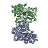 1l5wC  1l6iC 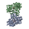 1qm5S S: Starting model for refinement C: citing same article ( |
|---|---|
| Similar structure data |


-Links
 PDBj
PDBj
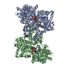
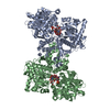
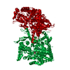
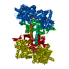
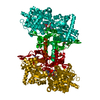
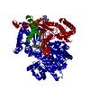
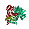
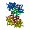
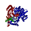
- Assembly
| Deposited unit | 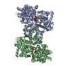
| ||||||||
|---|---|---|---|---|---|---|---|---|---|
| 1 |
| ||||||||
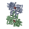
| Unit cell |
|
-Components
| #1: Protein | Mass: 90547.062 Da / Num. of mol.: 2 Source method: isolated from a genetically manipulated source Source: (gene. exp.) #2: Sugar | 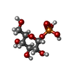  Type: D-saccharide / Mass: 260.136 Da / Num. of mol.: 2 Type: D-saccharide / Mass: 260.136 Da / Num. of mol.: 2Source method: isolated from a genetically manipulated source Formula: C6H13O9P #3: Chemical |   Mass: 122.143 Da / Num. of mol.: 2 / Source method: obtained synthetically / Formula: C4H12NO3 / Comment: pH buffer*YM Mass: 122.143 Da / Num. of mol.: 2 / Source method: obtained synthetically / Formula: C4H12NO3 / Comment: pH buffer*YM#4: Chemical |   Mass: 247.142 Da / Num. of mol.: 2 / Source method: obtained synthetically / Formula: C8H10NO6P Mass: 247.142 Da / Num. of mol.: 2 / Source method: obtained synthetically / Formula: C8H10NO6P#5: Water | ChemComp-HOH / |  Mass: 18.015 Da / Num. of mol.: 1235 / Source method: isolated from a natural source / Formula: H2O Mass: 18.015 Da / Num. of mol.: 1235 / Source method: isolated from a natural source / Formula: H2O |
|---|
-Experimental details
-Experiment
| Experiment | Method: X-RAY DIFFRACTION / Number of used crystals: 1 |
|---|
- Sample preparation
| Crystal | Density Matthews: 2.38 Å3/Da / Density % sol: 48 % | ||||||||||||||||||||||||||||||||||||||||||
|---|---|---|---|---|---|---|---|---|---|---|---|---|---|---|---|---|---|---|---|---|---|---|---|---|---|---|---|---|---|---|---|---|---|---|---|---|---|---|---|---|---|---|---|
| Crystal grow | Temperature: 291 K / Method: vapor diffusion, hanging drop / pH: 8.5 Details: PEG 4000, TRIS, Lithium Chloride, Glucose-1-phosphate., pH 8.5, VAPOR DIFFUSION, HANGING DROP, temperature 291K | ||||||||||||||||||||||||||||||||||||||||||
| Crystal grow | *PLUS | ||||||||||||||||||||||||||||||||||||||||||
| Components of the solutions | *PLUS
|
-Data collection
| Diffraction | Mean temperature: 100 K |
|---|---|
| Diffraction source | Source: SYNCHROTRON / Site: ELETTRA  / Beamline: 5.2R / Wavelength: 1 Å / Beamline: 5.2R / Wavelength: 1 Å |
| Detector | Type: MARRESEARCH / Detector: IMAGE PLATE / Date: Dec 10, 1999 |
| Radiation | Monochromator: Si 111 / Protocol: SINGLE WAVELENGTH / Monochromatic (M) / Laue (L): M / Scattering type: x-ray |
| Radiation wavelength | Wavelength: 1 Å / Relative weight: 1 |
| Reflection | Resolution: 2→40 Å / Num. all: 110577 / Num. obs: 110577 / % possible obs: 94.7 % / Redundancy: 3.8 % / Biso Wilson estimate: 17.386 Å2 / Rmerge(I) obs: 0.15 / Net I/σ(I): 7.2 |
| Reflection shell | Resolution: 2→2.1 Å / Redundancy: 2.8 % / Rmerge(I) obs: 0.55 / Mean I/σ(I) obs: 1.8 / Num. unique all: 13882 / % possible all: 82.4 |
| Reflection | *PLUS Lowest resolution: 40 Å / Num. measured all: 423852 |
| Reflection shell | *PLUS % possible obs: 82.4 % |
- Processing
| Software |
| ||||||||||||||||||||||||||||||||||||||||||||||||||||||||||||||||||||||||||||||||||||
|---|---|---|---|---|---|---|---|---|---|---|---|---|---|---|---|---|---|---|---|---|---|---|---|---|---|---|---|---|---|---|---|---|---|---|---|---|---|---|---|---|---|---|---|---|---|---|---|---|---|---|---|---|---|---|---|---|---|---|---|---|---|---|---|---|---|---|---|---|---|---|---|---|---|---|---|---|---|---|---|---|---|---|---|---|---|
| Refinement | Method to determine structure: FOURIER SYNTHESIS Starting model: 1QM5 Resolution: 2→20 Å / Cor.coef. Fo:Fc: 0.951 / Cor.coef. Fo:Fc free: 0.923 / SU B: 7.69621 / SU ML: 0.20349 / Cross valid method: THROUGHOUT / ESU R: 0.2014 / ESU R Free: 0.16739 / Stereochemistry target values: Engh & Huber
| ||||||||||||||||||||||||||||||||||||||||||||||||||||||||||||||||||||||||||||||||||||
| Solvent computation | Ion probe radii: 0.8 Å / Shrinkage radii: 0.8 Å / VDW probe radii: 1.4 Å | ||||||||||||||||||||||||||||||||||||||||||||||||||||||||||||||||||||||||||||||||||||
| Displacement parameters | Biso mean: 23.975 Å2
| ||||||||||||||||||||||||||||||||||||||||||||||||||||||||||||||||||||||||||||||||||||
| Refinement step | Cycle: LAST / Resolution: 2→20 Å
| ||||||||||||||||||||||||||||||||||||||||||||||||||||||||||||||||||||||||||||||||||||
| Refine LS restraints |
| ||||||||||||||||||||||||||||||||||||||||||||||||||||||||||||||||||||||||||||||||||||
| Refinement | *PLUS Rfactor obs: 0.182 / Rfactor Rfree: 0.22 / Rfactor Rwork: 0.18 | ||||||||||||||||||||||||||||||||||||||||||||||||||||||||||||||||||||||||||||||||||||
| Solvent computation | *PLUS | ||||||||||||||||||||||||||||||||||||||||||||||||||||||||||||||||||||||||||||||||||||
| Displacement parameters | *PLUS | ||||||||||||||||||||||||||||||||||||||||||||||||||||||||||||||||||||||||||||||||||||
| Refine LS restraints | *PLUS Type: p_angle_deg / Dev ideal: 1.8 |
