 Movie
Movie Controller

[English] 日本語
 Yorodumi
- PDB-1ahp: OLIGOSACCHARIDE SUBSTRATE BINDING IN ESCHERICHIA COLI MALTODEXTRI... -
+ Open data

- Basic information
| Entry | Database: PDB / ID: 1ahp | |||||||||
|---|---|---|---|---|---|---|---|---|---|---|
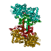
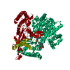
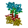
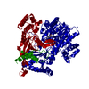
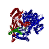
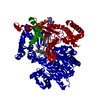
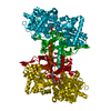
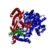
| Title | OLIGOSACCHARIDE SUBSTRATE BINDING IN ESCHERICHIA COLI MALTODEXTRIN PHSPHORYLASE | |||||||||
 Components Components | E.COLI MALTODEXTRIN PHOSPHORYLASE | |||||||||
 Keywords Keywords | ECOLI / PHOSPHORYLASE / OLIGOSACCHARIDE / INDUCED-FIT / SUBSTRATE / MALTODEXTRIN / STACKING | |||||||||
| Function / homology |  Function and homology information Function and homology informationmaltodextrin phosphorylase activity / alpha-glucan catabolic process / glycogen phosphorylase / glycogen phosphorylase activity / glycogen catabolic process / pyridoxal phosphate binding / protein homodimerization activity / cytosol / cytoplasm Similarity search - Function | |||||||||
| Biological species |  | |||||||||
| Method |  X-RAY DIFFRACTION / SYNCHROTRON / MOLECULAR REPLACEMENT / Resolution: 3 Å X-RAY DIFFRACTION / SYNCHROTRON / MOLECULAR REPLACEMENT / Resolution: 3 Å | |||||||||
 Authors Authors | O'Reilly, M. / Watson, K.A. / Schinzel, R. / Palm, D. / Johnson, L.N. | |||||||||
 Citation Citation | Journal: Nat.Struct.Biol. / Year: 1997 Title: Oligosaccharide substrate binding in Escherichia coli maltodextrin phosphorylase. Authors: O'Reilly, M. / Watson, K.A. / Schinzel, R. / Palm, D. / Johnson, L.N. | |||||||||
| History |
|
- Structure visualization
| Structure viewer | Molecule: MolmilJmol/JSmol |
|---|
- Downloads & links
-Download
| PDBx/mmCIF format | 1ahp.cif.gz | 291.9 KB | Display | PDBx/mmCIF format |
|---|---|---|---|---|
| PDB format | pdb1ahp.ent.gz | 238.5 KB | Display | PDB format |
| PDBx/mmJSON format | 1ahp.json.gz | Tree view | PDBx/mmJSON format | |
| Others |  Other downloads Other downloads |
-Validation report
| Arichive directory | https://data.pdbj.org/pub/pdb/validation_reports/ah/1ahpftp://data.pdbj.org/pub/pdb/validation_reports/ah/1ahp | HTTPS FTP |
|---|
-Related structure data
| Similar structure data |
|---|


-Links
 PDBj
PDBj


- Assembly
| Deposited unit | 
| |||||||||||||||
|---|---|---|---|---|---|---|---|---|---|---|---|---|---|---|---|---|
| 1 |
| |||||||||||||||
| Unit cell |
| |||||||||||||||
| Noncrystallographic symmetry (NCS) | NCS domain:
NCS oper: (Code: given Matrix: (-0.726932, -0.000378, -0.686708), Vector: |
-Components
| #1: Protein | Mass: 90365.008 Da / Num. of mol.: 2 / Mutation: N112A Source method: isolated from a genetically manipulated source Details: PYRIDOXAL PHOSPHATE COFACTOR ATTACHED TO LYS 645 / Source: (gene. exp.) #2: Polysaccharide |   Source method: isolated from a genetically manipulated source Details: oligosaccharide / References: alpha-maltose #3: Chemical |   Mass: 96.063 Da / Num. of mol.: 2 / Source method: obtained synthetically / Formula: SO4 Mass: 96.063 Da / Num. of mol.: 2 / Source method: obtained synthetically / Formula: SO4#4: Chemical |   Mass: 92.094 Da / Num. of mol.: 2 / Source method: obtained synthetically / Formula: C3H8O3 Mass: 92.094 Da / Num. of mol.: 2 / Source method: obtained synthetically / Formula: C3H8O3#5: Chemical |   Mass: 247.142 Da / Num. of mol.: 2 / Source method: obtained synthetically / Formula: C8H10NO6P Mass: 247.142 Da / Num. of mol.: 2 / Source method: obtained synthetically / Formula: C8H10NO6P |
|---|
-Experimental details
-Experiment
| Experiment | Method: X-RAY DIFFRACTION / Number of used crystals: 2 |
|---|
- Sample preparation
| Crystal | Density Matthews: 3.05 Å3/Da / Density % sol: 50 % | ||||||||||||||||||||||||||||||||||||||||||||||||||||||||||||||||||
|---|---|---|---|---|---|---|---|---|---|---|---|---|---|---|---|---|---|---|---|---|---|---|---|---|---|---|---|---|---|---|---|---|---|---|---|---|---|---|---|---|---|---|---|---|---|---|---|---|---|---|---|---|---|---|---|---|---|---|---|---|---|---|---|---|---|---|---|
| Crystal grow | Method: vapor diffusion, hanging drop / pH: 6.4 Details: 0.1M NATARTRATE, PH 6.4, 24% PEG 3350, 1MM SODIUM AZIDE, 16 DEG CELCIUS, 50MM MALTOHEXAOSE STOCK, 16.5MG/ML PROTEIN STOCK, HANGING DROPS 1MICRO LITRE WELL & 0.5 MICRO LITRE MALTOHEXAOSE 0.5 ...Details: 0.1M NATARTRATE, PH 6.4, 24% PEG 3350, 1MM SODIUM AZIDE, 16 DEG CELCIUS, 50MM MALTOHEXAOSE STOCK, 16.5MG/ML PROTEIN STOCK, HANGING DROPS 1MICRO LITRE WELL & 0.5 MICRO LITRE MALTOHEXAOSE 0.5 MICRO LITRE PROTEIN, vapor diffusion - hanging drop | ||||||||||||||||||||||||||||||||||||||||||||||||||||||||||||||||||
| Crystal grow | *PLUS Temperature: 16 ℃ / Method: vapor diffusion, hanging drop | ||||||||||||||||||||||||||||||||||||||||||||||||||||||||||||||||||
| Components of the solutions | *PLUS
|
-Data collection
| Diffraction | Mean temperature: 100 K |
|---|---|
| Diffraction source | Source: SYNCHROTRON / Site: SRS  / Beamline: PX9.6 / Wavelength: 0.87 / Beamline: PX9.6 / Wavelength: 0.87 |
| Detector | Type: MAR scanner 300 mm plate / Detector: IMAGE PLATE / Date: Feb 1, 1995 / Details: MIRRORS |
| Radiation | Monochromator: GRAPHITE(002) / Monochromatic (M) / Laue (L): M / Scattering type: x-ray |
| Radiation wavelength | Wavelength: 0.87 Å / Relative weight: 1 |
| Reflection | Resolution: 3→20 Å / Num. obs: 33108 / % possible obs: 81.7 % / Observed criterion σ(I): 1 / Redundancy: 1.6 % / Biso Wilson estimate: 57 Å2 / Rmerge(I) obs: 0.14 / Net I/σ(I): 5.4 |
| Reflection shell | Resolution: 3→3.08 Å / Redundancy: 1 % / Rmerge(I) obs: 0.318 / Mean I/σ(I) obs: 1.9 / % possible all: 80.3 |
| Reflection | *PLUS Num. measured all: 52324 |
- Processing
| Software |
| ||||||||||||||||||||||||||||||||||||||||||||||||||||||||||||
|---|---|---|---|---|---|---|---|---|---|---|---|---|---|---|---|---|---|---|---|---|---|---|---|---|---|---|---|---|---|---|---|---|---|---|---|---|---|---|---|---|---|---|---|---|---|---|---|---|---|---|---|---|---|---|---|---|---|---|---|---|---|
| Refinement | Method to determine structure: MOLECULAR REPLACEMENT Starting model: MALP NATIVE STRUCTURE Resolution: 3→10 Å / Rfactor Rfree error: 0.017 / Data cutoff high absF: 1000000 / Data cutoff low absF: 0.001 / Isotropic thermal model: GROUPED B / Cross valid method: THROUGHOUT / σ(F): 1 Details: COORDINATES FOR MOLECULE A WERE PROVIDED BY THE DEPOSITOR. MOLECULE B WAS GENERATED BY THE PDB USING NCS SYMMETRY.
| ||||||||||||||||||||||||||||||||||||||||||||||||||||||||||||
| Displacement parameters | Biso mean: 25.4 Å2 | ||||||||||||||||||||||||||||||||||||||||||||||||||||||||||||
| Refine analyze |
| ||||||||||||||||||||||||||||||||||||||||||||||||||||||||||||
| Refinement step | Cycle: LAST / Resolution: 3→10 Å
| ||||||||||||||||||||||||||||||||||||||||||||||||||||||||||||
| Refine LS restraints |
| ||||||||||||||||||||||||||||||||||||||||||||||||||||||||||||
| Refine LS restraints NCS | NCS model details: STRICT | ||||||||||||||||||||||||||||||||||||||||||||||||||||||||||||
| LS refinement shell | Resolution: 3→3.13 Å / Rfactor Rfree error: 0.042 / Total num. of bins used: 8
| ||||||||||||||||||||||||||||||||||||||||||||||||||||||||||||
| Xplor file |
| ||||||||||||||||||||||||||||||||||||||||||||||||||||||||||||
| Software | *PLUS Name: X-PLOR / Version: 3.1 / Classification: refinement | ||||||||||||||||||||||||||||||||||||||||||||||||||||||||||||
| Refine LS restraints | *PLUS
|
