 Movie
Movie Controller

[English] 日本語
 Yorodumi
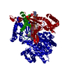
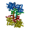
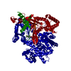
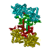
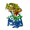
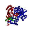
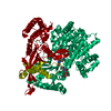
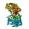
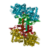
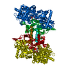
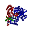
- PDB-2gpn: 100 K STRUCTURE OF GLYCOGEN PHOSPHORYLASE AT 2.0 ANGSTROMS RESOLUTION -
+ Open data

- Basic information
| Entry | Database: PDB / ID: 2gpn | ||||||
|---|---|---|---|---|---|---|---|
| Title | 100 K STRUCTURE OF GLYCOGEN PHOSPHORYLASE AT 2.0 ANGSTROMS RESOLUTION | ||||||
 Components Components | GLYCOGEN PHOSPHORYLASE B | ||||||
 Keywords Keywords | GLYCOGEN PHOSPHORYLASE / WATER STRUCTURE / 100 K X-RAY STRUCTURE / MPD / TRANSFERASE | ||||||
| Function / homology |  Function and homology information Function and homology informationglycogen phosphorylase / glycogen phosphorylase activity / glycogen catabolic process / skeletal muscle myofibril / pyridoxal phosphate binding / nucleotide binding Similarity search - Function | ||||||
| Biological species |  | ||||||
| Method |  X-RAY DIFFRACTION / SYNCHROTRON / DIFFERENCE FOURIER / Resolution: 1.99 Å X-RAY DIFFRACTION / SYNCHROTRON / DIFFERENCE FOURIER / Resolution: 1.99 Å | ||||||
 Authors Authors | Gregoriou, M. / Noble, M.E.M. / Watson, K.A. / Garman, E.F. / Krulle, T.M. / De La Fuente, C. / Fleet, G.W.J. / Oikonomakos, N.G. / Johnson, L.N. | ||||||
 Citation Citation | Journal: Protein Sci. / Year: 1998 Title: The structure of a glycogen phosphorylase glucopyranose spirohydantoin complex at 1.8 A resolution and 100 K: the role of the water structure and its contribution to binding. Authors: Gregoriou, M. / Noble, M.E. / Watson, K.A. / Garman, E.F. / Krulle, T.M. / de la Fuente, C. / Fleet, G.W. / Oikonomakos, N.G. / Johnson, L.N. #2: Journal: Biochemistry / Year: 1991Title: Glucose Analogue Inhibitors of Glycogen Phosphorylase: The Design of Potential Drugs for Diabetes Authors: Martin, J.L. / Veluraja, K. / Ross, K. / Johnson, L.N. / Fleet, G.W. / Ramsden, N.G. / Bruce, I. / Orchard, M.G. / Oikonomakos, N.G. / Papageorgiou, A.C. / Leonidas, D.D. / Tsitoura, H.S. | ||||||
| History |
|
- Structure visualization
| Structure viewer | Molecule: MolmilJmol/JSmol |
|---|
- Downloads & links
-Download
| PDBx/mmCIF format | 2gpn.cif.gz | 189.3 KB | Display | PDBx/mmCIF format |
|---|---|---|---|---|
| PDB format | pdb2gpn.ent.gz | 150.3 KB | Display | PDB format |
| PDBx/mmJSON format | 2gpn.json.gz | Tree view | PDBx/mmJSON format | |
| Others |  Other downloads Other downloads |
-Validation report
| Arichive directory | https://data.pdbj.org/pub/pdb/validation_reports/gp/2gpnftp://data.pdbj.org/pub/pdb/validation_reports/gp/2gpn | HTTPS FTP |
|---|
-Related structure data
| Related structure data | 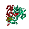 1a8iC 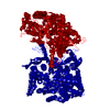 1gpbS S: Starting model for refinement C: citing same article ( |
|---|---|
| Similar structure data |


-Links
 PDBj
PDBj
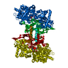
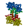
- Assembly
| Deposited unit | 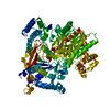
| ||||||||
|---|---|---|---|---|---|---|---|---|---|
| 1 | 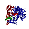
| ||||||||
| Unit cell |
|
-Components
| #1: Protein | Mass: 97519.320 Da / Num. of mol.: 1 / Source method: isolated from a natural source / Details: SCHIFF'S BASE LINK BETWEEN PLP AND LYS 680 / Source: (natural) |
|---|---|
| #2: Water | ChemComp-HOH /  Mass: 18.015 Da / Num. of mol.: 817 / Source method: isolated from a natural source / Formula: H2O Mass: 18.015 Da / Num. of mol.: 817 / Source method: isolated from a natural source / Formula: H2O |
-Experimental details
-Experiment
| Experiment | Method: X-RAY DIFFRACTION / Number of used crystals: 1 |
|---|
- Sample preparation
| Crystal | Density Matthews: 2.4 Å3/Da / Density % sol: 48.7 % | |||||||||||||||||||||||||||||||||||
|---|---|---|---|---|---|---|---|---|---|---|---|---|---|---|---|---|---|---|---|---|---|---|---|---|---|---|---|---|---|---|---|---|---|---|---|---|
| Crystal grow | Temperature: 289 K / pH: 6.7 Details: THE PROTEIN WAS CRYSTALLIZED FROM 0.01 M BES, PH 6.7, 0.003 M DTT, 0.001 M SPERMINE, 0.0001 M EDTA, 0.02 % (W/V) SODIUM AZIDE AT 16 DEGREES C. THE CRYSTALS WERE CRYOPROTECTED WITH 25% (V/V) ...Details: THE PROTEIN WAS CRYSTALLIZED FROM 0.01 M BES, PH 6.7, 0.003 M DTT, 0.001 M SPERMINE, 0.0001 M EDTA, 0.02 % (W/V) SODIUM AZIDE AT 16 DEGREES C. THE CRYSTALS WERE CRYOPROTECTED WITH 25% (V/V) MPD (2-METHYL-2,4-PENTANEDIOL)., temperature 289K | |||||||||||||||||||||||||||||||||||
| Crystal grow | *PLUS Temperature: 16 ℃ / Method: unknown | |||||||||||||||||||||||||||||||||||
| Components of the solutions | *PLUS
|
-Data collection
| Diffraction | Mean temperature: 100 K |
|---|---|
| Diffraction source | Source: SYNCHROTRON / Site: SRS  / Beamline: PX9.6 / Wavelength: 0.87 / Beamline: PX9.6 / Wavelength: 0.87 |
| Detector | Type: MARRESEARCH / Detector: IMAGE PLATE / Date: May 19, 1996 / Details: MIRROR |
| Radiation | Monochromator: SI(111) / Monochromatic (M) / Laue (L): M / Scattering type: x-ray |
| Radiation wavelength | Wavelength: 0.87 Å / Relative weight: 1 |
| Reflection | Resolution: 1.99→19.4 Å / Num. obs: 57703 / % possible obs: 89.7 % / Redundancy: 3.5 % / Rmerge(I) obs: 0.114 / Net I/σ(I): 10.1 |
| Reflection shell | Resolution: 1.99→2.14 Å / Redundancy: 2.1 % / Rmerge(I) obs: 0.373 / Mean I/σ(I) obs: 2.1 / % possible all: 89.5 |
| Reflection | *PLUS Num. measured all: 192914 |
| Reflection shell | *PLUS % possible obs: 89.5 % |
- Processing
| Software |
| ||||||||||||||||||||
|---|---|---|---|---|---|---|---|---|---|---|---|---|---|---|---|---|---|---|---|---|---|
| Refinement | Method to determine structure: DIFFERENCE FOURIER Starting model: AN UNPUBLISHED 1.5 ANGSTROMS MODEL (E.P.MITCHELL) AND PDB ENTRY 1GPB Resolution: 1.99→19.4 Å / Cross valid method: FREE R-FACTOR
| ||||||||||||||||||||
| Refine analyze | Luzzati sigma a obs: 0.18 Å | ||||||||||||||||||||
| Refinement step | Cycle: LAST / Resolution: 1.99→19.4 Å
| ||||||||||||||||||||
| Software | *PLUS Name: REFMAC / Classification: refinement | ||||||||||||||||||||
| Refinement | *PLUS Num. reflection all: 57703 / Rfactor all: 0.248 / Rfactor obs: 0.197 | ||||||||||||||||||||
| Solvent computation | *PLUS | ||||||||||||||||||||
| Displacement parameters | *PLUS | ||||||||||||||||||||
| Refine LS restraints | *PLUS
|
