 Movie
Movie Controller

[English] 日本語
 Yorodumi
- PDB-1bx3: EFFECTS OF COMMONLY USED CRYOPROTECTANTS ON GLYCOGEN PHOSPHORYLAS... -
+ Open data

- Basic information
| Entry | Database: PDB / ID: 1bx3 | ||||||
|---|---|---|---|---|---|---|---|
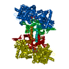
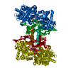
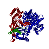
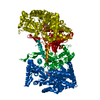
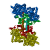
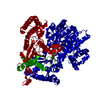
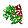
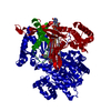
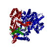
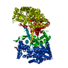
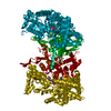
| Title | EFFECTS OF COMMONLY USED CRYOPROTECTANTS ON GLYCOGEN PHOSPHORYLASE ACTIVITY AND STRUCTURE | ||||||
 Components Components | PROTEIN (GLYCOGEN PHOSPHORYLASE B) | ||||||
 Keywords Keywords | TRANSFERASE / PHOSPHORYLASE / INHIBITOR / CRYOPROTECTANT / MPD / DMSO / CRYOCRYSTALLOGRAPHY | ||||||
| Function / homology |  Function and homology information Function and homology informationglycogen phosphorylase / glycogen phosphorylase activity / glycogen catabolic process / skeletal muscle myofibril / pyridoxal phosphate binding / nucleotide binding Similarity search - Function | ||||||
| Biological species |  | ||||||
| Method |  X-RAY DIFFRACTION / SYNCHROTRON / OTHER / Resolution: 2.3 Å X-RAY DIFFRACTION / SYNCHROTRON / OTHER / Resolution: 2.3 Å | ||||||
 Authors Authors | Tsitsanou, K.E. / Oikonomakos, N.G. / Zographos, S.E. / Skamnaki, V.T. / Gregoriou, M. / Watson, K.A. / Johnson, L.N. / Fleet, G.W.J. | ||||||
 Citation Citation | Journal: Protein Sci. / Year: 1999 Title: Effects of commonly used cryoprotectants on glycogen phosphorylase activity and structure. Authors: Tsitsanou, K.E. / Oikonomakos, N.G. / Zographos, S.E. / Skamnaki, V.T. / Gregoriou, M. / Watson, K.A. / Johnson, L.N. / Fleet, G.W. #1: Journal: Glycogen Phosphorylase B: Description of the Protein StructureYear: 1991 Title: Glycogen Phosphorylase B: Description of the Protein Structure Authors: Acharya, K.R. / Stuart, D.I. / Varvill, K.M. / Johnson, L.N. | ||||||
| History |
|
- Structure visualization
| Structure viewer | Molecule: MolmilJmol/JSmol |
|---|
- Downloads & links
-Download
| PDBx/mmCIF format | 1bx3.cif.gz | 188.8 KB | Display | PDBx/mmCIF format |
|---|---|---|---|---|
| PDB format | pdb1bx3.ent.gz | 147.4 KB | Display | PDB format |
| PDBx/mmJSON format | 1bx3.json.gz | Tree view | PDBx/mmJSON format | |
| Others |  Other downloads Other downloads |
-Validation report
| Arichive directory | https://data.pdbj.org/pub/pdb/validation_reports/bx/1bx3ftp://data.pdbj.org/pub/pdb/validation_reports/bx/1bx3 | HTTPS FTP |
|---|
-Related structure data
| Related structure data | 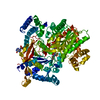 1b4dC  2gpnS S: Starting model for refinement C: citing same article ( |
|---|---|
| Similar structure data |


-Links
 PDBj
PDBj
- Assembly
| Deposited unit | 
| ||||||||||||
|---|---|---|---|---|---|---|---|---|---|---|---|---|---|
| 1 | 
| ||||||||||||
| Unit cell |
| ||||||||||||
| Components on special symmetry positions |
|
-Components
| #1: Protein | Mass: 97291.203 Da / Num. of mol.: 1 / Source method: isolated from a natural source / Details: SCHIFF'S BASE LINK BETWEEN PLP AND LYS 680 / Source: (natural) |
|---|---|
| #2: Chemical | ChemComp-PLP /   Mass: 247.142 Da / Num. of mol.: 1 / Source method: obtained synthetically / Formula: C8H10NO6P Mass: 247.142 Da / Num. of mol.: 1 / Source method: obtained synthetically / Formula: C8H10NO6P |
| #3: Water | ChemComp-HOH /  Mass: 18.015 Da / Num. of mol.: 673 / Source method: isolated from a natural source / Formula: H2O Mass: 18.015 Da / Num. of mol.: 673 / Source method: isolated from a natural source / Formula: H2O |
-Experimental details
-Experiment
| Experiment | Method: X-RAY DIFFRACTION / Number of used crystals: 1 |
|---|
- Sample preparation
| Crystal | Density Matthews: 2.35 Å3/Da / Density % sol: 47.66 % | |||||||||||||||||||||||||||||||||||
|---|---|---|---|---|---|---|---|---|---|---|---|---|---|---|---|---|---|---|---|---|---|---|---|---|---|---|---|---|---|---|---|---|---|---|---|---|
| Crystal grow | Temperature: 289 K / pH: 6.7 Details: THE PROTEIN WAS CRYSTALLIZED FROM 0.01 M BES, PH 6.7, 0.003 M DTT, 0.001 M SPERMINE, 0.0001 M EDTA, 0.02 % (W/V) SODIUM AZIDE AT 16 DEGREES C. THE CRYSTALS WERE CRYOPROTECTED WITH 30% (V/V) ...Details: THE PROTEIN WAS CRYSTALLIZED FROM 0.01 M BES, PH 6.7, 0.003 M DTT, 0.001 M SPERMINE, 0.0001 M EDTA, 0.02 % (W/V) SODIUM AZIDE AT 16 DEGREES C. THE CRYSTALS WERE CRYOPROTECTED WITH 30% (V/V) DMSO (DIMETHYLSULFOXIDE)., temperature 289K | |||||||||||||||||||||||||||||||||||
| Crystal grow | *PLUS Temperature: 16 ℃ / Method: unknownDetails: Oikonomakos, N.G., (1985) Biochim.Biophys.Acta., 832, 248. | |||||||||||||||||||||||||||||||||||
| Components of the solutions | *PLUS
|
-Data collection
| Diffraction | Mean temperature: 100 K |
|---|---|
| Diffraction source | Source: SYNCHROTRON / Site: EMBL/DESY, HAMBURG  / Beamline: X11 / Wavelength: 0.928 / Beamline: X11 / Wavelength: 0.928 |
| Detector | Type: MARRESEARCH / Detector: IMAGE PLATE / Date: Jul 29, 1996 |
| Radiation | Protocol: SINGLE WAVELENGTH / Monochromatic (M) / Laue (L): M / Scattering type: x-ray |
| Radiation wavelength | Wavelength: 0.928 Å / Relative weight: 1 |
| Reflection | Resolution: 2.3→36.8 Å / Num. obs: 41713 / % possible obs: 99.9 % / Redundancy: 5.9 % / Rmerge(I) obs: 0.042 / Net I/σ(I): 20.59 |
| Reflection shell | Resolution: 2.3→2.34 Å / Rmerge(I) obs: 0.103 / Mean I/σ(I) obs: 10.48 / % possible all: 99.5 |
| Reflection | *PLUS Num. measured all: 244996 |
| Reflection shell | *PLUS % possible obs: 99.5 % |
- Processing
| Software |
| ||||||||||||||||||||||||||||||||||||||||||||||||||||||||||||
|---|---|---|---|---|---|---|---|---|---|---|---|---|---|---|---|---|---|---|---|---|---|---|---|---|---|---|---|---|---|---|---|---|---|---|---|---|---|---|---|---|---|---|---|---|---|---|---|---|---|---|---|---|---|---|---|---|---|---|---|---|---|
| Refinement | Method to determine structure: OTHER Starting model: PDB ENTRY 2GPN Resolution: 2.3→36.8 Å / Cross valid method: FREE R-FACTOR / σ(F): 0
| ||||||||||||||||||||||||||||||||||||||||||||||||||||||||||||
| Refinement step | Cycle: LAST / Resolution: 2.3→36.8 Å
| ||||||||||||||||||||||||||||||||||||||||||||||||||||||||||||
| Refine LS restraints |
|
