 Movie
Movie Controller

[English] 日本語
 Yorodumi
- PDB-1p2d: Crystal Structure of Glycogen Phosphorylase B in complex with Bet... -
+ Open data

- Basic information
| Entry | Database: PDB / ID: 1p2d | |||||||||
|---|---|---|---|---|---|---|---|---|---|---|
| Title | Crystal Structure of Glycogen Phosphorylase B in complex with Beta Cyclodextrin | |||||||||
 Components Components | Glycogen phosphorylase, muscle form | |||||||||
 Keywords Keywords | TRANSFERASE | |||||||||
| Function / homology |  Function and homology information Function and homology informationglycogen phosphorylase / glycogen phosphorylase activity / glycogen catabolic process / skeletal muscle myofibril / pyridoxal phosphate binding / nucleotide binding Similarity search - Function | |||||||||
| Biological species |  | |||||||||
| Method |  X-RAY DIFFRACTION / SYNCHROTRON / FOURIER SYNTHESIS / Resolution: 1.94 Å X-RAY DIFFRACTION / SYNCHROTRON / FOURIER SYNTHESIS / Resolution: 1.94 Å | |||||||||
 Authors Authors | Pinotsis, N. / Leonidas, D.D. / Chrysina, E.D. / Oikonomakos, N.G. / Mavridis, I.M. | |||||||||
 Citation Citation | Journal: Protein Sci. / Year: 2003 Title: The binding of beta- and gamma-cyclodextrins to glycogen phosphorylase b: Kinetic and crystallographic studies. Authors: Pinotsis, N. / Leonidas, D.D. / Chrysina, E.D. / Oikonomakos, N.G. / Mavridis, I.M. | |||||||||
| History |
|
- Structure visualization
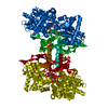
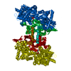
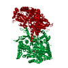
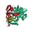
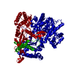
| Structure viewer | Molecule: MolmilJmol/JSmol |
|---|
- Downloads & links
-Download
| PDBx/mmCIF format | 1p2d.cif.gz | 182.4 KB | Display | PDBx/mmCIF format |
|---|---|---|---|---|
| PDB format | pdb1p2d.ent.gz | 142.5 KB | Display | PDB format |
| PDBx/mmJSON format | 1p2d.json.gz | Tree view | PDBx/mmJSON format | |
| Others |  Other downloads Other downloads |
-Validation report
| Arichive directory | https://data.pdbj.org/pub/pdb/validation_reports/p2/1p2dftp://data.pdbj.org/pub/pdb/validation_reports/p2/1p2d | HTTPS FTP |
|---|
-Related structure data
| Related structure data | 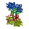 1p29C  1p2bC 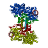 1p2gC 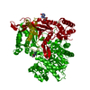 1gfzS C: citing same article ( S: Starting model for refinement |
|---|---|
| Similar structure data |


-Links
 PDBj
PDBj


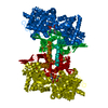
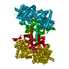
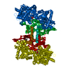
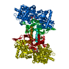
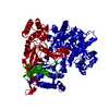
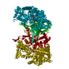
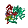
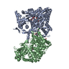
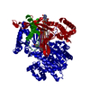
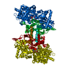
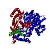
- Assembly
| Deposited unit | 
| ||||||||
|---|---|---|---|---|---|---|---|---|---|
| 1 | 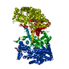
| ||||||||
| Unit cell |
| ||||||||
| Components on special symmetry positions |
| ||||||||
| Details | THIS ENTRY CONTAINS THE CRYSTALLOGRAPHIC ASYMMETRIC UNIT WHICH CONSISTS OF 1 CHAIN. COORDINATES FOR A COMPLETE DIMER REPRESENTING THE KNOWN BIOLOGICALLY SIGNIFICANT OLIGOMERIZATION STATE OF THE MOLECULE CAN BE GENERATED BY APPLYING THE TWO FOLD AXIS : y,x,1-z |
-Components
| #1: Protein | Mass: 97291.203 Da / Num. of mol.: 1 / Source method: isolated from a natural source / Source: (natural) |
|---|---|
| #2: Polysaccharide | alpha-D-glucopyranose-(1-4)-alpha-D-glucopyranose / alpha-maltose  Source method: isolated from a genetically manipulated source Details: oligosaccharide / References: alpha-maltose |
| #3: Chemical | ChemComp-PLP /   Mass: 247.142 Da / Num. of mol.: 1 / Source method: obtained synthetically / Formula: C8H10NO6P Mass: 247.142 Da / Num. of mol.: 1 / Source method: obtained synthetically / Formula: C8H10NO6P |
| #4: Water | ChemComp-HOH /  Mass: 18.015 Da / Num. of mol.: 324 / Source method: isolated from a natural source / Formula: H2O Mass: 18.015 Da / Num. of mol.: 324 / Source method: isolated from a natural source / Formula: H2O |
-Experimental details
-Experiment
| Experiment | Method: X-RAY DIFFRACTION / Number of used crystals: 1 |
|---|
- Sample preparation
| Crystal | Density Matthews: 2.48 Å3/Da / Density % sol: 50.37 % | ||||||||||||||||||||||||||||||||||||||||||||||||||||||
|---|---|---|---|---|---|---|---|---|---|---|---|---|---|---|---|---|---|---|---|---|---|---|---|---|---|---|---|---|---|---|---|---|---|---|---|---|---|---|---|---|---|---|---|---|---|---|---|---|---|---|---|---|---|---|---|
| Crystal grow | Temperature: 289 K / Method: small tubes / pH: 6.7 / Details: BES, EDTA, pH 6.7, SMALL TUBES, temperature 289K | ||||||||||||||||||||||||||||||||||||||||||||||||||||||
| Crystal grow | *PLUS pH: 6.7 / Method: unknownDetails: Oikonomakos, N.G., (2000) J.Biol.Chem., 275, 34566. | ||||||||||||||||||||||||||||||||||||||||||||||||||||||
| Components of the solutions | *PLUS
|
-Data collection
| Diffraction | Mean temperature: 293 K |
|---|---|
| Diffraction source | Source: SYNCHROTRON / Site: EMBL/DESY, HAMBURG  / Beamline: X11 / Wavelength: 0.8068 Å / Beamline: X11 / Wavelength: 0.8068 Å |
| Detector | Type: MARRESEARCH / Detector: IMAGE PLATE / Date: Nov 22, 2002 |
| Radiation | Protocol: SINGLE WAVELENGTH / Monochromatic (M) / Laue (L): M / Scattering type: x-ray |
| Radiation wavelength | Wavelength: 0.8068 Å / Relative weight: 1 |
| Reflection | Resolution: 1.94→30 Å / Num. all: 68504 / Num. obs: 68504 / % possible obs: 93.4 % / Observed criterion σ(F): 0 / Observed criterion σ(I): -3 / Redundancy: 10.4 % / Biso Wilson estimate: 17.9 Å2 / Rsym value: 0.076 / Net I/σ(I): 13.3 |
| Reflection shell | Resolution: 1.94→1.96 Å / Mean I/σ(I) obs: 2.1 / Rsym value: 0.499 / % possible all: 85 |
| Reflection | *PLUS Lowest resolution: 30 Å / Num. measured all: 714419 / Rmerge(I) obs: 0.076 |
| Reflection shell | *PLUS % possible obs: 85 % / Rmerge(I) obs: 0.499 |
- Processing
| Software |
| ||||||||||||||||||||||||||||||||||||
|---|---|---|---|---|---|---|---|---|---|---|---|---|---|---|---|---|---|---|---|---|---|---|---|---|---|---|---|---|---|---|---|---|---|---|---|---|---|
| Refinement | Method to determine structure: FOURIER SYNTHESIS Starting model: 1GFZ Resolution: 1.94→29.51 Å / Rfactor Rfree error: 0.004 / Data cutoff high absF: 3573624.95 / Data cutoff low absF: 0 / Isotropic thermal model: RESTRAINED / Cross valid method: THROUGHOUT / σ(F): 0 / Stereochemistry target values: Engh & Huber
| ||||||||||||||||||||||||||||||||||||
| Solvent computation | Solvent model: FLAT MODEL / Bsol: 63.2621 Å2 / ksol: 0.380243 e/Å3 | ||||||||||||||||||||||||||||||||||||
| Displacement parameters | Biso mean: 29.7 Å2
| ||||||||||||||||||||||||||||||||||||
| Refine analyze |
| ||||||||||||||||||||||||||||||||||||
| Refinement step | Cycle: LAST / Resolution: 1.94→29.51 Å
| ||||||||||||||||||||||||||||||||||||
| Refine LS restraints |
| ||||||||||||||||||||||||||||||||||||
| LS refinement shell | Resolution: 1.94→2.06 Å / Rfactor Rfree error: 0.013 / Total num. of bins used: 6
| ||||||||||||||||||||||||||||||||||||
| Xplor file |
| ||||||||||||||||||||||||||||||||||||
| Refinement | *PLUS Lowest resolution: 30 Å / % reflection Rfree: 5 % | ||||||||||||||||||||||||||||||||||||
| Solvent computation | *PLUS | ||||||||||||||||||||||||||||||||||||
| Displacement parameters | *PLUS | ||||||||||||||||||||||||||||||||||||
| Refine LS restraints | *PLUS
|
