 Movie
Movie Controller


+ Open data

- Basic information
| Entry | Database: PDB / ID: 1b4d | ||||||
|---|---|---|---|---|---|---|---|
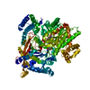
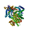
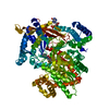
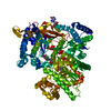
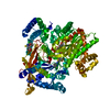
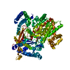
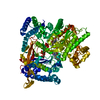
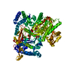
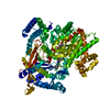
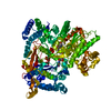
| Title | AMIDOCARBAMATE INHIBITOR OF GLYCOGEN PHOSPHORYLASE | ||||||
 Components Components | PROTEIN (GLYCOGEN PHOSPHORYLASE B) | ||||||
 Keywords Keywords | TRANSFERASE / GLYCOGEN PHOSPHORYLASE / INHIBITOR BINDING / AMIDOCARBAMATE | ||||||
| Function / homology |  Function and homology information Function and homology informationglycogen phosphorylase / glycogen phosphorylase activity / glycogen catabolic process / skeletal muscle myofibril / pyridoxal phosphate binding / nucleotide binding Similarity search - Function | ||||||
| Biological species |  | ||||||
| Method |  X-RAY DIFFRACTION / SYNCHROTRON / OTHER / Resolution: 2 Å X-RAY DIFFRACTION / SYNCHROTRON / OTHER / Resolution: 2 Å | ||||||
 Authors Authors | Tsitsanou, K.E. / Oikonomakos, N.G. / Zographos, S.E. / Skamnaki, V.T. / Gregoriou, M. / Watson, K.A. / Johnson, L.N. / Fleet, G.W.J. | ||||||
 Citation Citation | Journal: Protein Sci. / Year: 1999 Title: Effects of commonly used cryoprotectants on glycogen phosphorylase activity and structure. Authors: Tsitsanou, K.E. / Oikonomakos, N.G. / Zographos, S.E. / Skamnaki, V.T. / Gregoriou, M. / Watson, K.A. / Johnson, L.N. / Fleet, G.W. #1: Journal: Synlett / Year: 1997Title: Stereospecific Synthesis of Spirohydantoins of Beta-Glucopyranose:Inhibitors of Glycogen Phosphorylase Authors: Krulle, T.M. / De La Fuente, C. / Watson, K.A. / Gregoriou, M. / Johnson, L.N. / Tsitsanou, K.E. / Zographos, S.E. / Oikonomakos, N.G. / Fleet, G.W.J. #2: Journal: Protein Sci. / Year: 1995Title: N-Acetyl-Beta-D-Glucopyranosylamine:A Potent T-State Inhibitor of Glycogen Phosphorylase. A Comparison with Alpha-D-Glucose Authors: Oikonomakos, N.G. / Kontou, M. / Zographos, S.E. / Watson, K.A. / Johnson, L.N. / Bichard, C.J.F. / Fleet, G.W.J. / Acharya, K.R. #3: Journal: Biochemistry / Year: 1991Title: Glucose Analogue Inhibitors of Glycogen Phosphorylase: The Design of Potential Drugs for Diabetes Authors: Martin, J.L. / Veluraja, K. / Ross, K. / Johnson, L.N. / Fleet, G.W. / Ramsden, N.G. / Bruce, I. / Orchard, M.G. / Oikonomakos, N.G. / Papageorgiou, A.C. / Leonidas, D.D. / Tsitoura, H.S. | ||||||
| History |
|
- Structure visualization
| Structure viewer | Molecule: MolmilJmol/JSmol |
|---|
- Downloads & links
-Download
| PDBx/mmCIF format | 1b4d.cif.gz | 196.2 KB | Display | PDBx/mmCIF format |
|---|---|---|---|---|
| PDB format | pdb1b4d.ent.gz | 151.6 KB | Display | PDB format |
| PDBx/mmJSON format | 1b4d.json.gz | Tree view | PDBx/mmJSON format | |
| Others |  Other downloads Other downloads |
-Validation report
| Arichive directory | https://data.pdbj.org/pub/pdb/validation_reports/b4/1b4dftp://data.pdbj.org/pub/pdb/validation_reports/b4/1b4d | HTTPS FTP |
|---|
-Related structure data
| Related structure data | 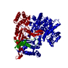 1bx3C 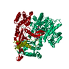 1a8iS S: Starting model for refinement C: citing same article ( |
|---|---|
| Similar structure data |


-Links
 PDBj
PDBj
- Assembly
| Deposited unit | 
| ||||||||
|---|---|---|---|---|---|---|---|---|---|
| 1 |
| ||||||||
| Unit cell |
|
-Components
| #1: Protein | Mass: 97291.203 Da / Num. of mol.: 1 / Source method: isolated from a natural source / Source: (natural) |
|---|---|
| #2: Sugar | ChemComp-CRA /   Type: D-saccharide / Mass: 280.232 Da / Num. of mol.: 1 Type: D-saccharide / Mass: 280.232 Da / Num. of mol.: 1Source method: isolated from a genetically manipulated source Formula: C9H16N2O8 |
| #3: Chemical | ChemComp-PLP /   Mass: 247.142 Da / Num. of mol.: 1 / Source method: obtained synthetically / Formula: C8H10NO6P Mass: 247.142 Da / Num. of mol.: 1 / Source method: obtained synthetically / Formula: C8H10NO6P |
| #4: Chemical | ChemComp-IMP /   Mass: 348.206 Da / Num. of mol.: 1 / Source method: obtained synthetically / Formula: C10H13N4O8P Mass: 348.206 Da / Num. of mol.: 1 / Source method: obtained synthetically / Formula: C10H13N4O8P |
| #5: Water | ChemComp-HOH /  Mass: 18.015 Da / Num. of mol.: 820 / Source method: isolated from a natural source / Formula: H2O Mass: 18.015 Da / Num. of mol.: 820 / Source method: isolated from a natural source / Formula: H2O |
-Experimental details
-Experiment
| Experiment | Method: X-RAY DIFFRACTION / Number of used crystals: 1 |
|---|
- Sample preparation
| Crystal | Density Matthews: 2.38 Å3/Da / Density % sol: 48.26 % |
|---|---|
| Crystal grow | Temperature: 289 K / pH: 6.7 Details: CO-CRYSTALLIZED COMPLEX WAS OBTAINED FROM 0.01 M BES, PH 6.7, 0.003 M DTT, 0.001 M SPERMINE, 0.0001 M SODIUM EDTA, 0.02 % SODIUM AZIDE AND 0.01 M AMIDOCARBAMATE AT 16 DEGREES C. JUST BEFORE ...Details: CO-CRYSTALLIZED COMPLEX WAS OBTAINED FROM 0.01 M BES, PH 6.7, 0.003 M DTT, 0.001 M SPERMINE, 0.0001 M SODIUM EDTA, 0.02 % SODIUM AZIDE AND 0.01 M AMIDOCARBAMATE AT 16 DEGREES C. JUST BEFORE DATA COLLECTION, THE CRYSTALS WERE TRANSFERRED TO A FRESH SOLUTION OF THE ABOVE BUFFER CONTAINING 30% GLYCEROL FOR 30-60 SEC., temperature 289K |
| Crystal grow | *PLUS Method: unknown |
-Data collection
| Diffraction | Mean temperature: 100 K |
|---|---|
| Diffraction source | Source: SYNCHROTRON / Site: EMBL/DESY, HAMBURG  / Beamline: X11 / Wavelength: 0.928 / Beamline: X11 / Wavelength: 0.928 |
| Detector | Type: MAR scanner 180 mm plate / Detector: IMAGE PLATE / Date: Oct 18, 1995 |
| Radiation | Protocol: SINGLE WAVELENGTH / Monochromatic (M) / Laue (L): M / Scattering type: x-ray |
| Radiation wavelength | Wavelength: 0.928 Å / Relative weight: 1 |
| Reflection | Resolution: 2→20 Å / Num. obs: 62995 / % possible obs: 99.4 % / Redundancy: 6.6 % / Rmerge(I) obs: 0.06 / Net I/σ(I): 15.67 |
| Reflection shell | Resolution: 2→2.07 Å / Rmerge(I) obs: 0.219 / Mean I/σ(I) obs: 6.2 / % possible all: 99.6 |
- Processing
| Software |
| ||||||||||||||||||||||||||||||||||||||||||||||||||||||||||||
|---|---|---|---|---|---|---|---|---|---|---|---|---|---|---|---|---|---|---|---|---|---|---|---|---|---|---|---|---|---|---|---|---|---|---|---|---|---|---|---|---|---|---|---|---|---|---|---|---|---|---|---|---|---|---|---|---|---|---|---|---|---|
| Refinement | Method to determine structure: OTHER Starting model: PDB ENTRY 1A8I Resolution: 2→20 Å / Cross valid method: FREE R-FACTOR / σ(F): 0
| ||||||||||||||||||||||||||||||||||||||||||||||||||||||||||||
| Refinement step | Cycle: LAST / Resolution: 2→20 Å
| ||||||||||||||||||||||||||||||||||||||||||||||||||||||||||||
| Refine LS restraints |
| ||||||||||||||||||||||||||||||||||||||||||||||||||||||||||||
| Refinement | *PLUS Rfactor obs: 0.182 / Rfactor Rfree: 0.229 / Rfactor Rwork: 0.182 | ||||||||||||||||||||||||||||||||||||||||||||||||||||||||||||
| Solvent computation | *PLUS | ||||||||||||||||||||||||||||||||||||||||||||||||||||||||||||
| Displacement parameters | *PLUS | ||||||||||||||||||||||||||||||||||||||||||||||||||||||||||||
| Refine LS restraints | *PLUS
|
