 Movie
Movie Controller

[English] 日本語
 Yorodumi
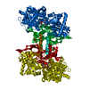
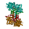
- PDB-2ecp: THE CRYSTAL STRUCTURE OF THE E. COLI MALTODEXTRIN PHOSPHORYLASE C... -
+ Open data

- Basic information
| Entry | Database: PDB / ID: 2ecp | |||||||||
|---|---|---|---|---|---|---|---|---|---|---|
| Title | THE CRYSTAL STRUCTURE OF THE E. COLI MALTODEXTRIN PHOSPHORYLASE COMPLEX | |||||||||
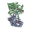
 Components Components | MALTODEXTRIN PHOSPHORYLASE | |||||||||
 Keywords Keywords | ACARBOSE / DIABETES / PHOSPHORYLASE / MALP / GLYCOSYLTRANSFERASE | |||||||||
| Function / homology |  Function and homology information Function and homology informationmaltodextrin phosphorylase activity / alpha-glucan catabolic process / glycogen phosphorylase / glycogen phosphorylase activity / glycogen catabolic process / pyridoxal phosphate binding / protein homodimerization activity / cytoplasm / cytosol Similarity search - Function | |||||||||
| Biological species |  | |||||||||
| Method |  X-RAY DIFFRACTION / SYNCHROTRON / MOLECULAR REPLACEMENT / Resolution: 2.95 Å X-RAY DIFFRACTION / SYNCHROTRON / MOLECULAR REPLACEMENT / Resolution: 2.95 Å | |||||||||
 Authors Authors | O'Reilly, M. / Watson, K.A. / Johnson, L.N. | |||||||||
 Citation Citation | Journal: Biochemistry / Year: 1999 Title: The crystal structure of the Escherichia coli maltodextrin phosphorylase-acarbose complex. Authors: O'Reilly, M. / Watson, K.A. / Johnson, L.N. | |||||||||
| History |
|
- Structure visualization
| Structure viewer | Molecule: MolmilJmol/JSmol |
|---|
- Downloads & links
-Download
| PDBx/mmCIF format | 2ecp.cif.gz | 333.4 KB | Display | PDBx/mmCIF format |
|---|---|---|---|---|
| PDB format | pdb2ecp.ent.gz | 262.4 KB | Display | PDB format |
| PDBx/mmJSON format | 2ecp.json.gz | Tree view | PDBx/mmJSON format | |
| Others |  Other downloads Other downloads |
-Validation report
| Arichive directory | https://data.pdbj.org/pub/pdb/validation_reports/ec/2ecpftp://data.pdbj.org/pub/pdb/validation_reports/ec/2ecp | HTTPS FTP |
|---|
-Related structure data
| Similar structure data |
|---|


-Links
 PDBj
PDBj

- Assembly
| Deposited unit | 
| ||||||||
|---|---|---|---|---|---|---|---|---|---|
| 1 |
| ||||||||
| Unit cell |
| ||||||||
| Noncrystallographic symmetry (NCS) | NCS oper: (Code: given Matrix: (-0.666, 0.745, 0.038), Vector: |
-Components
| #1: Protein | Mass: 90276.836 Da / Num. of mol.: 2 Source method: isolated from a genetically manipulated source Source: (gene. exp.) #2: Polysaccharide | 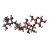  Type: oligosaccharide, Oligosaccharide / Class: Inhibitor / Mass: 645.606 Da / Num. of mol.: 2 Type: oligosaccharide, Oligosaccharide / Class: Inhibitor / Mass: 645.606 Da / Num. of mol.: 2Source method: isolated from a genetically manipulated source Details: oligosaccharide / References: alpha-acarbose #3: Chemical |   Mass: 92.094 Da / Num. of mol.: 2 / Source method: obtained synthetically / Formula: C3H8O3 Mass: 92.094 Da / Num. of mol.: 2 / Source method: obtained synthetically / Formula: C3H8O3#4: Chemical |   Mass: 247.142 Da / Num. of mol.: 2 / Source method: obtained synthetically / Formula: C8H10NO6P Mass: 247.142 Da / Num. of mol.: 2 / Source method: obtained synthetically / Formula: C8H10NO6P#5: Water | ChemComp-HOH / |  Mass: 18.015 Da / Num. of mol.: 93 / Source method: isolated from a natural source / Formula: H2O Mass: 18.015 Da / Num. of mol.: 93 / Source method: isolated from a natural source / Formula: H2OHas protein modification | N | |
|---|
-Experimental details
-Experiment
| Experiment | Method: X-RAY DIFFRACTION / Number of used crystals: 1 |
|---|
- Sample preparation
| Crystal | Density Matthews: 2.4 Å3/Da / Density % sol: 55 % Description: MALP NATIVE STRUCTURE USED AS STARTING MODEL FOR MOLECULAR REPLACEMENT. | ||||||||||||||||||||||||||||||||||||
|---|---|---|---|---|---|---|---|---|---|---|---|---|---|---|---|---|---|---|---|---|---|---|---|---|---|---|---|---|---|---|---|---|---|---|---|---|---|
| Crystal grow | pH: 8.5 / Details: pH 8.5 | ||||||||||||||||||||||||||||||||||||
| Crystal grow | *PLUS Temperature: 18 ℃ / Method: vapor diffusion, hanging drop | ||||||||||||||||||||||||||||||||||||
| Components of the solutions | *PLUS
|
-Data collection
| Diffraction | Mean temperature: 100 K |
|---|---|
| Diffraction source | Source: SYNCHROTRON / Site: ELETTRA  / Beamline: 5.2R / Wavelength: 0.9 / Beamline: 5.2R / Wavelength: 0.9 |
| Detector | Type: MAR scanner 180 mm plate / Detector: IMAGE PLATE / Details: MIRRORS |
| Radiation | Monochromator: DIAMOND C(111) / Monochromatic (M) / Laue (L): M / Scattering type: x-ray |
| Radiation wavelength | Wavelength: 0.9 Å / Relative weight: 1 |
| Reflection | Resolution: 2.95→33.6 Å / Num. obs: 33657 / % possible obs: 88.9 % / Redundancy: 2.6 % / Biso Wilson estimate: 44.7 Å2 / Rmerge(I) obs: 0.068 / Net I/σ(I): 12.5 |
| Reflection shell | Resolution: 2.95→3.11 Å / Redundancy: 2.1 % / Rmerge(I) obs: 0.157 / Mean I/σ(I) obs: 5.3 / % possible all: 78.2 |
| Reflection shell | *PLUS % possible obs: 78.2 % |
- Processing
| Software |
| ||||||||||||||||||||||||||||||||||||||||||||||||||||||||||||||||||||||||||||||||||||
|---|---|---|---|---|---|---|---|---|---|---|---|---|---|---|---|---|---|---|---|---|---|---|---|---|---|---|---|---|---|---|---|---|---|---|---|---|---|---|---|---|---|---|---|---|---|---|---|---|---|---|---|---|---|---|---|---|---|---|---|---|---|---|---|---|---|---|---|---|---|---|---|---|---|---|---|---|---|---|---|---|---|---|---|---|---|
| Refinement | Method to determine structure: MOLECULAR REPLACEMENT / Resolution: 2.95→15 Å / Cross valid method: THROUGHOUT / σ(F): 0 / ESU R Free: 0.57 / Details: TIGHT NCS RESTRAINTS APPLIED
| ||||||||||||||||||||||||||||||||||||||||||||||||||||||||||||||||||||||||||||||||||||
| Displacement parameters | Biso mean: 53.6 Å2 | ||||||||||||||||||||||||||||||||||||||||||||||||||||||||||||||||||||||||||||||||||||
| Refinement step | Cycle: LAST / Resolution: 2.95→15 Å
| ||||||||||||||||||||||||||||||||||||||||||||||||||||||||||||||||||||||||||||||||||||
| Refine LS restraints |
| ||||||||||||||||||||||||||||||||||||||||||||||||||||||||||||||||||||||||||||||||||||
| Software | *PLUS Name: REFMAC / Classification: refinement | ||||||||||||||||||||||||||||||||||||||||||||||||||||||||||||||||||||||||||||||||||||
| Refinement | *PLUS Rfactor obs: 0.241 | ||||||||||||||||||||||||||||||||||||||||||||||||||||||||||||||||||||||||||||||||||||
| Solvent computation | *PLUS | ||||||||||||||||||||||||||||||||||||||||||||||||||||||||||||||||||||||||||||||||||||
| Displacement parameters | *PLUS | ||||||||||||||||||||||||||||||||||||||||||||||||||||||||||||||||||||||||||||||||||||
| Refine LS restraints | *PLUS
|
