[English] 日本語
Yorodumi
- EMDB-9788: Structure of SUR1 subunit bound with repaglinide -

+
Open data


ID or keywords:

Loading...

-
Basic information

Entry
Database: EMDB / ID: EMD-9788
TitleStructure of SUR1 subunit bound with repaglinide
Map data
Sample
  • Complex: SUR1ABCC8
    • Protein or peptide: ATP-binding cassette sub-family C member 8 isoform X2
  • Ligand: Repaglinide
  • Ligand: Digitonin
Function / homology
Function and homology information


glutamate secretion, neurotransmission / inward rectifying potassium channel / sulfonylurea receptor activity / ATPase-coupled monoatomic cation transmembrane transporter activity / inorganic cation transmembrane transport / neuromuscular process / positive regulation of insulin secretion involved in cellular response to glucose stimulus / potassium channel activity / negative regulation of insulin secretion / ABC-type transporter activity ...glutamate secretion, neurotransmission / inward rectifying potassium channel / sulfonylurea receptor activity / ATPase-coupled monoatomic cation transmembrane transporter activity / inorganic cation transmembrane transport / neuromuscular process / positive regulation of insulin secretion involved in cellular response to glucose stimulus / potassium channel activity / negative regulation of insulin secretion / ABC-type transporter activity / ADP binding / presynapse / transmembrane transporter binding / ATP hydrolysis activity / ATP binding
Similarity search - Function
ATP-binding cassette subfamily C member 8 / Sulphonylurea receptor / ABC transporter transmembrane region / ABC transporter type 1, transmembrane domain / ABC transporter integral membrane type-1 fused domain profile. / ABC transporter type 1, transmembrane domain superfamily / ABC transporter-like, conserved site / ABC transporters family signature. / ABC transporter / ABC transporter-like, ATP-binding domain ...ATP-binding cassette subfamily C member 8 / Sulphonylurea receptor / ABC transporter transmembrane region / ABC transporter type 1, transmembrane domain / ABC transporter integral membrane type-1 fused domain profile. / ABC transporter type 1, transmembrane domain superfamily / ABC transporter-like, conserved site / ABC transporters family signature. / ABC transporter / ABC transporter-like, ATP-binding domain / ATP-binding cassette, ABC transporter-type domain profile. / ATPases associated with a variety of cellular activities / AAA+ ATPase domain / P-loop containing nucleoside triphosphate hydrolase
Similarity search - Domain/homology
ATP-binding cassette sub-family C member 8 isoform X2
Similarity search - Component
Biological speciesMus musculus (house mouse) / Mesocricetus auratus (golden hamster)
Methodsingle particle reconstruction / cryo EM / Resolution: 3.53 Å
AuthorsChen L / Ding D / Wang M / Wu J-X / Kang Y
Funding support China, 4 items
OrganizationGrant numberCountry
Ministry of Science and Technology (China)2016YFA0502004 China
National Natural Science Foundation of China31622021 China
National Natural Science Foundation of China31821091 China
National Natural Science Foundation of China31870833 China
Citation
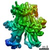
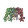
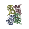
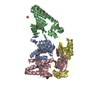
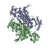
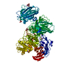
Journal: Cell Rep / Year: 2019
Title: The Structural Basis for the Binding of Repaglinide to the Pancreatic K Channel.
Authors: Dian Ding / Mengmeng Wang / Jing-Xiang Wu / Yunlu Kang / Lei Chen /
Abstract: Repaglinide (RPG) is a short-acting insulin secretagogue widely prescribed for the treatment of type 2 diabetes. It boosts insulin secretion by inhibiting the pancreatic ATP-sensitive potassium ...Repaglinide (RPG) is a short-acting insulin secretagogue widely prescribed for the treatment of type 2 diabetes. It boosts insulin secretion by inhibiting the pancreatic ATP-sensitive potassium channel (K). However, the mechanisms by which RPG binds to the K channel are poorly understood. Here, we describe two cryo-EM structures: the pancreatic K channel in complex with inhibitory RPG and adenosine-5'-(γ-thio)-triphosphate (ATPγS) at 3.3 Å and a medium-resolution structure of a RPG-bound mini SUR1 protein in which the N terminus of the inward-rectifying potassium channel 6.1 (Kir6.1) is fused to the ABC transporter module of the sulfonylurea receptor 1 (SUR1). These structures reveal the binding site of RPG in the SUR1 subunit. Furthermore, the high-resolution structure reveals the complex architecture of the ATP binding site, which is formed by both Kir6.2 and SUR1 subunits, and the domain-domain interaction interfaces.
History
DepositionJan 25, 2019-
Header (metadata) releaseMay 22, 2019-
Map releaseMay 22, 2019-
UpdateSep 29, 2021-
Current statusSep 29, 2021Processing site: PDBj / Status: Released

-
Structure visualization

Movie
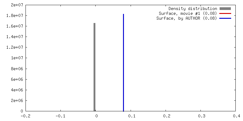
  • Surface view with section colored by density value
  • Surface level: 0.08
  • Imaged by UCSF Chimera
  • Download
  • Surface view colored by cylindrical radius
  • Surface level: 0.08
  • Imaged by UCSF Chimera
  • Download
  • Surface view with fitted model
  • Atomic models: PDB-6jb3
  • Surface level: 0.08
  • Imaged by UCSF Chimera
  • Download
Movie viewer
Structure viewerEM map:
SurfViewMolmilJmol/JSmol
Supplemental images

Downloads & links

-
Map

FileDownload / File: emd_9788.map.gz / Format: CCP4 / Size: 64 MB / Type: IMAGE STORED AS FLOATING POINT NUMBER (4 BYTES)
Projections & slices

Image control

Size
Brightness
Contrast
Others
AxesZ (Sec.)Y (Row.)X (Col.)
1.32 Å/pix.
x 256 pix.
= 338.944 Å
1.32 Å/pix.
x 256 pix.
= 338.944 Å
1.32 Å/pix.
x 256 pix.
= 338.944 Å

Surface

Projections

Slices (1/3)

Slices (1/2)

Slices (2/3)

Images are generated by Spider.

Voxel sizeX=Y=Z: 1.324 Å
Density
Contour LevelBy AUTHOR: 0.08 / Movie #1: 0.08
Minimum - Maximum-0.18529668 - 0.34005585
Average (Standard dev.)0.00027882177 (±0.005020385)
SymmetrySpace group: 1
Details

EMDB XML:

Map geometry
Axis orderXYZ
Origin000
Dimensions256256256
Spacing256256256
CellA=B=C: 338.944 Å
α=β=γ: 90.0 °

CCP4 map header:

modeImage stored as Reals
Å/pix. X/Y/Z1.3241.3241.324
M x/y/z256256256
origin x/y/z0.0000.0000.000
length x/y/z338.944338.944338.944
α/β/γ90.00090.00090.000
start NX/NY/NZ000
NX/NY/NZ513513513
MAP C/R/S123
start NC/NR/NS000
NC/NR/NS256256256
D min/max/mean-0.1850.3400.000

-
Supplemental data

-
Additional map: #1

Fileemd_9788_additional_1.map
Projections & Slices
AxesZYX

Projections

Slices (1/2)
Density Histograms

-
Additional map: sharpened map with a bfactor -100

Fileemd_9788_additional_2.map
Annotationsharpened map with a bfactor -100
Projections & Slices
AxesZYX

Projections

Slices (1/2)
Density Histograms

-
Sample components

-
Entire : SUR1

EntireName: SUR1ABCC8
Components
  • Complex: SUR1ABCC8
    • Protein or peptide: ATP-binding cassette sub-family C member 8 isoform X2
  • Ligand: Repaglinide
  • Ligand: Digitonin

-
Supramolecule #1: SUR1

SupramoleculeName: SUR1 / type: complex / ID: 1 / Parent: 0 / Macromolecule list: #1
Source (natural)Organism: Mus musculus (house mouse)
Recombinant expressionOrganism: Homo sapiens (human)

-
Macromolecule #1: ATP-binding cassette sub-family C member 8 isoform X2

MacromoleculeName: ATP-binding cassette sub-family C member 8 isoform X2 / type: protein_or_peptide / ID: 1 / Number of copies: 1 / Enantiomer: LEVO
Source (natural)Organism: Mesocricetus auratus (golden hamster)
Molecular weightTheoretical: 177.295516 KDa
Recombinant expressionOrganism: Homo sapiens (human)
SequenceString: MPLAFCGTEN HSAAYRVDQG VLNNGCFVDA LNVVPHVFLL FITFPILFIG WGSQSSKVHI HHSTWLHFPG HNLRWILTFI LLFVLVCEI AEGILSDGVT ESRHLHLYMP AGMAFMAAIT SVVYYHNIET SNFPKLLIAL LIYWTLAFIT KTIKFVKFYD H AIGFSQLR ...String:
MPLAFCGTEN HSAAYRVDQG VLNNGCFVDA LNVVPHVFLL FITFPILFIG WGSQSSKVHI HHSTWLHFPG HNLRWILTFI LLFVLVCEI AEGILSDGVT ESRHLHLYMP AGMAFMAAIT SVVYYHNIET SNFPKLLIAL LIYWTLAFIT KTIKFVKFYD H AIGFSQLR FCLTGLLVIL YGMLLLVEVN VIRVRRYIFF KTPREVKPPE DLQDLGVRFL QPFVNLLSKG TYWWMNAFIK TA HKKPIDL RAIGKLPIAM RALTNYQRLC VAFDAQARKD TQSPQGARAI WRALCHAFGR RLILSSTFRI LADLLGFAGP LCI FGIVDH LGKENHVFQP KTQFLGVYFV SSQEFLGNAY VLAVLLFLAL LLQRTFLQAS YYVAIETGIN LRGAIQTKIY NKIM HLSTS NLSMGEMTAG QICNLVAIDT NQLMWFFFLC PNLWAMPVQI IVGVILLYYI LGVSALIGAA VIILLAPVQY FVATK LSQA QRSTLEHSNE RLKQTNEMLR GMKLLKLYAW ESIFCSRVEV TRRKEMTSLR AFAVYTSISI FMNTAIPIAA VLITFV GHV SFFKESDLSP SVAFASLSLF HILVTPLFLL SSVVRSTVKA LVSVQKLSEF LSSAEIREEQ CAPREPAPQG QAGKYQA VP LKVVNRKRPA REEVRDLLGP LQRLAPSMDG DADNFCVQII GGFFTWTPDG IPTLSNITIR IPRGQLTMIV GQVGCGKS S LLLATLGEMQ KVSGAVFWNS NLPDSEGEDP SSPERETAAG SDIRSRGPVA YASQKPWLLN ATVEENITFE SPFNKQRYK MVIEACSLQP DIDILPHGDQ TQIGERGINL SGGQRQRISV ARALYQQTNV VFLDDPFSAL DVHLSDHLMQ AGILELLRDD KRTVVLVTH KLQYLPHADW IIAMKDGTIQ REGTLKDFQR SECQLFEHWK TLMNRQDQEL EKETVMERKA SEPSQGLPRA M SSRDGLLL DEEEEEEEAA ESEEDDNLSS VLHQRAKIPW RACTKYLSSA GILLLSLLVF SQLLKHMVLV AIDYWLAKWT DS ALVLSPA ARNCSLSQEC DLDQSVYAMV FTLLCSLGIV LCLVTSVTVE WTGLKVAKRL HRSLLNRIIL APMRFFETTP LGS ILNRFS SDCNTIDQHI PSTLECLSRS TLLCVSALTV ISYVTPVFLV ALLPLAVVCY FIQKYFRVAS RDLQQLDDTT QLPL LSHFA ETVEGLTTIR AFRYEARFQQ KLLEYTDSNN IASLFLTAAN RWLEVRMEYI GACVVLIAAA TSISNSLHRE LSAGL VGLG LTYALMVSNY LNWMVRNLAD MEIQLGAVKR IHALLKTEAE SYEGLLAPSL IPKNWPDQGK IQIQNLSVRY DSSLKP VLK HVNALISPGQ KIGICGRTGS GKSSFSLAFF RMVDMFEGRI IIDGIDIAKL PLHTLRSRLS IILQDPVLFS GTIRFNL DP EKKCSDSTLW EALEIAQLKL VVKALPGGLD AIITEGGENF SQGQRQLFCL ARAFVRKTSI FIMDEATASI DMATENIL Q KVVMTAFADR TVVTIAHRVH TILSADLVMV LKRGAILEFD KPETLLSQKD SVFASFVRAD K

-
Macromolecule #2: Repaglinide

MacromoleculeName: Repaglinide / type: ligand / ID: 2 / Number of copies: 1 / Formula: BJX
Molecular weightTheoretical: 452.586 Da
Chemical component information

ChemComp-BJX:
Repaglinide / medication*YM / Repaglinide

-
Macromolecule #3: Digitonin

MacromoleculeName: Digitonin / type: ligand / ID: 3 / Number of copies: 1 / Formula: AJP
Molecular weightTheoretical: 1.229312 KDa
Chemical component information

ChemComp-AJP:
Digitonin / detergent*YM / Digitonin

-
Experimental details

-
Structure determination

Methodcryo EM
Processingsingle particle reconstruction
Aggregation stateparticle

-
Sample preparation

BufferpH: 7.5
VitrificationCryogen name: ETHANE

-
Electron microscopy

MicroscopeFEI TITAN KRIOS
Electron beamAcceleration voltage: 300 kV / Electron source: FIELD EMISSION GUN
Electron opticsIllumination mode: FLOOD BEAM / Imaging mode: BRIGHT FIELDBright-field microscopy
Image recordingFilm or detector model: GATAN K2 QUANTUM (4k x 4k) / Average electron dose: 50.0 e/Å2
Experimental equipment
Model: Titan Krios / Image courtesy: FEI Company

-
Image processing

Startup modelType of model: EMDB MAP
EMDB ID:
Initial angle assignmentType: OTHER
Final angle assignmentType: OTHER
Final reconstructionResolution.type: BY AUTHOR / Resolution: 3.53 Å / Resolution method: FSC 0.143 CUT-OFF / Software - Name: RELION (ver. 2.0) / Number images used: 653991
FSC plot (resolution estimation)

+
About Yorodumi

-
News

-
Feb 9, 2022. New format data for meta-information of EMDB entries

New format data for meta-information of EMDB entries

  • Version 3 of the EMDB header file is now the official format.
  • The previous official version 1.9 will be removed from the archive.

Related info.:EMDB header

External links:wwPDB to switch to version 3 of the EMDB data model

-
Aug 12, 2020. Covid-19 info

Covid-19 info

URL: https://pdbj.org/emnavi/covid19.php

New page: Covid-19 featured information page in EM Navigator.

Related info.:Covid-19 info / Mar 5, 2020. Novel coronavirus structure data

+
Mar 5, 2020. Novel coronavirus structure data

Novel coronavirus structure data

Related info.:Yorodumi Speices / Aug 12, 2020. Covid-19 info

External links:COVID-19 featured content - PDBj / Molecule of the Month (242):Coronavirus Proteases

+
Jan 31, 2019. EMDB accession codes are about to change! (news from PDBe EMDB page)

EMDB accession codes are about to change! (news from PDBe EMDB page)

  • The allocation of 4 digits for EMDB accession codes will soon come to an end. Whilst these codes will remain in use, new EMDB accession codes will include an additional digit and will expand incrementally as the available range of codes is exhausted. The current 4-digit format prefixed with “EMD-” (i.e. EMD-XXXX) will advance to a 5-digit format (i.e. EMD-XXXXX), and so on. It is currently estimated that the 4-digit codes will be depleted around Spring 2019, at which point the 5-digit format will come into force.
  • The EM Navigator/Yorodumi systems omit the EMD- prefix.

Related info.:Q: What is EMD? / ID/Accession-code notation in Yorodumi/EM Navigator

External links:EMDB Accession Codes are Changing Soon! / Contact to PDBj

+
Jul 12, 2017. Major update of PDB

Major update of PDB

  • wwPDB released updated PDB data conforming to the new PDBx/mmCIF dictionary.
  • This is a major update changing the version number from 4 to 5, and with Remediation, in which all the entries are updated.
  • In this update, many items about electron microscopy experimental information are reorganized (e.g. em_software).
  • Now, EM Navigator and Yorodumi are based on the updated data.

External links:wwPDB Remediation / Enriched Model Files Conforming to OneDep Data Standards Now Available in the PDB FTP Archive

-
Yorodumi

Thousand views of thousand structures

  • Yorodumi is a browser for structure data from EMDB, PDB, SASBDB, etc.
  • This page is also the successor to EM Navigator detail page, and also detail information page/front-end page for Omokage search.
  • The word "yorodu" (or yorozu) is an old Japanese word meaning "ten thousand". "mi" (miru) is to see.

Related info.:EMDB / PDB / SASBDB / Comparison of 3 databanks / Yorodumi Search / Aug 31, 2016. New EM Navigator & Yorodumi / Yorodumi Papers / Jmol/JSmol / Function and homology information / Changes in new EM Navigator and Yorodumi

Read more