 ムービー
ムービー コントローラー


+ データを開く

- 基本情報
基本情報
| 登録情報 | データベース: EMDB / ID: EMD-9225 | |||||||||
|---|---|---|---|---|---|---|---|---|---|---|
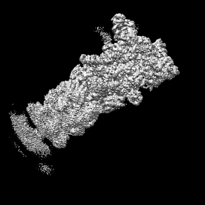
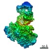
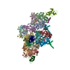
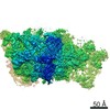
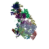
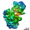
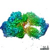
| タイトル | Cryo-EM structures and dynamics of substrate-engaged human 26S proteasome | |||||||||
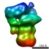
 マップデータ マップデータ | State EB map of substrate-engaged human proteasome low pass-filtered to 3 Angstrom, without amplitude correction | |||||||||
 試料 試料 |
| |||||||||
| 生物種 |  Homo sapiens (ヒト) Homo sapiens (ヒト) | |||||||||
| 手法 | 単粒子再構成法 / クライオ電子顕微鏡法 / 解像度: 3.3 Å | |||||||||
 データ登録者 データ登録者 | Mao YD | |||||||||
 引用 引用 | ジャーナル: Nature / 年: 2019 タイトル: Cryo-EM structures and dynamics of substrate-engaged human 26S proteasome. 著者: Yuanchen Dong / Shuwen Zhang / Zhaolong Wu / Xuemei Li / Wei Li Wang / Yanan Zhu / Svetla Stoilova-McPhie / Ying Lu / Daniel Finley / Youdong Mao /   要旨: The proteasome is an ATP-dependent, 2.5-megadalton molecular machine that is responsible for selective protein degradation in eukaryotic cells. Here we present cryo-electron microscopy structures of ...The proteasome is an ATP-dependent, 2.5-megadalton molecular machine that is responsible for selective protein degradation in eukaryotic cells. Here we present cryo-electron microscopy structures of the substrate-engaged human proteasome in seven conformational states at 2.8-3.6 Å resolution, captured during breakdown of a polyubiquitylated protein. These structures illuminate a spatiotemporal continuum of dynamic substrate-proteasome interactions from ubiquitin recognition to substrate translocation, during which ATP hydrolysis sequentially navigates through all six ATPases. There are three principal modes of coordinated hydrolysis, featuring hydrolytic events in two oppositely positioned ATPases, in two adjacent ATPases and in one ATPase at a time. These hydrolytic modes regulate deubiquitylation, initiation of translocation and processive unfolding of substrates, respectively. Hydrolysis of ATP powers a hinge-like motion in each ATPase that regulates its substrate interaction. Synchronization of ATP binding, ADP release and ATP hydrolysis in three adjacent ATPases drives rigid-body rotations of substrate-bound ATPases that are propagated unidirectionally in the ATPase ring and unfold the substrate. | |||||||||
| 履歴 |
|
- 構造の表示
構造の表示
| ムービー |
 ムービービューア ムービービューア |
|---|---|
| 構造ビューア | EMマップ: SurfViewMolmilJmol/JSmol |
| 添付画像 |
- ダウンロードとリンク
-EMDBアーカイブ
| マップデータ | emd_9225.map.gz | 745.6 MB | EMDBマップデータ形式 | |
|---|---|---|---|---|
| ヘッダ (付随情報) | emd-9225-v30.xmlemd-9225.xml | 15.1 KB 15.1 KB | 表示 表示 | EMDBヘッダ |
| 画像 |  emd_9225.png emd_9225.png | 39.2 KB | ||
| その他 | emd_9225_additional_1.map.gzemd_9225_additional_2.map.gz | 752.5 MB 730.9 MB | ||
| アーカイブディレクトリ |  http://ftp.pdbj.org/pub/emdb/structures/EMD-9225ftp://ftp.pdbj.org/pub/emdb/structures/EMD-9225 http://ftp.pdbj.org/pub/emdb/structures/EMD-9225ftp://ftp.pdbj.org/pub/emdb/structures/EMD-9225 | HTTPS FTP |
-検証レポート
| 文書・要旨 | emd_9225_validation.pdf.gz | 77.9 KB | 表示 | EMDB検証レポート |
|---|---|---|---|---|
| 文書・詳細版 | emd_9225_full_validation.pdf.gz | 77 KB | 表示 | |
| XML形式データ | emd_9225_validation.xml.gz | 492 B | 表示 | |
| アーカイブディレクトリ | https://ftp.pdbj.org/pub/emdb/validation_reports/EMD-9225ftp://ftp.pdbj.org/pub/emdb/validation_reports/EMD-9225 | HTTPS FTP |
-関連構造データ
| 関連構造データ |  9215C  9216C  9217C 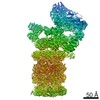 9218C 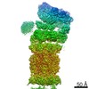 9219C 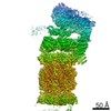 9220C 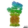 9221C 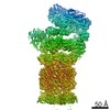 9222C 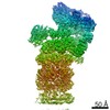 9223C  9224C 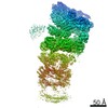 9226C 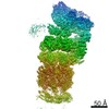 9227C 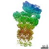 9228C  9229C  6msbC 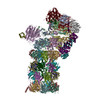 6msdC 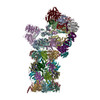 6mseC 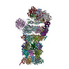 6msgC 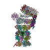 6mshC 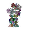 6msjC 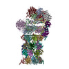 6mskC C: 同じ文献を引用 ( |
|---|---|
| 類似構造データ | |
| 電子顕微鏡画像生データ | EMPIAR-10669 (タイトル: Cryo-EM dataset of the substrate-engaged human 26S proteasome Data size: 13.9 TB Data #1: Drift-corrected frame-averaged super-counting mode micrographs and extracted particles of substrate-engaged human 26S proteasome [micrographs - single frame]) |

-リンク
| EMDBのページ | EMDB (EBI/PDBe) / EMDataResource |
|---|---|
| 「今月の分子」の関連する項目 |

-マップ
| ファイル | ダウンロード / ファイル: emd_9225.map.gz / 形式: CCP4 / 大きさ: 824 MB / タイプ: IMAGE STORED AS FLOATING POINT NUMBER (4 BYTES) | ||||||||||||||||||||||||||||||||||||||||||||||||||||||||||||
|---|---|---|---|---|---|---|---|---|---|---|---|---|---|---|---|---|---|---|---|---|---|---|---|---|---|---|---|---|---|---|---|---|---|---|---|---|---|---|---|---|---|---|---|---|---|---|---|---|---|---|---|---|---|---|---|---|---|---|---|---|---|
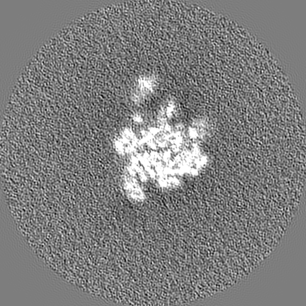
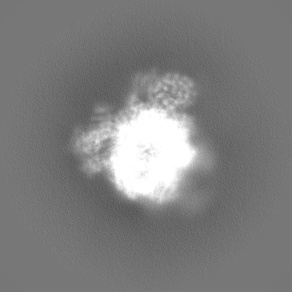
| 注釈 | State EB map of substrate-engaged human proteasome low pass-filtered to 3 Angstrom, without amplitude correction | ||||||||||||||||||||||||||||||||||||||||||||||||||||||||||||
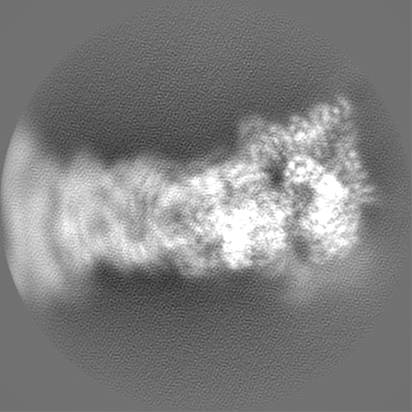
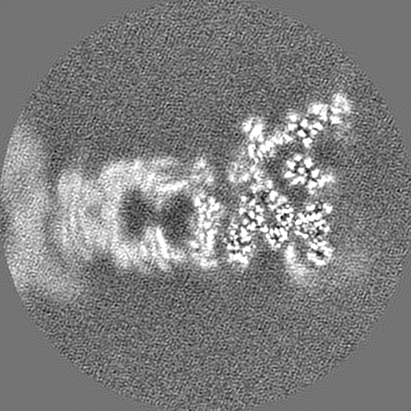
| 投影像・断面図 | 画像のコントロール
画像は Spider により作成 | ||||||||||||||||||||||||||||||||||||||||||||||||||||||||||||
| ボクセルのサイズ | X=Y=Z: 0.685 Å | ||||||||||||||||||||||||||||||||||||||||||||||||||||||||||||
| 密度 |
| ||||||||||||||||||||||||||||||||||||||||||||||||||||||||||||
| 対称性 | 空間群: 1 | ||||||||||||||||||||||||||||||||||||||||||||||||||||||||||||
| 詳細 | EMDB XML:
CCP4マップ ヘッダ情報:
| ||||||||||||||||||||||||||||||||||||||||||||||||||||||||||||
 Z (Sec.)
 Y (Row.)
 X (Col.)


-添付データ
-追加マップ: State EB map of substrate-engaged human proteasome low...
| ファイル | emd_9225_additional_1.map | ||||||||||||
|---|---|---|---|---|---|---|---|---|---|---|---|---|---|
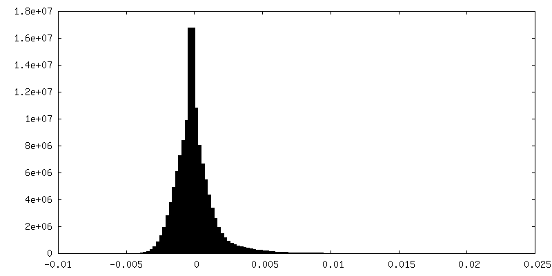
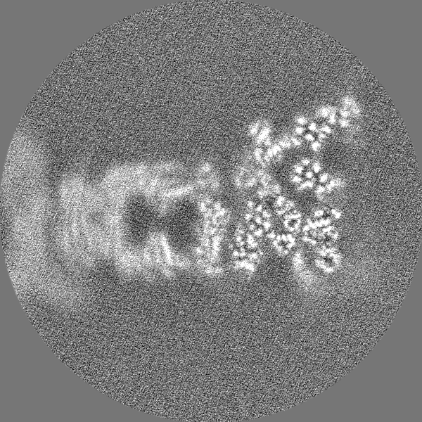
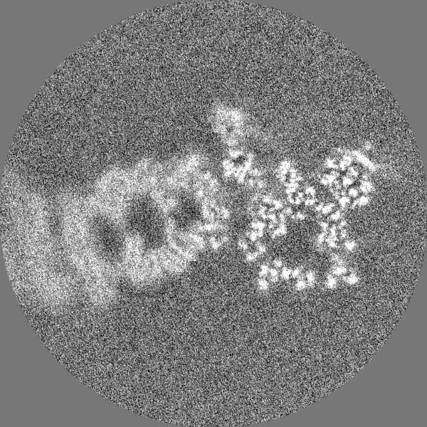
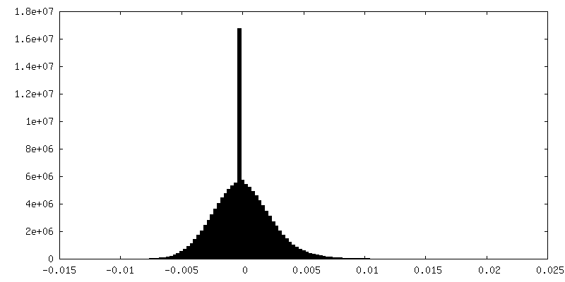
| 注釈 | State EB map of substrate-engaged human proteasome low pass-filtered to 3 Angstrom, with amplitude corrected by a B-factor of -35 | ||||||||||||
| 投影像・断面図 |
| ||||||||||||
| 密度ヒストグラム |


-追加マップ: Unfiltered, uncorrected raw EB map
| ファイル | emd_9225_additional_2.map | ||||||||||||
|---|---|---|---|---|---|---|---|---|---|---|---|---|---|
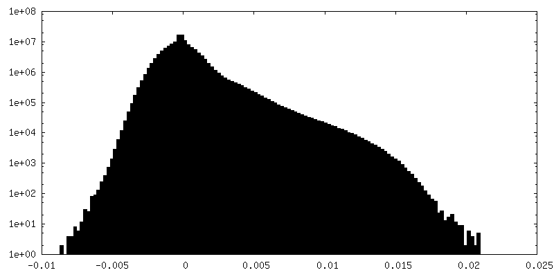
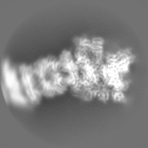
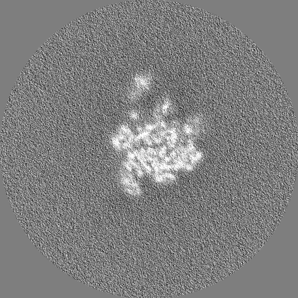
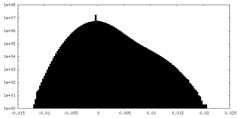
| 注釈 | Unfiltered, uncorrected raw EB map | ||||||||||||
| 投影像・断面図 |
| ||||||||||||
| 密度ヒストグラム |

- 試料の構成要素
-全体 : Proteasome
| 全体 | 名称: Proteasome |
|---|---|
| 要素 |
|
-超分子 #1: Proteasome
| 超分子 | 名称: Proteasome / タイプ: complex / ID: 1 / 親要素: 0 |
|---|---|
| 由来(天然) | 生物種: Homo sapiens (ヒト) |
| 組換発現 | 生物種: Homo sapiens (ヒト) |
-実験情報
-構造解析
| 手法 | クライオ電子顕微鏡法 |
|---|---|
 解析 解析 | 単粒子再構成法 |
| 試料の集合状態 | particle |
-試料調製
| 緩衝液 | pH: 7.5 |
|---|---|
| グリッド | 詳細: unspecified |
| 凍結 | 凍結剤: ETHANE |
- 電子顕微鏡法
| 顕微鏡 | FEI TITAN KRIOS |
|---|---|
| 撮影 | フィルム・検出器のモデル: GATAN K2 SUMMIT (4k x 4k) 平均電子線量: 44.0 e/Å2 |
| 電子線 | 加速電圧: 300 kV / 電子線源:  FIELD EMISSION GUN FIELD EMISSION GUN |
| 電子光学系 | 照射モード: FLOOD BEAM / 撮影モード: BRIGHT FIELD |
| 実験機器 |  モデル: Titan Krios / 画像提供: FEI Company |
-画像解析
| 最終 再構成 | 想定した対称性 - 点群: C1 (非対称) / 解像度のタイプ: BY AUTHOR / 解像度: 3.3 Å / 解像度の算出法: FSC 0.143 CUT-OFF / 使用した粒子像数: 242965 |
|---|---|
| 初期 角度割当 | タイプ: COMMON LINE |
| 最終 角度割当 | タイプ: PROJECTION MATCHING |
